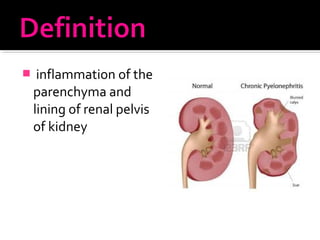
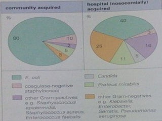
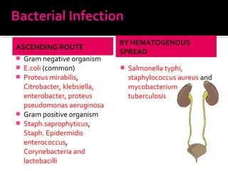
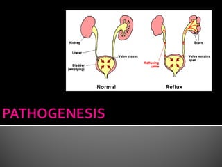
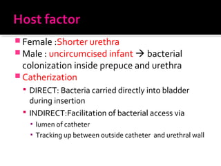
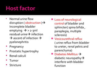
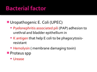
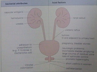
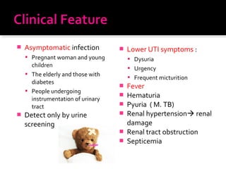
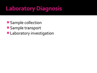
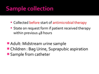
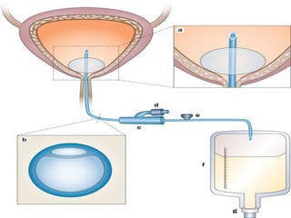
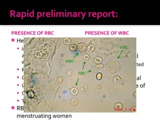
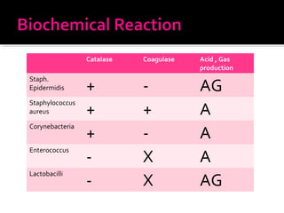
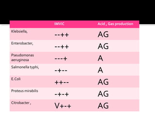
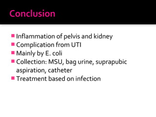
Nur Amalina Aminuddin Baki
60
 Definition
 Etiology
 Pathogenesis
 Clinical feature
 Lab diagnosis
 Treatment
 inflammation of the
parenchyma and
lining of renal pelvis
of kidney
 Epidemiological viewpoint
 Community acquired ( common)
 hospital acquired (catherization)(40%)
ASCENDING ROUTE
 Gram negative organism
 E.coli (common)
 Proteus mirabilis,
Citrobacter, klebsiella,
enterobacter, proteus
pseudomonas aeruginosa
 Gram positive organism
 Staph.saprophyticus,
Staph. Epidermidis
enterococcus,
Corynebacteria and
lactobacilli
BY HEMATOGENOUS
SPREAD
 Salmonella typhi,
staphylococcus aureus and
mycobacterium
tuberculosis
VIRUS
 Rare
 Virus Human
polymaviruses , JC and BK
 Cytomegalovirus and
rubella
 Korean hemorrhagic fever
virus
 Mumps and HIV
 Recovered in urine in
absence of UTI
PARASITE
 Fungi : candida spp and
histoplasma capsulatum
 Protozoa : trichomonas
vaginalis
 Helminth: schistosoma
haematobium
 Female :Shorter urethra
 Male : uncircumcised infant  bacterial
colonization inside prepuce and urethra
 Catherization
 DIRECT: Bacteria carried directly into bladder
during insertion
 INDIRECT:Facilitation of bacterial access via
▪ lumen of catheter
▪ Tracking up between outside catheter and urethral wall
• Normal urine flow
disruption ( obstruction )
Incomplete bladder
emptying  > 2-3ml
residual urine infection
 ascent of infection 
pyelonephritis
 Pregnancy
 Prostatic hypertrophy
 Renal calculi
 Tumor
 Stricture
 Loss of neurological
control of bladder and
sphincter( spina bifida ,
paraplegia, multiple
sclerosis)
 Vesicourethral reflux
( urine reflux from bladder
to ureter, renal pelvis and
parenchyma)
 Diabetes Mellitus 
diabetic neuropathy
interfere with bladder
function
 Uropathogenic E. Coli (UPEC)
 Pyelonephritis associated pili (PAP) adhesion to
urethral and bladder epithellium in
 K antigen that help E coli to be phagocytosis-
resistant
 Hemolysin ( membrane damaging toxin)
 Proteus spp
 Urease
 Asymptomatic infection
 Pregnant woman and young
children
 The elderly and those with
diabetes
 People undergoing
instrumentation of urinary
tract
 Detect only by urine
screening
 Lower UTI symptoms :
 Dysuria
 Urgency
 Frequent micturition
 Fever
 Hematuria
 Pyuria ( M. TB)
 Renal hypertension renal
damage
 Renal tract obstruction
 Septicemia
 Sample collection
 Sample transport
 Laboratory investigation
 Collected before start of antimicrobial therapy
 State on request form if patient received therapy
within previous 48 hours
 Adult: Midstream urine sample
 Children : Bag Urine, Suprapubic aspiration
 Sample from catheter
 MidStream Urine
 Clean labia/ glans with non-antiseptic soap and
water
 Void first part of urine stream
 Collect midstream sample into sterile wide
mouthed container
 difficult in babies and young children
 Bacteriuria considered SIGNIFICANT when
 Properly collected
 > 105 organism /ml
 Only a single bacterial species
 Not apply to urine specimen not contaminated by
periurethral flora
 Contaminated urine of MSU
 < 104 organism /ml
 Contain > 1 bacterial species
 BAG URINE :
 Stick plastic bag to perineum/ penis
 Frequent heavy contamination
 Suprapubic aspiration
 Collected directly from bladder by needle
insertion
 No contamination
 Significant bacteruria not applied
 M. Tuberculosis
 3 early morning urine sample
 Do not require precaution as MSU
 Culture technique inhibit other organism’s growth
 S. haematobium
 Last few ml of late morning sample after exercise
 Only in patient undergoing catherization
 Withdraw using a syringe and needle from
catheter tube
 Do not take urine from catheter drainage bag
( bacterial multiplication)
 Significant bacteruria not applied
 Immediate with minimum delay
 Multiplication of organism in specimen alters
result
 Macroscopic :
 appearance
 colour
 turbidity
 Microscopic : enable Rapid preliminary report
 Presence of
▪ RBC
▪ WBC
▪ pus
PRESENCE OF RBC
 Hematuria:
 Infection of urinary tract
and elsewhere ( bacterial
endocarditis)
 Renal trauma
 Calculi
 Urinary tract carcinoma
 Clotting disorder
 Thrombocytopenia
 RBC contamination of
menstruating women
PRESENCE OF WBC
 Normal : <10/ml
 Abnormal : > 10/ml
 Not always associated
with bacteruria
 Confused with renal
tubular cell in urine of
aspirin-misuser
 An important finding and may reflect :
 Concurrent antibiotic therapy
 Other diseases( neoplasm, urinary calculi)
 Infection with organism not detected by routine
urine culture method
 Based on factor:
 Collection- carried out properly
 Storage
▪ cultured within one hour of collection
▪ Stored at 4 Celsius not > 18 hour before culture
 Antibiotic treatment – smaller number of
organism may be significant
 Fluid intake - influence quantitative result
 Specimen – for MSU specimen
 Colony Morphology on Mc Conkey agar:
 LF : E.coli, Klebsiella, Enterobacter
 NLF: Proteus mirabilis, Pseudomonas, Staph.
Aureus, S. Typhi
 Colony morphology on Blood agar:
 Gamma: E.coli, proteus mirabilis
 Beta: Staph. Aureus, Pseudomonas
 Gram Stain
 Positive:
▪ Staphylococcus saprophyticus ,Staph. Epidermidis and
enterococcus, Corynebacteria and lactobacilli
 Negative:
▪ E.coli,Proteus mirabilis, Citrobacter ,Klebsiella,
enterobacter, proteus and pseudomonas aeruginosa
Catalase Coagulase Acid , Gas
production
Staph.
Epidermidis + - AG
Staphylococcus
aureus + + A
Corynebacteria
+ - A
Enterococcus
- X A
Lactobacilli
- X AG
IMViC Acid , Gas production
Klebsiella,
--++ AG
Enterobacter,
--++ AG
Pseudomonas
aeruginosa ---+ A
Salmonella typhi,
-+-- A
E.Coli
++-- AG
Proteus mirabilis
-+-+ AG
Citrobacter ,
V+-+ AG
 Specific antibacterial therapy
 Drink large volume of fluid
 Continued systemic treatment till sign and
symptom subsides
 Usually 10 days
 > 10 days to sterilize kidney
 Regularly emptying bladder in healthy
women
 Prevention of hospital-acquired infection in
catheterized patient by :
 Avoid catherization whenever possible
 Minimize duration of catheterization
 Insert catheter with good aseptic technique
 Use closed sterile drainage system
 Maintain a gravity drain
 Use topical antiseptic around meatus in women
 Wash hand before and after inserting catheter,
collecting specimen and emptying drainage bags
 Inflammation of pelvis and kidney
 Complication from UTI
 Mainly by E. coli
 Collection: MSU, bag urine, suprapubic
aspiration, catheter
 Treatment based on infection
 Textbook of Microbiology, 4th
Edition, Dir. Prof. C
P Baveja, 2013, Arya Publications
 Mims’ Medical Microbiology, 4th
edition, Mims,
2008 , MOSBY Elsevier
Mellss microb renal pyelonephritis

Mellss microb renal pyelonephritis

  • 1.
  • 2.
     Definition  Etiology Pathogenesis  Clinical feature  Lab diagnosis  Treatment
  • 3.
     inflammation ofthe parenchyma and lining of renal pelvis of kidney
  • 4.
     Epidemiological viewpoint Community acquired ( common)  hospital acquired (catherization)(40%)
  • 6.
    ASCENDING ROUTE  Gramnegative organism  E.coli (common)  Proteus mirabilis, Citrobacter, klebsiella, enterobacter, proteus pseudomonas aeruginosa  Gram positive organism  Staph.saprophyticus, Staph. Epidermidis enterococcus, Corynebacteria and lactobacilli BY HEMATOGENOUS SPREAD  Salmonella typhi, staphylococcus aureus and mycobacterium tuberculosis
  • 7.
    VIRUS  Rare  VirusHuman polymaviruses , JC and BK  Cytomegalovirus and rubella  Korean hemorrhagic fever virus  Mumps and HIV  Recovered in urine in absence of UTI PARASITE  Fungi : candida spp and histoplasma capsulatum  Protozoa : trichomonas vaginalis  Helminth: schistosoma haematobium
  • 9.
     Female :Shorterurethra  Male : uncircumcised infant  bacterial colonization inside prepuce and urethra  Catherization  DIRECT: Bacteria carried directly into bladder during insertion  INDIRECT:Facilitation of bacterial access via ▪ lumen of catheter ▪ Tracking up between outside catheter and urethral wall
  • 11.
    • Normal urineflow disruption ( obstruction ) Incomplete bladder emptying  > 2-3ml residual urine infection  ascent of infection  pyelonephritis  Pregnancy  Prostatic hypertrophy  Renal calculi  Tumor  Stricture  Loss of neurological control of bladder and sphincter( spina bifida , paraplegia, multiple sclerosis)  Vesicourethral reflux ( urine reflux from bladder to ureter, renal pelvis and parenchyma)  Diabetes Mellitus  diabetic neuropathy interfere with bladder function
  • 12.
     Uropathogenic E.Coli (UPEC)  Pyelonephritis associated pili (PAP) adhesion to urethral and bladder epithellium in  K antigen that help E coli to be phagocytosis- resistant  Hemolysin ( membrane damaging toxin)  Proteus spp  Urease
  • 14.
     Asymptomatic infection Pregnant woman and young children  The elderly and those with diabetes  People undergoing instrumentation of urinary tract  Detect only by urine screening  Lower UTI symptoms :  Dysuria  Urgency  Frequent micturition  Fever  Hematuria  Pyuria ( M. TB)  Renal hypertension renal damage  Renal tract obstruction  Septicemia
  • 15.
     Sample collection Sample transport  Laboratory investigation
  • 16.
     Collected beforestart of antimicrobial therapy  State on request form if patient received therapy within previous 48 hours  Adult: Midstream urine sample  Children : Bag Urine, Suprapubic aspiration  Sample from catheter
  • 17.
     MidStream Urine Clean labia/ glans with non-antiseptic soap and water  Void first part of urine stream  Collect midstream sample into sterile wide mouthed container  difficult in babies and young children
  • 18.
     Bacteriuria consideredSIGNIFICANT when  Properly collected  > 105 organism /ml  Only a single bacterial species  Not apply to urine specimen not contaminated by periurethral flora  Contaminated urine of MSU  < 104 organism /ml  Contain > 1 bacterial species
  • 19.
     BAG URINE:  Stick plastic bag to perineum/ penis  Frequent heavy contamination  Suprapubic aspiration  Collected directly from bladder by needle insertion  No contamination  Significant bacteruria not applied
  • 21.
     M. Tuberculosis 3 early morning urine sample  Do not require precaution as MSU  Culture technique inhibit other organism’s growth  S. haematobium  Last few ml of late morning sample after exercise
  • 22.
     Only inpatient undergoing catherization  Withdraw using a syringe and needle from catheter tube  Do not take urine from catheter drainage bag ( bacterial multiplication)  Significant bacteruria not applied
  • 24.
     Immediate withminimum delay  Multiplication of organism in specimen alters result
  • 25.
     Macroscopic : appearance  colour  turbidity  Microscopic : enable Rapid preliminary report  Presence of ▪ RBC ▪ WBC ▪ pus
  • 26.
    PRESENCE OF RBC Hematuria:  Infection of urinary tract and elsewhere ( bacterial endocarditis)  Renal trauma  Calculi  Urinary tract carcinoma  Clotting disorder  Thrombocytopenia  RBC contamination of menstruating women PRESENCE OF WBC  Normal : <10/ml  Abnormal : > 10/ml  Not always associated with bacteruria  Confused with renal tubular cell in urine of aspirin-misuser
  • 27.
     An importantfinding and may reflect :  Concurrent antibiotic therapy  Other diseases( neoplasm, urinary calculi)  Infection with organism not detected by routine urine culture method
  • 28.
     Based onfactor:  Collection- carried out properly  Storage ▪ cultured within one hour of collection ▪ Stored at 4 Celsius not > 18 hour before culture  Antibiotic treatment – smaller number of organism may be significant  Fluid intake - influence quantitative result  Specimen – for MSU specimen
  • 29.
     Colony Morphologyon Mc Conkey agar:  LF : E.coli, Klebsiella, Enterobacter  NLF: Proteus mirabilis, Pseudomonas, Staph. Aureus, S. Typhi  Colony morphology on Blood agar:  Gamma: E.coli, proteus mirabilis  Beta: Staph. Aureus, Pseudomonas
  • 30.
     Gram Stain Positive: ▪ Staphylococcus saprophyticus ,Staph. Epidermidis and enterococcus, Corynebacteria and lactobacilli  Negative: ▪ E.coli,Proteus mirabilis, Citrobacter ,Klebsiella, enterobacter, proteus and pseudomonas aeruginosa
  • 31.
    Catalase Coagulase Acid, Gas production Staph. Epidermidis + - AG Staphylococcus aureus + + A Corynebacteria + - A Enterococcus - X A Lactobacilli - X AG
  • 32.
    IMViC Acid ,Gas production Klebsiella, --++ AG Enterobacter, --++ AG Pseudomonas aeruginosa ---+ A Salmonella typhi, -+-- A E.Coli ++-- AG Proteus mirabilis -+-+ AG Citrobacter , V+-+ AG
  • 33.
     Specific antibacterialtherapy  Drink large volume of fluid  Continued systemic treatment till sign and symptom subsides  Usually 10 days  > 10 days to sterilize kidney
  • 34.
     Regularly emptyingbladder in healthy women  Prevention of hospital-acquired infection in catheterized patient by :  Avoid catherization whenever possible  Minimize duration of catheterization  Insert catheter with good aseptic technique  Use closed sterile drainage system  Maintain a gravity drain  Use topical antiseptic around meatus in women  Wash hand before and after inserting catheter, collecting specimen and emptying drainage bags
  • 35.
     Inflammation ofpelvis and kidney  Complication from UTI  Mainly by E. coli  Collection: MSU, bag urine, suprapubic aspiration, catheter  Treatment based on infection
  • 36.
     Textbook ofMicrobiology, 4th Edition, Dir. Prof. C P Baveja, 2013, Arya Publications  Mims’ Medical Microbiology, 4th edition, Mims, 2008 , MOSBY Elsevier