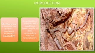
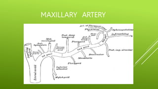
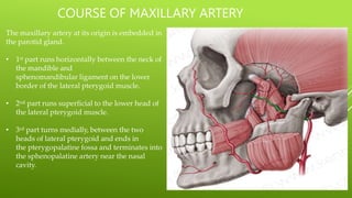
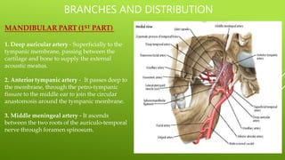
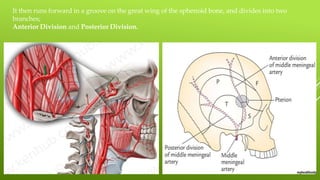
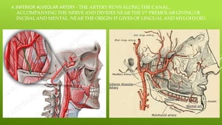
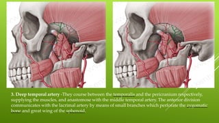
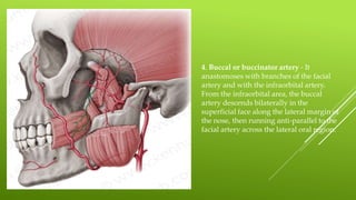
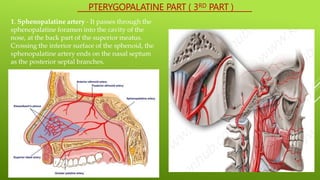
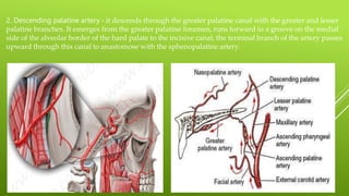
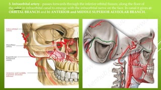
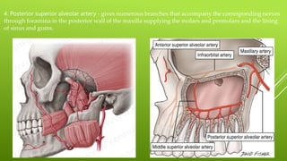
MAXILLARY ARTERY
Dr.Pragya, Dr.Karthik Raja,Dr.Muskan
CONTENT
OVERVIEW ON
CAROTID
ARTERIES
INTRODUCTION COURSE
BRANCHES
AND
DISTRIBUTIONS
OVERVIEW ON CAROTID ARTERIES
 The common carotid and internal carotid are
slightly dilated in an area known as
the carotid sinus, and is a baroreceptor that
reacts to changes in arterial blood pressure.
 The artery ends within the parotid gland by
dividing into the superficial temporal artery
and the maxillary artery.
Maxillary artery
is one of the two
terminal
branches of
the external
carotid artery.
It supplies blood
to maxilla and
mandibular
bones, deep facial
areas,
cerebral dura
mater and
the nasal cavity.
INTRODUCTION
MAXILLARY ARTERY
Main trunk divides into three parts:
Mandibular part (1st part) – It winds around deep to the neck of the mandible.
Pterygoid part (2nd part) – It travels between the two heads of the lateral pterygoid muscle.
Pterygopalatine part (3rd part) – Enters into the pterygopalatine fossa.
COURSE OF MAXILLARY ARTERY
The maxillary artery at its origin is embedded in
the parotid gland.
• 1st part runs horizontally between the neck of
the mandible and
sphenomandibular ligament on the lower
border of the lateral pterygoid muscle.
• 2nd part runs superficial to the lower head of
the lateral pterygoid muscle.
• 3rd part turns medially, between the two
heads of lateral pterygoid and ends in
the pterygopalatine fossa and terminates into
the sphenopalatine artery near the nasal
cavity.
MANDIBULAR PART (1ST PART)
1. Deep auricular artery - Superficially to the
tympanic membrane, passing between the
cartilage and bone to supply the external
acoustic meatus.
2. Anterior tympanic artery - It passes deep to
the membrane, through the petro-tympanic
fissure to the middle ear to join the circular
anastomosis around the tympanic membrane.
3. Middle meningeal artery - It ascends
between the two roots of the auriculo-temporal
nerve through foramen spinosum.
BRANCHES AND DISTRIBUTION
It then runs forward in a groove on the great wing of the sphenoid bone, and divides into two
branches;
Anterior Division and Posterior Division.
4. INFERIOR ALVEOLAR ARTERY - THE ARTERY RUNS ALONG THE CANAL ,
ACCOMPANYING THE NERVE AND DIVIDES NEAR THE 1ST PREMOLAR GIVING OF
INCISALAND MENTAL. NEAR THE ORIGIN IT GIVES OF LINGUAL AND MYLOHYOID.
5. Accessory meningeal artery - It
passes upwards through the
foramen ovale to supply the dura
mater of the floor of the middle
fossa and of the trigeminal cave
(Meckel’s cave).
1. Masseteric artery - accompanies the
lingual nerve. It is small, and passes
laterally through the mandibular
notch to the deep surface of
the masseter muscle, which it
supplies.
PTERYGOID PART ( 2ND PART )
2. Pterygoid artery - It supplies
the lateral pterygoid
muscle and medial pterygoid muscle.
3. Deep temporal artery -They course between the temporalis and the pericranium respectively,
supplying the muscles, and anastomose with the middle temporal artery. The anterior division
communicates with the lacrimal artery by means of small branches which perforate the zygomatic
bone and great wing of the sphenoid.
4. Buccal or buccinator artery - It
anastomoses with branches of the facial
artery and with the infraorbital artery.
From the infraorbital area, the buccal
artery descends bilaterally in the
superficial face along the lateral margin of
the nose, then running anti-parallel to the
facial artery across the lateral oral region.
PTERYGOPALATINE PART ( 3RD PART )
1. Sphenopalatine artery - It passes through the
sphenopalatine foramen into the cavity of the
nose, at the back part of the superior meatus.
Crossing the inferior surface of the sphenoid, the
sphenopalatine artery ends on the nasal septum
as the posterior septal branches.
2. Descending palatine artery - it descends through the greater palatine canal with the greater and lesser
palatine branches. It emerges from the greater palatine foramen, runs forward in a groove on the medial
side of the alveolar border of the hard palate to the incisive canal; the terminal branch of the artery passes
upward through this canal to anastomose with the sphenopalatine artery.
3. Infraorbital artery - passes forwards through the inferior orbital fissure, along the floor of
the orbit in infraorbital canal to emerge with the infraorbital nerve on the face. In canal it gives a)
ORBITAL BRANCH and b) ANTERIOR and MIDDLE SUPERIOR ALVEOLAR BRANCH.
4. Posterior superior alveolar artery - gives numerous branches that accompany the corresponding nerves
through foramina in the posterior wall of the maxilla supplying the molars and premolars and the lining
of sinus and gums.
5. Pharyngeal artery - It runs
backward through the pharyngeal
canal with the pharyngeal nerve,
and supplies structures such as
the pharynx, the posterior aspect of
the roof of the nasal cavity,
sphenoid sinus, and Eustachian
tube.
6. Artery of the pterygoid canal - It
passes backwards along the pterygoid
canal and supplies the upper part of
the pharynx, and auditory tube and sends
a small division into the tympanic
cavity to anastomose with the tympanic
arteries.
THANK YOU

maxillary artery presentation for anatomy

  • 1.
  • 2.
  • 3.
  • 4.
     The commoncarotid and internal carotid are slightly dilated in an area known as the carotid sinus, and is a baroreceptor that reacts to changes in arterial blood pressure.  The artery ends within the parotid gland by dividing into the superficial temporal artery and the maxillary artery.
  • 5.
    Maxillary artery is oneof the two terminal branches of the external carotid artery. It supplies blood to maxilla and mandibular bones, deep facial areas, cerebral dura mater and the nasal cavity. INTRODUCTION
  • 6.
  • 7.
    Main trunk dividesinto three parts: Mandibular part (1st part) – It winds around deep to the neck of the mandible. Pterygoid part (2nd part) – It travels between the two heads of the lateral pterygoid muscle. Pterygopalatine part (3rd part) – Enters into the pterygopalatine fossa.
  • 8.
    COURSE OF MAXILLARYARTERY The maxillary artery at its origin is embedded in the parotid gland. • 1st part runs horizontally between the neck of the mandible and sphenomandibular ligament on the lower border of the lateral pterygoid muscle. • 2nd part runs superficial to the lower head of the lateral pterygoid muscle. • 3rd part turns medially, between the two heads of lateral pterygoid and ends in the pterygopalatine fossa and terminates into the sphenopalatine artery near the nasal cavity.
  • 9.
    MANDIBULAR PART (1STPART) 1. Deep auricular artery - Superficially to the tympanic membrane, passing between the cartilage and bone to supply the external acoustic meatus. 2. Anterior tympanic artery - It passes deep to the membrane, through the petro-tympanic fissure to the middle ear to join the circular anastomosis around the tympanic membrane. 3. Middle meningeal artery - It ascends between the two roots of the auriculo-temporal nerve through foramen spinosum. BRANCHES AND DISTRIBUTION
  • 10.
    It then runsforward in a groove on the great wing of the sphenoid bone, and divides into two branches; Anterior Division and Posterior Division.
  • 11.
    4. INFERIOR ALVEOLARARTERY - THE ARTERY RUNS ALONG THE CANAL , ACCOMPANYING THE NERVE AND DIVIDES NEAR THE 1ST PREMOLAR GIVING OF INCISALAND MENTAL. NEAR THE ORIGIN IT GIVES OF LINGUAL AND MYLOHYOID.
  • 12.
    5. Accessory meningealartery - It passes upwards through the foramen ovale to supply the dura mater of the floor of the middle fossa and of the trigeminal cave (Meckel’s cave).
  • 13.
    1. Masseteric artery- accompanies the lingual nerve. It is small, and passes laterally through the mandibular notch to the deep surface of the masseter muscle, which it supplies. PTERYGOID PART ( 2ND PART )
  • 14.
    2. Pterygoid artery- It supplies the lateral pterygoid muscle and medial pterygoid muscle.
  • 15.
    3. Deep temporalartery -They course between the temporalis and the pericranium respectively, supplying the muscles, and anastomose with the middle temporal artery. The anterior division communicates with the lacrimal artery by means of small branches which perforate the zygomatic bone and great wing of the sphenoid.
  • 16.
    4. Buccal orbuccinator artery - It anastomoses with branches of the facial artery and with the infraorbital artery. From the infraorbital area, the buccal artery descends bilaterally in the superficial face along the lateral margin of the nose, then running anti-parallel to the facial artery across the lateral oral region.
  • 17.
    PTERYGOPALATINE PART (3RD PART ) 1. Sphenopalatine artery - It passes through the sphenopalatine foramen into the cavity of the nose, at the back part of the superior meatus. Crossing the inferior surface of the sphenoid, the sphenopalatine artery ends on the nasal septum as the posterior septal branches.
  • 18.
    2. Descending palatineartery - it descends through the greater palatine canal with the greater and lesser palatine branches. It emerges from the greater palatine foramen, runs forward in a groove on the medial side of the alveolar border of the hard palate to the incisive canal; the terminal branch of the artery passes upward through this canal to anastomose with the sphenopalatine artery.
  • 19.
    3. Infraorbital artery- passes forwards through the inferior orbital fissure, along the floor of the orbit in infraorbital canal to emerge with the infraorbital nerve on the face. In canal it gives a) ORBITAL BRANCH and b) ANTERIOR and MIDDLE SUPERIOR ALVEOLAR BRANCH.
  • 20.
    4. Posterior superioralveolar artery - gives numerous branches that accompany the corresponding nerves through foramina in the posterior wall of the maxilla supplying the molars and premolars and the lining of sinus and gums.
  • 21.
    5. Pharyngeal artery- It runs backward through the pharyngeal canal with the pharyngeal nerve, and supplies structures such as the pharynx, the posterior aspect of the roof of the nasal cavity, sphenoid sinus, and Eustachian tube.
  • 22.
    6. Artery ofthe pterygoid canal - It passes backwards along the pterygoid canal and supplies the upper part of the pharynx, and auditory tube and sends a small division into the tympanic cavity to anastomose with the tympanic arteries.
  • 23.