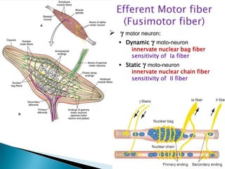
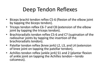
Presented by
   Dr Sadatinejad, Seyyed Mohsen,student of Medicine
   from Iran,Kashan 19/12/2012
   Middle ear muscles : without any MS
              Muscle                             Number of MS
              Latissimus dorsi                       400
              soleus                                 400
              abductor pollicis brevis               80
              quadriceps                            1300
              medial gastrocnemius                   150
              extensor digitorum longus (EDL)        190
              lumbrical

        Muscle                            Density of MS (per gram)
        lumbrical                                   12
        soleus                                      0.4
        EDL                                         3.7

   Motor units
   kind of extrafusal muscle fibers : “red” or slow fiber (Type I)
Afferent nerve fiber
     (Proprioception)
   Ia: primery ending (annulospiral)
          end to nuclear bag fibers
          or Dynamic fiber
   II : secendary ending (flower-spray)
          end to nuclear chain fibers
          or Static fiber
Fig. 8.6. Responses of afferent fibers from a muscle spindle. A,
Length of muscle containing muscle spindle. Muscle length is
changed with various waveforms of stretch. B, Response of type
Ia afferent fibers from a primary ending of the muscle spindle. C,
Response of type II afferent fibers from a secondary ending of the
muscle spindle. Type Ia afferents respond to rapid stretch; type II
afferents respond to length.
Efferent Motor fiber
   (Fusimotor fiber)
 γ motor neuron:
   Dynamic γ moto-neuron
       innervate nuclear bag fiber
       sensitivity of Ia fiber
    Static γ moto-neuron
       innervate nuclear chain fiber
       sensitivity of II fiber
(Negative )
Lenght Feedback
Strech Reflex
(Myotatic or Tendon Reflex)
Deep Tendon Reflexes
• Biceps brachii tendon reflex C5-6 (flexion of the elbow joint
  by tapping the biceps tendon).
• Triceps tendon reflex C6-7 and C8 (extension of the elbow
  joint by tapping the triceps tendon).
• Brachioradialis tendon reflex C5-6 and C7 (supination of the
  radioulnar joints by tapping the insertion of the
  brachioradialis tendon).
• Patellar tendon reflex (knee jerk) L2, L3, and L4 (extension
  of knee joint on tapping the patellar tendon).
• Achilles tendon reflex (ankle jerk) S1 and 2 (plantar flexion
  of ankle joint on tapping the Achilles tendon—tendo
  calcaneus).
Muscle strength reflexes
Monosynaptic arch and Strech Reflex
     (Ascending & Descending branch)
Unconscious Propericeotion
         Cerebellum
conscious Propericeotion
         Cerebral Cortec and Basal Ganglia
Conscious              Propericeotion
Afferent Fiber
                                Post horn of   1
from Muscle          DRG
Spindle          1          1   gray matter


                                           Medulla
                                           oblongata
Conscious              Propericeotion
Afferent Fiber
                                Post horn of   1
from Muscle          DRG
Spindle          1          1   gray matter


                                                   Medulla
                                                   oblongata
Unconscious                Propericeotion
Afferent Fiber
                                 Post horn of   1
from Muscle          DRG
Spindle          1         1     gray matter


                                 Medulla
                                 oblongata
Sup peduncle


 Cerebral Corex
  Basal Ganglia    RED Nucleus
Subsantial Nigra


                   RubroSpinal
                      Tract


                   Ant Horn Of
                   spinal cord
                   γ motor neuron
Sup peduncle

     Dentothalamic tract

                                   Controlateral
                                  Ventrolteral (VL)
                                nucleus of Thalamus
         Internal Capsule
         (corona radiata)
                                       Cerebral Cortex
                                    (Primery Motor Area)


                                    CorticoSpinal Tract
          Ant.CorticoSpinal          (Pyramidal Fiber)
                              20%
                                                  80%

Dicussation in spinal Segment                 Lat.
                                             Cortico
              Dicussation Of Pyramids        Spinal
Inferior C e r e b e l l a r peduncle


  Reticular        Lat.Vestibular     Med.Vestibular
 Formation            nucleus           nucleus


Reticulospinal   VestibuloSpinal       MLF
     Tract            Tract      (med.longitudinal
                                    Fasiculus)


                                    ↑Cranial III,IV,VI
                                    ↓Cranial XI to SCM
                                      and Trapezious
CorticoTectal tract
 SpinoTectal Tract
 Substantial nigra
Med & Lat Geniculate            RED Nucleus
                               Spinolivary Tract


   TectoSpinal          Dicussation    OlivoSpinal Tract
      Tract            in brain Stem     (Dicussation
                                        in brain Stem)

   Ant. Horn of
   Spinal Cord
Common Terminal Moto r Pat hway
(A) The common terminal pathway of all centers involve d in moto r activity is the large
anterior horn cell ( A1) and its a xon (!-motoneuron. The central regions that influence
motor activity via descending pathways a re interconnecte d in many ways. The most
important af fe rent pathways ste m from the cereb ellum, which receives the impulses
of muscle receptors via the spinocereb ellar tracts (A2) and the stimuli of the cortex via
the corticopontine tracts (A3). The cereb ellar impulses are transmitte d via the
parvocellular part of the dentate nucleus ( A4) and the ventral lateral nucleus of thalamus
( A5) to the precentral corte x (area 4) (A6). The corticospinal (pyramidal) tract (A7 )
descends from area 4 to the anterior horn and gives of f collaterals in the pons ( A8) that
return to the cereb ellum. Additional cereb ellar impulses are transmitte d via the
emboliform nucleus ( A9 ) and the centromedian nucleus of the thalamus (A10) to the
striatum ( A11) and via the magnocellular part of the dentate nucleus (A12) to the red
nucleus (A13). From here f ib ers run in the central tegmental tract (A14) via the olive
(A15) back to the cereb ellum and in the rubroreticulospinal tract (A16) to the anterior
horn. Fib ers from the globose nucleus (A17) run to the interstitial nucleus of Cajal ( A18)
and from there i n the interstitiospinal fasciculus (A19) to the anterior horn. Finally, cere
b e llofugal f ib ers are relaye d in the ve stibular nuclei (A20) and in the reticular
formation (A21 ) to the vestibulospinal tract (A22) and the reticulospinal tract (A23),
respectively.The descending pathways can b e divide d into two groups according to their
ef fect on the muscles: one group stimulates the fle xor muscles , and another group
stimulates the e xtensor muscles. The corticospinal tract and the rubroret iculospinal
tract activate mainly the neurons of the flexor muscles and inhibit the neurons of the
extensor muscles. This cor responds to the functional importance of the corticospinal
tract for delicate and precise movements, especially those of hand and f inger muscles
where flexor muscles play an important role. In contrast, the f i b ers of the ve st
ibulospinal tract and the f i b ers from the pontine reticular formation inhibit the flexors
and activate the ex tensors. They b elong to a p hylogenetically old motor system that is
directe d against the ef fect of gravity and, thus, is of special importance for body posture
and balance. The peripheral f ib ers that run through the posterior ro ot into the anterior
horn originate from the muscle receptors. The af ferent f i b ers of the annulospiral
endings (A24 ) terminate with their collaterals directly on the !-motoneurons, while the f
ib ers of the tendon organs (A25) terminate o n interneurons. Many descending
pathways influence the !-neurons via the spinal reflex apparatus. They terminate on the
large !neurons and on the small "-neurons ( A26). Since the "-neurons have a lower
threshold of stimulation, they a re stimulate d f irst, which results i n the activation of
muscle spindles. The latter send their impulses to the !-neurons. Thus, the "-neurons and
muscle spindles have a starter function for voluntar y movements.
A27 Accessor y olive . A28 Skeletal muscles. A29 Muscle spindle.
Mascle spindle دوک عضلانی عصبی

Mascle spindle دوک عضلانی عصبی

  • 1.
    Presented by Dr Sadatinejad, Seyyed Mohsen,student of Medicine from Iran,Kashan 19/12/2012
  • 3.
    Middle ear muscles : without any MS Muscle Number of MS Latissimus dorsi 400 soleus 400 abductor pollicis brevis 80 quadriceps 1300 medial gastrocnemius 150 extensor digitorum longus (EDL) 190 lumbrical Muscle Density of MS (per gram) lumbrical 12 soleus 0.4 EDL 3.7  Motor units  kind of extrafusal muscle fibers : “red” or slow fiber (Type I)
  • 4.
    Afferent nerve fiber (Proprioception)  Ia: primery ending (annulospiral) end to nuclear bag fibers or Dynamic fiber  II : secendary ending (flower-spray) end to nuclear chain fibers or Static fiber
  • 5.
    Fig. 8.6. Responsesof afferent fibers from a muscle spindle. A, Length of muscle containing muscle spindle. Muscle length is changed with various waveforms of stretch. B, Response of type Ia afferent fibers from a primary ending of the muscle spindle. C, Response of type II afferent fibers from a secondary ending of the muscle spindle. Type Ia afferents respond to rapid stretch; type II afferents respond to length.
  • 6.
    Efferent Motor fiber (Fusimotor fiber)  γ motor neuron:  Dynamic γ moto-neuron innervate nuclear bag fiber sensitivity of Ia fiber  Static γ moto-neuron innervate nuclear chain fiber sensitivity of II fiber
  • 7.
  • 8.
  • 9.
    Deep Tendon Reflexes •Biceps brachii tendon reflex C5-6 (flexion of the elbow joint by tapping the biceps tendon). • Triceps tendon reflex C6-7 and C8 (extension of the elbow joint by tapping the triceps tendon). • Brachioradialis tendon reflex C5-6 and C7 (supination of the radioulnar joints by tapping the insertion of the brachioradialis tendon). • Patellar tendon reflex (knee jerk) L2, L3, and L4 (extension of knee joint on tapping the patellar tendon). • Achilles tendon reflex (ankle jerk) S1 and 2 (plantar flexion of ankle joint on tapping the Achilles tendon—tendo calcaneus).
  • 10.
  • 12.
    Monosynaptic arch andStrech Reflex (Ascending & Descending branch) Unconscious Propericeotion Cerebellum conscious Propericeotion Cerebral Cortec and Basal Ganglia
  • 13.
    Conscious Propericeotion Afferent Fiber Post horn of 1 from Muscle DRG Spindle 1 1 gray matter Medulla oblongata
  • 14.
    Conscious Propericeotion Afferent Fiber Post horn of 1 from Muscle DRG Spindle 1 1 gray matter Medulla oblongata
  • 15.
    Unconscious Propericeotion Afferent Fiber Post horn of 1 from Muscle DRG Spindle 1 1 gray matter Medulla oblongata
  • 17.
    Sup peduncle CerebralCorex Basal Ganglia RED Nucleus Subsantial Nigra RubroSpinal Tract Ant Horn Of spinal cord γ motor neuron
  • 18.
    Sup peduncle Dentothalamic tract Controlateral Ventrolteral (VL) nucleus of Thalamus Internal Capsule (corona radiata) Cerebral Cortex (Primery Motor Area) CorticoSpinal Tract Ant.CorticoSpinal (Pyramidal Fiber) 20% 80% Dicussation in spinal Segment Lat. Cortico Dicussation Of Pyramids Spinal
  • 19.
    Inferior C er e b e l l a r peduncle Reticular Lat.Vestibular Med.Vestibular Formation nucleus nucleus Reticulospinal VestibuloSpinal MLF Tract Tract (med.longitudinal Fasiculus) ↑Cranial III,IV,VI ↓Cranial XI to SCM and Trapezious
  • 20.
    CorticoTectal tract SpinoTectalTract Substantial nigra Med & Lat Geniculate RED Nucleus Spinolivary Tract TectoSpinal Dicussation OlivoSpinal Tract Tract in brain Stem (Dicussation in brain Stem) Ant. Horn of Spinal Cord
  • 21.
    Common Terminal Motor Pat hway (A) The common terminal pathway of all centers involve d in moto r activity is the large anterior horn cell ( A1) and its a xon (!-motoneuron. The central regions that influence motor activity via descending pathways a re interconnecte d in many ways. The most important af fe rent pathways ste m from the cereb ellum, which receives the impulses of muscle receptors via the spinocereb ellar tracts (A2) and the stimuli of the cortex via the corticopontine tracts (A3). The cereb ellar impulses are transmitte d via the parvocellular part of the dentate nucleus ( A4) and the ventral lateral nucleus of thalamus ( A5) to the precentral corte x (area 4) (A6). The corticospinal (pyramidal) tract (A7 ) descends from area 4 to the anterior horn and gives of f collaterals in the pons ( A8) that return to the cereb ellum. Additional cereb ellar impulses are transmitte d via the emboliform nucleus ( A9 ) and the centromedian nucleus of the thalamus (A10) to the striatum ( A11) and via the magnocellular part of the dentate nucleus (A12) to the red nucleus (A13). From here f ib ers run in the central tegmental tract (A14) via the olive (A15) back to the cereb ellum and in the rubroreticulospinal tract (A16) to the anterior horn. Fib ers from the globose nucleus (A17) run to the interstitial nucleus of Cajal ( A18) and from there i n the interstitiospinal fasciculus (A19) to the anterior horn. Finally, cere b e llofugal f ib ers are relaye d in the ve stibular nuclei (A20) and in the reticular formation (A21 ) to the vestibulospinal tract (A22) and the reticulospinal tract (A23), respectively.The descending pathways can b e divide d into two groups according to their ef fect on the muscles: one group stimulates the fle xor muscles , and another group stimulates the e xtensor muscles. The corticospinal tract and the rubroret iculospinal tract activate mainly the neurons of the flexor muscles and inhibit the neurons of the extensor muscles. This cor responds to the functional importance of the corticospinal tract for delicate and precise movements, especially those of hand and f inger muscles where flexor muscles play an important role. In contrast, the f i b ers of the ve st ibulospinal tract and the f i b ers from the pontine reticular formation inhibit the flexors and activate the ex tensors. They b elong to a p hylogenetically old motor system that is directe d against the ef fect of gravity and, thus, is of special importance for body posture and balance. The peripheral f ib ers that run through the posterior ro ot into the anterior horn originate from the muscle receptors. The af ferent f i b ers of the annulospiral endings (A24 ) terminate with their collaterals directly on the !-motoneurons, while the f ib ers of the tendon organs (A25) terminate o n interneurons. Many descending pathways influence the !-neurons via the spinal reflex apparatus. They terminate on the large !neurons and on the small "-neurons ( A26). Since the "-neurons have a lower threshold of stimulation, they a re stimulate d f irst, which results i n the activation of muscle spindles. The latter send their impulses to the !-neurons. Thus, the "-neurons and muscle spindles have a starter function for voluntar y movements. A27 Accessor y olive . A28 Skeletal muscles. A29 Muscle spindle.

Editor's Notes

  • #3 دوک عضلانی ساختاری دوکی شکل که در عضلات اسکلتی وجود دارد و حاوی 5 تا 10 رشته عضلانی باریک تخصص یافته است و توسط کپسولی از بافت همبند متراکم پوشیده شده و این کپسول متصل می شود به اندومیوزیوم رشته های عضلانی کناری یا اینکه خودش ادامه یافته و به تاندون متصل میشود. طول دوک چیزی حدود 10 میلی متر است. فضای داخل دوک توسط مایع لنف پرشده و رشته های اینترافیوزال در داخل دوک قرار دارند. Fusiform یعنی دوکی شکل پس اینترا فیوزال یعنی داخل دوک و رشته های بیرون از دوک را اکسترافیوزال می نامند.رشته های اینترا فیوزال بسیار باریک تر از رشته های خارج دوکی اند و برخلاف آنها فقط دو انتهای فیبرهای اینترافیوزال مخطط است است یعنی دو قطب رشته ها قابلیت انقباض دارد و در منطقه وسط یا استوایی فیبرها تعدادی هسته جمع شده و رشته های انقباضی قرار ندارد. دوک عضلانی به طور موازی با رشته های اکسترافیوزال قرارا دارد و همین عامل در عملکرد آن تاثیر دارد(در مقایسه با اندام گلژی تاندون که به طور سری با رشته ها قرار دارد).
  • #4 به طور کلی عملکرد دوک عضلانی در تنظیم میزان انقباض و کشش سلول است (که به طور کامل در آینده بحث می کنیم) و همین عملکرد سبب می شود تعداد آن در عضلات مختلف متفاوت باشد.به طور میانگین تعداد دوک در عضلات بدن حدود 200 عدد است ولی این مقدار در هر عضله فرق می کند که در جدول بالا امده است و این تعداد متناسب با اندازه عضلات است’. در همه عضلات دوک وجود دارد به جز بعضی استثنائات مثل عضلات گوش میانی که دوک ندارد.اما با توجه به کار و دقت عضلات دانسیته یا چگالی دوک ها متفاوت است. نسبت موتور یونیت ها به دوک های عضلانی در عضلات خیلی متغیر نیست و هر چند موتور یونیت معادل یک دوک است اما متناسب کار و دقت عضله تعداد موتور یونیت های مربوط به یک دوک کمتر می شود و تداد دوک ها زیاد می شود. مثلا در عضلات حنجره، انگشتان دست، چشم این چگالی خیلی زیاد است ولی در عضلاتی مثل عضلات شکم و تنه (حرکات غیردقیق)این تعداد کمتر است.تعداد و دانسیته دوک ها به نوع رشته های عضله هم بستگی دارد. گفته می شود که تعداد دوک ها در فاسیکل های حاوی رشته های قرمز بیشتر است.
  • #5 اما در مورد رشته های اینترافیوزال و عصب گیری آنهادر داخل دوک عضلانی حدود 5 تا 10 فیبرعضلانی وجود دارد که در کل دو نوع هستند. رشته های nuclear bag و nuclear chain. رشته های نوکلیربگ در وسط دوک واقع شده و تعداد کمتری نسبت به نوکلیرچین ها که در اطراف واقع شده اند دارند. رشته های نوکلیر بگ در وسط که قسمت استوایی نامیده می شود برآمده شده و تجمعی ازهسته ها وجود دراد.رشته های نوکلیرچین کوتاه تر و باریک تر از رشته های کیسه هسته ای هستند و در وسط آنها هسته ها بصورت یک خط ردیف شده اند. در دوقطب هر دو نوع رشته رشته های انقباضی وجود دارد و دوسر رشته ها قابلیت انقباض دارد. انواع رشته های آوران یا حسی (حس عمقی عضلات) : توضیحرشته های مارپیچی حلقوی که عمدتا درناحیه ی استوایی رشته های کیسه هسته ای درزیر غشای پایه یا اکسترنال لامینا فیبر به طور مارپیچی قرار می گیرند که اصطلاحا گفته می شود بصورت هایپولمال قرار می گیرد. (رشته های یک بی Ib هم وجود دارد که مربوط به حس عمقی تاندون است) بعضی کتب : این اعصاب حسی به هر دو نوع رشته می روندرشته های 2 حسی در کمی خارج تر از موقعیت رشته های حلقوی به رشته های نوکلیرچین می رسند و روی غشای آن بخش می شوند. توجه کنید که رشته های آوران وقتی از فیبرفاصله می گیرند میلینه می شوند و وارد عصب ورودی دوک می شوند تا به مراکز عصبی بروند. و سرعت انتقال ایمپالس در فیبرهای یک بیش از دو است.رشته های نوکلیر بگ مسئول پاسخ دینامیک هستند و تغییر وضعیت عضله و سرعت تغییر طول عضله را گزارش می کنند. درحالت عادی یک فرکانس ایمپالس پایه وجود دارد(چون عضله دارای یک تون انقباضی است).وقتی طول عضله زیاد شود و کشیده شود چون این رشته های داخل دوک نیز کشیده می شوند، و تعداد ایمپالس های ارسالی زیاد می شود و اصطلاحا دوک پیام مثبت می فرستد. وقتی وضعیت عضله به گونه ای شود که طول عضله کم شود و دوک شل یا ریلکس شود یعنی کشش فیبرهای اینترافیوزال کم شود، فرکانس امپالس های حسی کم می شود(اصطلاحا پیام منفی است البته نه به این معنی که مهاری باشد یعنی تحریک کم شده) این رشته ها به تغییرات سریع حتی لرزش ها حساسند و ضربه به تاندون چون سبب کشیدگی عضله می شود سبب پاسخ دینامیک می شود.رشته های زجیر هسته ای مسئول پاسخ استاتیک هستند یعنی در زمانی که طول عضله ثابت و بدون تغییر است ایمپالس صادر می کنند یعنی در طول تغییر پاسخ دینامیک صادر می شه و وقتی طول ثابت شد پیام استاتیک صادر می شه تا مرکز عصبی از پایداری وضعیت و طول عضله مطلع شوند.بعضی از مراجع پاسخ استاتیک را به هر دو فیبر منتسب می دانند.
  • #7 موتورها به قسمت های انتهایی فیبرهای داخل دوکی سیناپس می کنند. گاما موتور نورون بصورت اپی لمال سیناپس می شود با فیبر عضله در موتور end plateموتور گامای استاتیک هم فیبر نوکلیرچین و هم نوکلیر بگ را عصب دهی می کند و پاسخ استاتیک را تنظیم می کنند چون بعضی کتب معتقدند پاسخ استاتیک از هر دو نوع رشته حاصل می شودبه طور کلی عضلات دارای دو نوع موتور نورون هستند. گاما و آلقا برای اینترا و اکسترا فیوزال فایبر. یک سوم رشته ها گام هستند و دو سوم آلفا و هر دو از شاخ جلویی نخاع منشا می گیرند..
  • #8 کشش = تخلیه ایمپالس بالا می رود.نتیجه : انقباض—در انقباض : دوک شل می شود پس اثرفیدبک منفی دارد.حالا اینکه رشته های گاما تنظیم کننده اند یعنی چی ؟ یعنی برای انقباض ارادی یک عضله از مراکز موتور بالا تحریک به نورون های آلفا برای انقباض و به موتورهای گاما می رسد. علت این است که گاما بر دوک اثر کرده و دوک را که باید شل شده باشد از دو طرف می کشد و مجدداد حساسیت آنرا بالا می برد و درحقیقت سبب حفظ حساسیت دوک می شود و لذا دوک با صادر کردن ایمپتالس بیشتر و تحریک نورون های آلفا به انقباض کمک می کند. اصطلاحا گفته می شود که نورون های گاما coactivationبرای نورون های آلفا دارند.
  • #9 در اختلالاتی مثل هایپررفلکسیا یا هایپورفلکسیا یا آرفلکسیا ممکن است در تخلیه ایمپالس حس عمقی اشکال ایجاد شده.با فازایش سن هم عملکرد حس دینامیک پایین مآمده و رفلکس کم می شود
  • #11 1 At the National Institutes of Health, the reflexes aregraded on the following scale: 0, absent; 1, reduced, traceresponse, or present only with reinforcement; 2 and 3, inlower and upper half of normal range, respectively; 4, en-hanced, with or without clonus.
  • #16 اسپاینوسربلار قدامی دوباره در بالا قبل از ورود از پایه فوقانی تقاطع می کند. لذا حس عمقی ناگهانه هر طرف در همان طرف مخچه بررسی می شود.رشته ها به موسی فایبر ها در کورتکس مخچه و به هسته های عمقی می روند.
  • #20 هسته های وستیبولار در بصل النخاع
  • #21 هسته های وستیبولار در بصل النخاع