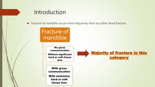
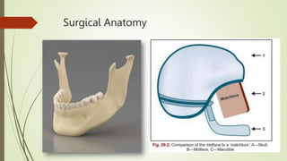
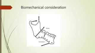
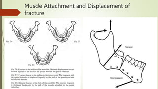
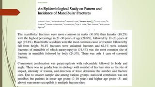
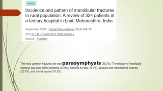
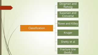
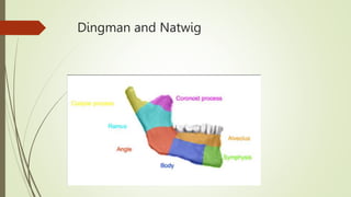
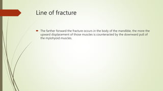
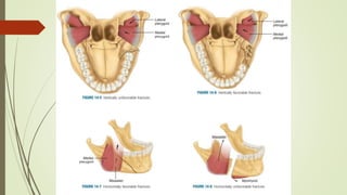
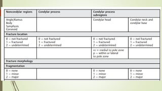
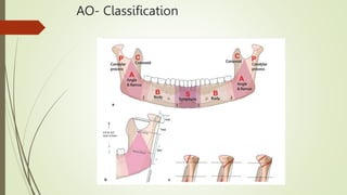
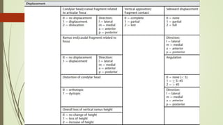
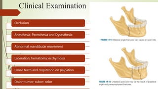
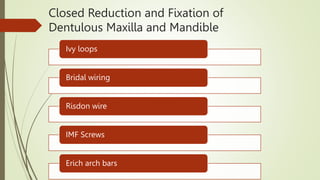
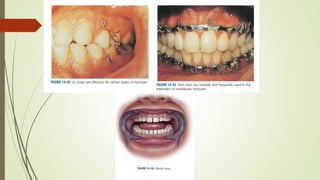
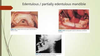
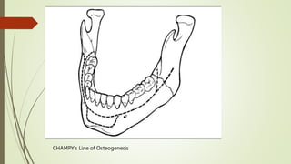
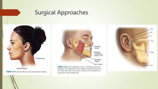
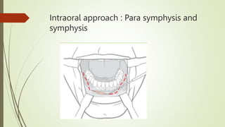
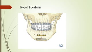
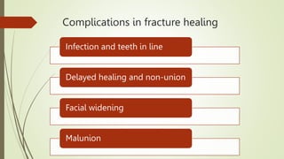
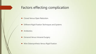
Mandibular Fracture: Symphysis
; Para-symphysis and Body
Moderated By: Dr. Aviral Verma
Made by: Gauri Bargoti
Contents
 Introduction
 Surgical anatomy
 Biomechanical consideration
 Incidence
 Etiology
 Classification
 Diagnosis
 Treatment
 Complications
 References
Introduction
 Fracture of mandible occurs more frequently than any other facial fracture.
Fracture of
mandible
No gross
communication
Without significant
hard or soft tissue
loss
With gross
communication
With extensive
hard or soft
tissue loss
Surgical Anatomy
Biomechanical consideration
Muscle Attachment and Displacement of
fracture
Incidence
The most common fractures site was parasymphysis (39.3%). The etiology of mandibular
fractures was road traffic accidents (42.9%), followed by falls (25.9%), assaults and interpersonal violence
(20.7%), and animal injuries (10.5%)
Etiology
Classification
Classification
Dingman and
Natwig
Kazanian and
Converse
Rowe and Killey
Kruger
Shetty et al
Fracture line
(body)
Dingman and Natwig
Kazanian and
Converse
Class I Class II Class III
Rowe and
Killey
Those not
involving basal
bone
Those
involving basal
bone
Kruger Classification
Relation to external environment
localization Type of fracture
Dentition of jaw
with reference
to splints
Shetty
et
al
F
L
O
S
I
D
Line of fracture
 The farther forward the fracture occurs in the body of the mandible, the more the
upward displacement of those muscles is counteracted by the downward pull of
the mylohyoid muscles.
AO- Classification
Diagnosis
PATIENT HISTORY
A thorough history
Patient’s health history
History Of TMJ disfunction
Object causing trauma
Direction of trauma
Clinical Examination
Occlusion
Anesthesia; Paresthesia and Dysesthesia
Abnormal mandibular movement
Laceration; hematoma; ecchymosis
Loose teeth and crepitation on palpation
Dolor; tumor; rubor; color
Radiological Examination
Panoramic Examination
Lateral Oblique
PA view
Occlusal view
Periapical
Towne’s view
CT and TMJ
General Principles of treatment
 The patient’s general physical status should be carefully evaluated and monitored before any
consideration of treating mandibular fracture.
 Diagnosis and treatment of mandibular fractures should be approached methodically, not with an
emergency-type of mentality.
 Dental injuries should be evaluated and treated concurrently with treatment of mandibular fracture.
 Reestablishment of occlusion is the primary goal in the treatment of mandibular fractures.
 With multiple facial fractures, mandibular fractures should be treated first.
 Intermaxillary fixation time should vary according to the type, location, number, and severity of the
mandibular fractures, the patient’s age and health, and the method used for reduction and
immobilization.
 Prophylactic antibiotics should be used for compound fractures.
 Nutritional needs should be closely monitored postoperatively.
 Mandibular fractures can be treated by closed reduction
Teeth are often injured with mandibular
fractures
 Fractured teeth can become infected and jeopardize bone union; however, an intact
tooth in the line of fracture that is maintaining bone fragments can be protected with
antibiotic coverage.
 A second molar on an otherwise edentulous posterior fracture segment should be
maintained to prevent superior displacement of the fragment in intermaxillary fixation.
 Mandibular canines are the cornerstone of occlusion and should be maintained at all
costs.
 Some teeth are not critical to restoration and can be removed when their prognosis is
doubtful and when maintenance may adversely affect fracture treatment.
 Some fractured teeth cannot be salvaged, no matter how critical they may be
Indication for closed reduction
Non displaced favorable fracture
Grossly comminuted fracture
Fracture exposed by significant loss of tissue
Edentulous mandibular fracture
Children with developing dentition
Indications for open reduction
Unfavorable Displaced fracture
Multiple Facial Fracture
Edentulous mandible with Severe displaced fragments
Edentulous maxilla opposing mandible
Malunion
Delay of treatment and interposition of soft tissue between mandibular fragments
Medical condition contraindicating IMF
Treatment
History
Closed Reduction and Fixation of
Dentulous Maxilla and Mandible
Ivy loops
Bridal wiring
Risdon wire
IMF Screws
Erich arch bars
Edentulous / partially edentulous mandible
CHAMPY’s Line of Osteogenesis
Surgical Approaches
Intraoral approach : Para symphysis and
symphysis
Intraoral approach: body
 The mucosa is incised to the bone with the blade positioned perpendicular to the
bone to avoid the mental nerve.
 The incision is made approximately 5 mm from the mucogingival junction to allow
adequate mobile tissue for closure. The proximal portion of the incision should be
carried along the external oblique ridge, only as high as the mandibular occlusal
plane.
 Extending the incision higher predisposes the buccal fat pad to prolapsing onto the
surgical field. The anterior surface of the ramus can then be exposed by stripping the
buccinator and temporal tendon with a notched angled retractor and periosteal
elevator.
 Once the coronoid is exposed, a curved Kocher clamp can be applied, which will act
as a self-retaining retractor
Rigid Fixation
Complications in fracture healing
Infection and teeth in line
Delayed healing and non-union
Facial widening
Malunion
Factors effecting complication
 Closed Versus Open Reduction.
 Different Rigid Fixation Techniques and Systems
 Antibiotics
 Extraoral Versus Intraoral Surgery
 Wire Osteosynthesis Versus Rigid Fixation
Summary
 From this review, it is apparent that infection is the most common type of complication
arising from the treatment of mandibular fractures. Although the incidence varies, it
appears to range from 5% to 10% (average).
 Although it would be expected that open reduction would contribute to infection, this
relationship is not evident in a review of the literature.
 Rigid fixation techniques are initially seen to result in a higher complication rate, but as
surgeons become more proficient with the procedures, complication rates fall.
 There are few studies on the incidence of permanent nerve damage associated with
mandibular fractures to allow any definitive conclusions to be reached.
References
 Killey’s fracture of mandible : 4th edition
 Trauma: Fonseca
 Peter wardbooth

Mandibular Fracture.pptx

  • 1.
    Mandibular Fracture: Symphysis ;Para-symphysis and Body Moderated By: Dr. Aviral Verma Made by: Gauri Bargoti
  • 2.
    Contents  Introduction  Surgicalanatomy  Biomechanical consideration  Incidence  Etiology  Classification  Diagnosis  Treatment  Complications  References
  • 3.
    Introduction  Fracture ofmandible occurs more frequently than any other facial fracture. Fracture of mandible No gross communication Without significant hard or soft tissue loss With gross communication With extensive hard or soft tissue loss
  • 4.
  • 5.
  • 6.
    Muscle Attachment andDisplacement of fracture
  • 7.
  • 9.
    The most commonfractures site was parasymphysis (39.3%). The etiology of mandibular fractures was road traffic accidents (42.9%), followed by falls (25.9%), assaults and interpersonal violence (20.7%), and animal injuries (10.5%)
  • 10.
  • 11.
  • 12.
    Classification Dingman and Natwig Kazanian and Converse Roweand Killey Kruger Shetty et al Fracture line (body)
  • 13.
  • 14.
  • 15.
    Rowe and Killey Those not involvingbasal bone Those involving basal bone
  • 16.
    Kruger Classification Relation toexternal environment localization Type of fracture Dentition of jaw with reference to splints
  • 17.
  • 18.
    Line of fracture The farther forward the fracture occurs in the body of the mandible, the more the upward displacement of those muscles is counteracted by the downward pull of the mylohyoid muscles.
  • 21.
  • 23.
  • 24.
    PATIENT HISTORY A thoroughhistory Patient’s health history History Of TMJ disfunction Object causing trauma Direction of trauma
  • 25.
    Clinical Examination Occlusion Anesthesia; Paresthesiaand Dysesthesia Abnormal mandibular movement Laceration; hematoma; ecchymosis Loose teeth and crepitation on palpation Dolor; tumor; rubor; color
  • 26.
    Radiological Examination Panoramic Examination LateralOblique PA view Occlusal view Periapical Towne’s view CT and TMJ
  • 27.
    General Principles oftreatment  The patient’s general physical status should be carefully evaluated and monitored before any consideration of treating mandibular fracture.  Diagnosis and treatment of mandibular fractures should be approached methodically, not with an emergency-type of mentality.  Dental injuries should be evaluated and treated concurrently with treatment of mandibular fracture.  Reestablishment of occlusion is the primary goal in the treatment of mandibular fractures.  With multiple facial fractures, mandibular fractures should be treated first.  Intermaxillary fixation time should vary according to the type, location, number, and severity of the mandibular fractures, the patient’s age and health, and the method used for reduction and immobilization.  Prophylactic antibiotics should be used for compound fractures.  Nutritional needs should be closely monitored postoperatively.  Mandibular fractures can be treated by closed reduction
  • 28.
    Teeth are ofteninjured with mandibular fractures  Fractured teeth can become infected and jeopardize bone union; however, an intact tooth in the line of fracture that is maintaining bone fragments can be protected with antibiotic coverage.  A second molar on an otherwise edentulous posterior fracture segment should be maintained to prevent superior displacement of the fragment in intermaxillary fixation.  Mandibular canines are the cornerstone of occlusion and should be maintained at all costs.  Some teeth are not critical to restoration and can be removed when their prognosis is doubtful and when maintenance may adversely affect fracture treatment.  Some fractured teeth cannot be salvaged, no matter how critical they may be
  • 29.
    Indication for closedreduction Non displaced favorable fracture Grossly comminuted fracture Fracture exposed by significant loss of tissue Edentulous mandibular fracture Children with developing dentition
  • 30.
    Indications for openreduction Unfavorable Displaced fracture Multiple Facial Fracture Edentulous mandible with Severe displaced fragments Edentulous maxilla opposing mandible Malunion Delay of treatment and interposition of soft tissue between mandibular fragments Medical condition contraindicating IMF
  • 31.
  • 32.
  • 35.
    Closed Reduction andFixation of Dentulous Maxilla and Mandible Ivy loops Bridal wiring Risdon wire IMF Screws Erich arch bars
  • 37.
    Edentulous / partiallyedentulous mandible
  • 38.
    CHAMPY’s Line ofOsteogenesis
  • 39.
  • 40.
    Intraoral approach :Para symphysis and symphysis
  • 41.
    Intraoral approach: body The mucosa is incised to the bone with the blade positioned perpendicular to the bone to avoid the mental nerve.  The incision is made approximately 5 mm from the mucogingival junction to allow adequate mobile tissue for closure. The proximal portion of the incision should be carried along the external oblique ridge, only as high as the mandibular occlusal plane.  Extending the incision higher predisposes the buccal fat pad to prolapsing onto the surgical field. The anterior surface of the ramus can then be exposed by stripping the buccinator and temporal tendon with a notched angled retractor and periosteal elevator.  Once the coronoid is exposed, a curved Kocher clamp can be applied, which will act as a self-retaining retractor
  • 42.
  • 48.
    Complications in fracturehealing Infection and teeth in line Delayed healing and non-union Facial widening Malunion
  • 49.
    Factors effecting complication Closed Versus Open Reduction.  Different Rigid Fixation Techniques and Systems  Antibiotics  Extraoral Versus Intraoral Surgery  Wire Osteosynthesis Versus Rigid Fixation
  • 51.
    Summary  From thisreview, it is apparent that infection is the most common type of complication arising from the treatment of mandibular fractures. Although the incidence varies, it appears to range from 5% to 10% (average).  Although it would be expected that open reduction would contribute to infection, this relationship is not evident in a review of the literature.  Rigid fixation techniques are initially seen to result in a higher complication rate, but as surgeons become more proficient with the procedures, complication rates fall.  There are few studies on the incidence of permanent nerve damage associated with mandibular fractures to allow any definitive conclusions to be reached.
  • 52.
    References  Killey’s fractureof mandible : 4th edition  Trauma: Fonseca  Peter wardbooth