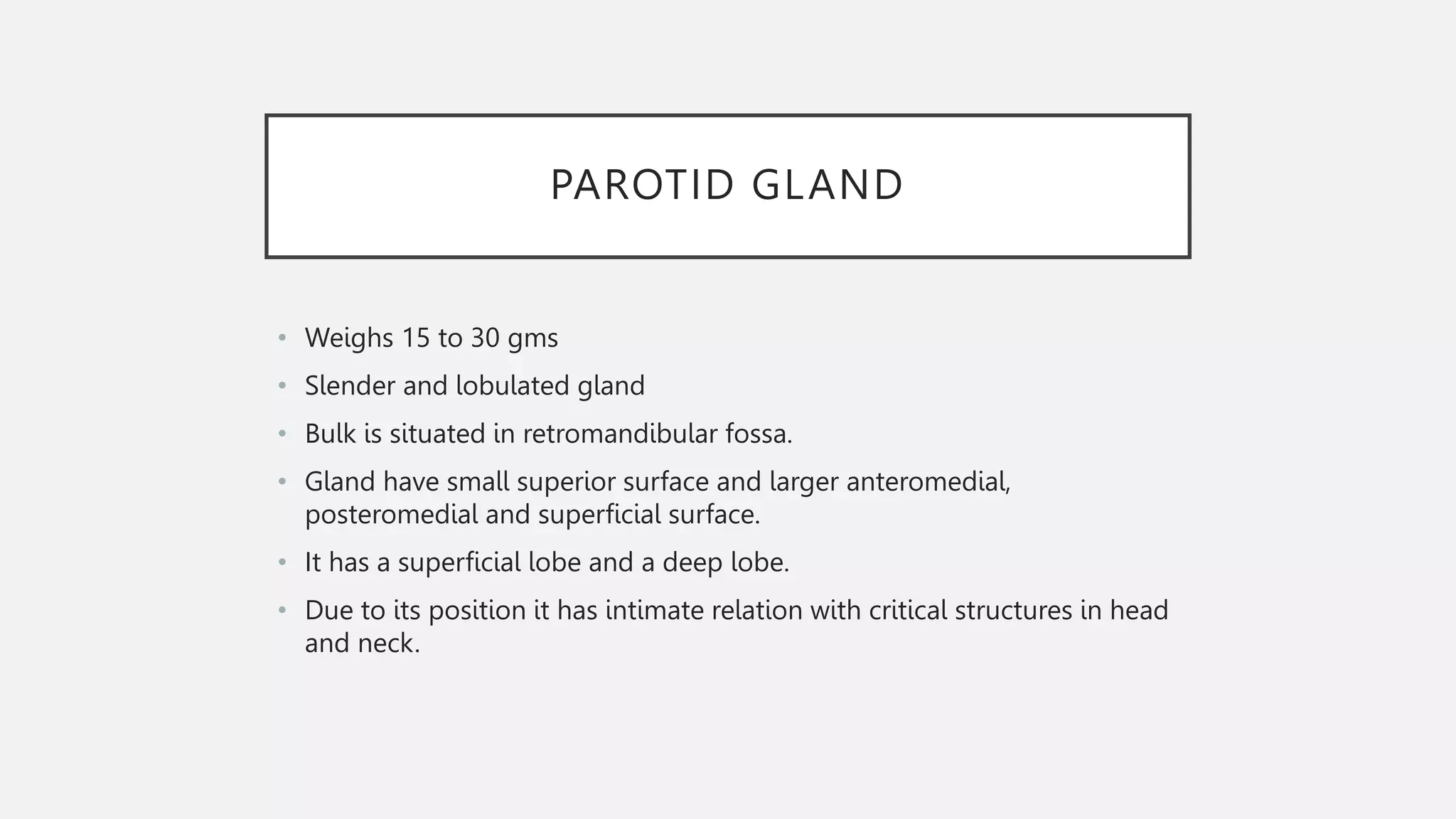
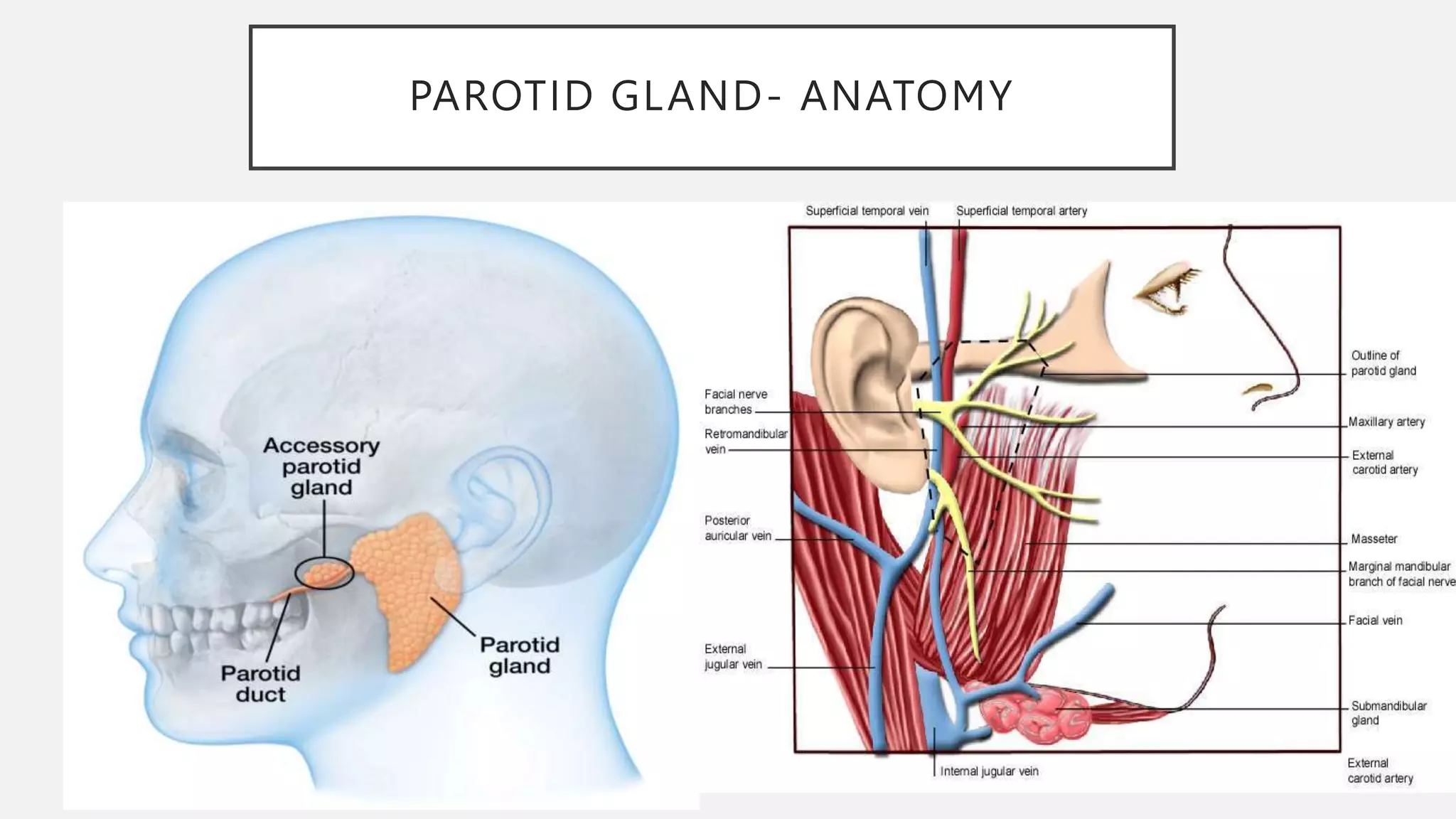
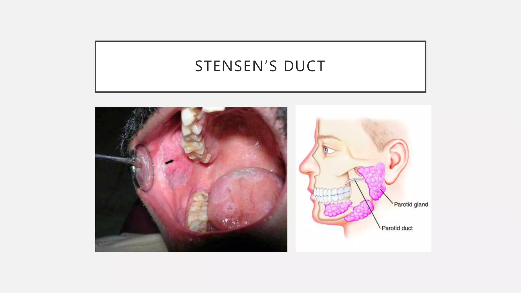
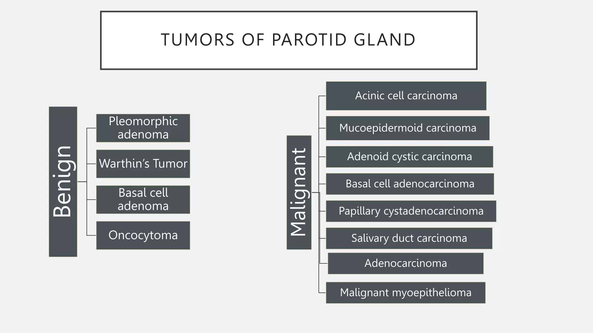
PAROTID GLAND- APPLIED
ANATOMY
Moderated by: Dr. Vidhi Rathi
Presented by: Gauri Bargoti
CONTENTS
• Introduction
• Types Of Glands
• Parotid Gland
• Parotidectomy
• References
INTRODUCTION
• The salivary gland system of the upper aerodigestive tract plays a critical
role in the functions of digestion, respiration, communication and overall
hemostasis.
• Some of these glands are small isolated or more densely packed bodies;
others form rather large organs.
• Smaller glands are in submucous layer and open with narrow ducts
while larger glands are in inner lining of oral cavity and open in larger
ducts.
TYPES OF GLANDS
• According to their secretion, they are divided into:
• A) Serous
• B) Mucous
• C) Mucous
• Anatomically into:
• A) Major
• B) Minor
PAROTID GLAND
• Weighs 15 to 30 gms
• Slender and lobulated gland
• Bulk is situated in retromandibular fossa.
• Gland have small superior surface and larger anteromedial,
posteromedial and superficial surface.
• It has a superficial lobe and a deep lobe.
• Due to its position it has intimate relation with critical structures in head
and neck.
PAROTID GLAND- ANATOMY
STENSEN’S DUCT
• It is formed from ductules that arises from either superficial or deep lobe
or both.
• It arises from the apex of parotid gland around masseter muscle
• Found along an imaginary line between oral commissure and lobule of
ear, measuring 4 to 7 cm.
STENSEN’S DUCT
SHAPE AND POSITION OF PAROTID
GLAND
• Upper end is grooved by external acoustic meatus and is wedged
shaped between it and the back of TMJ.
• The lower end lies between sternocleidomastoid and angle of mandible,
on the posterior belly of digastric
• The deep anteromedially grooves to fit over posterior margin of ramus
of mandible and posteromedial fits the mastoid process.
ARTERIES WITHIN PAROTID GLAND
VEINS RELATED TO PAROTID GLAND
VESSELS AND NERVES
• Sensory supply to gland is through greater auricular and
auriculotemporal nerves
• Postganglionic parasympathetic fibers come from otic ganglion through
auriculotemporal nerve.
• Preganglionic parasympathetic fibers come from glossopharyngeal
nerve
• Sympathetic fibers come from plexus on ECA or middle meningeal
arteries.
FACIAL NERVE AND PAROTID GLAND
SPACES OF PAROTID GLAND
• It encloses the parotid gland and its associated lymph nodes and facial
nerve and great vessels traversing it.
• Grondinsky and Holyoke and Coller and Yglesias concluded in separate
studies that infection may pass readily deep into parotid space and
lateral pharyngeal space lying deep to parotid gland
• Since parotid gland is attached to its surrounding fascia and it is not of
so much anatomical importance.
• Infections within this are related to gland and no cellulitis of connective
tissues
LYMPH NODES
• Parotid nodes drains lymphatic vessels from forehead, larger lateral
parts of both eyelids and from external nose.
ACCESSORY PAROTID GLAND
• The accessory parotid gland is a collection of salivary tissue separate from
the main parotid gland. When present, it may complicate parotidectomies,
promote parotitis, and serve as a potential site for benign and malignant
lesions to arise.
• In our meta-analysis of 3115 subjects, the overall pooled prevalence of an
APG was 32.1%, which renders it a likely anatomical variant to encounter in
the general population.
• References : Rosa MA, Lazarz DP, Pekala JR, Skinningsrud B, Lauritzen SS,
Solewski B, Pekala PA, Walocha JA, Tomaszewski KA. The accessory
parotid gland and its clinical significance. Journal of Craniofacial Surgery.
2020 May 1;31(3):856-60.
Applied Anatomy of parotid gland
INJURY TO FACIAL NERVE AND
PREVENTION
• In retromandibular approach to mandible, when incision is placed 2cm
posterior to ramus: INJURY TO FACIAL NERVE IS AVOIDED.
• When incision is placed at posterior border of ramus below earlobe:
BRANCHES OF FACIAL NERVE IS EXPOSED.
NON NEOPLASTIC INFLAMATION
• Sialolithiasis
TUMORS OF PAROTID GLAND
Benign
Pleomorphic
adenoma
Warthin’s Tumor
Basal cell
adenoma
Oncocytoma
Malignant
Acinic cell carcinoma
Mucoepidermoid carcinoma
Adenoid cystic carcinoma
Basal cell adenocarcinoma
Papillary cystadenocarcinoma
Salivary duct carcinoma
Adenocarcinoma
Malignant myoepithelioma
PAINFUL BILATERAL SWELLING
Mumps
EBV
Viral
CMV
PAROTIDECTOMY
• Superficial parotidectomy
Deep lobe parotidectomy
COMPLICATION AFTER PAROTIDECTOMY
Freys
syndrome
Facial nerve
injury
Scar
formation
Cosmetic
defects
IMAGING MODALITIES
Plain film radiography
Sialography
CT; MRI; USG
Sialoendography
REFERENCES
 Sicher and DuBRUL’S Oral anatomy by E.LLOYD DuBRUL (8th edition)
 Anatomy for surgeons: Volume 1 The head and neck by W. Henry
Hollinshead (3rd edition)
 Cunningham’s Manual of Practical Anatomy
 Surgical approaches to facial skeleton by Edward Ellis (3rd edition)
 Rosa MA, Lazarz DP, Pekala JR, Skinningsrud B, Lauritzen SS,
Solewski B, Pekala PA, Walocha JA, Tomaszewski KA. The accessory
parotid gland and its clinical significance. Journal of Craniofacial
Surgery. 2020 May 1;31(3):856-60.

Parotid Gland- Applied anatomy.pptx

  • 1.
    PAROTID GLAND- APPLIED ANATOMY Moderatedby: Dr. Vidhi Rathi Presented by: Gauri Bargoti
  • 2.
    CONTENTS • Introduction • TypesOf Glands • Parotid Gland • Parotidectomy • References
  • 3.
    INTRODUCTION • The salivarygland system of the upper aerodigestive tract plays a critical role in the functions of digestion, respiration, communication and overall hemostasis. • Some of these glands are small isolated or more densely packed bodies; others form rather large organs. • Smaller glands are in submucous layer and open with narrow ducts while larger glands are in inner lining of oral cavity and open in larger ducts.
  • 4.
    TYPES OF GLANDS •According to their secretion, they are divided into: • A) Serous • B) Mucous • C) Mucous • Anatomically into: • A) Major • B) Minor
  • 5.
    PAROTID GLAND • Weighs15 to 30 gms • Slender and lobulated gland • Bulk is situated in retromandibular fossa. • Gland have small superior surface and larger anteromedial, posteromedial and superficial surface. • It has a superficial lobe and a deep lobe. • Due to its position it has intimate relation with critical structures in head and neck.
  • 6.
  • 7.
    STENSEN’S DUCT • Itis formed from ductules that arises from either superficial or deep lobe or both. • It arises from the apex of parotid gland around masseter muscle • Found along an imaginary line between oral commissure and lobule of ear, measuring 4 to 7 cm.
  • 8.
  • 9.
    SHAPE AND POSITIONOF PAROTID GLAND • Upper end is grooved by external acoustic meatus and is wedged shaped between it and the back of TMJ. • The lower end lies between sternocleidomastoid and angle of mandible, on the posterior belly of digastric • The deep anteromedially grooves to fit over posterior margin of ramus of mandible and posteromedial fits the mastoid process.
  • 10.
  • 11.
    VEINS RELATED TOPAROTID GLAND
  • 12.
    VESSELS AND NERVES •Sensory supply to gland is through greater auricular and auriculotemporal nerves • Postganglionic parasympathetic fibers come from otic ganglion through auriculotemporal nerve. • Preganglionic parasympathetic fibers come from glossopharyngeal nerve • Sympathetic fibers come from plexus on ECA or middle meningeal arteries.
  • 14.
    FACIAL NERVE ANDPAROTID GLAND
  • 15.
    SPACES OF PAROTIDGLAND • It encloses the parotid gland and its associated lymph nodes and facial nerve and great vessels traversing it. • Grondinsky and Holyoke and Coller and Yglesias concluded in separate studies that infection may pass readily deep into parotid space and lateral pharyngeal space lying deep to parotid gland • Since parotid gland is attached to its surrounding fascia and it is not of so much anatomical importance. • Infections within this are related to gland and no cellulitis of connective tissues
  • 16.
    LYMPH NODES • Parotidnodes drains lymphatic vessels from forehead, larger lateral parts of both eyelids and from external nose.
  • 17.
    ACCESSORY PAROTID GLAND •The accessory parotid gland is a collection of salivary tissue separate from the main parotid gland. When present, it may complicate parotidectomies, promote parotitis, and serve as a potential site for benign and malignant lesions to arise. • In our meta-analysis of 3115 subjects, the overall pooled prevalence of an APG was 32.1%, which renders it a likely anatomical variant to encounter in the general population. • References : Rosa MA, Lazarz DP, Pekala JR, Skinningsrud B, Lauritzen SS, Solewski B, Pekala PA, Walocha JA, Tomaszewski KA. The accessory parotid gland and its clinical significance. Journal of Craniofacial Surgery. 2020 May 1;31(3):856-60.
  • 18.
    Applied Anatomy ofparotid gland
  • 19.
    INJURY TO FACIALNERVE AND PREVENTION • In retromandibular approach to mandible, when incision is placed 2cm posterior to ramus: INJURY TO FACIAL NERVE IS AVOIDED. • When incision is placed at posterior border of ramus below earlobe: BRANCHES OF FACIAL NERVE IS EXPOSED.
  • 20.
  • 21.
    TUMORS OF PAROTIDGLAND Benign Pleomorphic adenoma Warthin’s Tumor Basal cell adenoma Oncocytoma Malignant Acinic cell carcinoma Mucoepidermoid carcinoma Adenoid cystic carcinoma Basal cell adenocarcinoma Papillary cystadenocarcinoma Salivary duct carcinoma Adenocarcinoma Malignant myoepithelioma
  • 22.
  • 23.
  • 24.
  • 25.
    COMPLICATION AFTER PAROTIDECTOMY Freys syndrome Facialnerve injury Scar formation Cosmetic defects
  • 26.
    IMAGING MODALITIES Plain filmradiography Sialography CT; MRI; USG Sialoendography
  • 27.
    REFERENCES  Sicher andDuBRUL’S Oral anatomy by E.LLOYD DuBRUL (8th edition)  Anatomy for surgeons: Volume 1 The head and neck by W. Henry Hollinshead (3rd edition)  Cunningham’s Manual of Practical Anatomy  Surgical approaches to facial skeleton by Edward Ellis (3rd edition)  Rosa MA, Lazarz DP, Pekala JR, Skinningsrud B, Lauritzen SS, Solewski B, Pekala PA, Walocha JA, Tomaszewski KA. The accessory parotid gland and its clinical significance. Journal of Craniofacial Surgery. 2020 May 1;31(3):856-60.