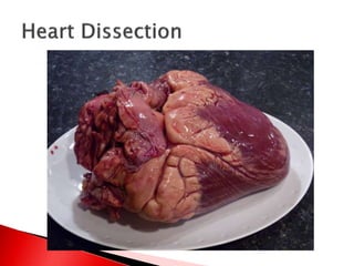
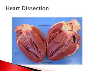
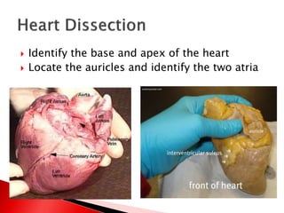
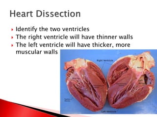
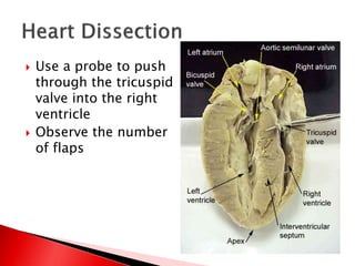
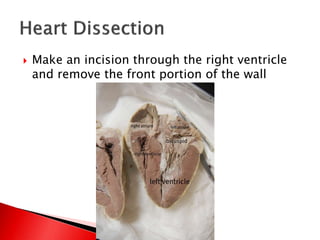
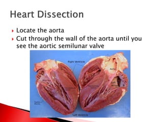
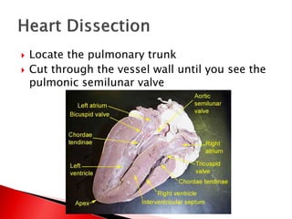
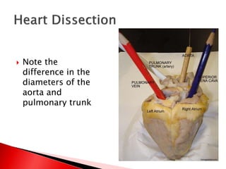
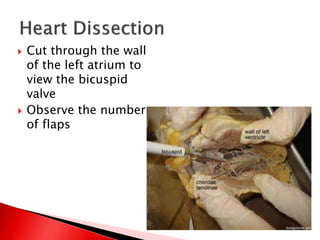
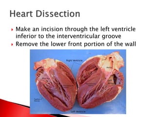
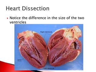
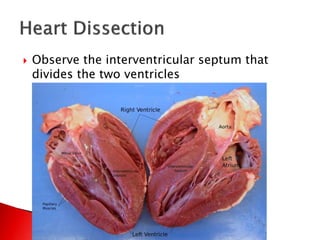
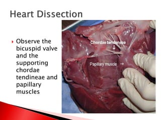
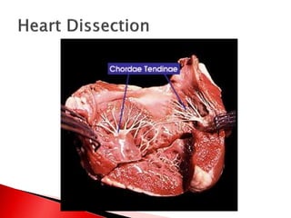
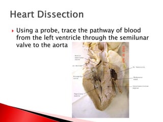
 Identify the base and apex of the heart
 Locate the auricles and identify the two atria
 Identify the two ventricles
 The right ventricle will have thinner walls
 The left ventricle will have thicker, more
muscular walls
 Cut through the wall
of the right atrium
and remove a
portion of the wall
 Be careful not to cut
the right ventricle
 Observe the tricuspid
valve
 Use a probe to push
through the tricuspid
valve into the right
ventricle
 Observe the number
of flaps
 Make an incision through the right ventricle
and remove the front portion of the wall
 Locate the aorta
 Cut through the wall of the aorta until you
see the aortic semilunar valve
 Locate the pulmonary trunk
 Cut through the vessel wall until you see the
pulmonic semilunar valve
 Note the
difference in the
diameters of the
aorta and
pulmonary trunk
 Cut through the wall
of the left atrium to
view the bicuspid
valve
 Observe the number
of flaps
 Make an incision through the left ventricle
inferior to the interventricular groove
 Remove the lower front portion of the wall
 Notice the difference in the size of the two
ventricles
 Observe the interventricular septum that
divides the two ventricles
 Observe the
bicuspid valve
and the
supporting
chordae
tendineae and
papillary
muscles
 Using a probe, trace the pathway of blood
from the left ventricle through the semilunar
valve to the aorta
 Repeat this action for the right ventricle,
tracing the pathway from the ventricle
through the pulmonic semilunar valve to the
pulmonary trunk

Mammal Heart Dissection

  • 4.
     Identify thebase and apex of the heart  Locate the auricles and identify the two atria
  • 5.
     Identify thetwo ventricles  The right ventricle will have thinner walls  The left ventricle will have thicker, more muscular walls
  • 6.
     Cut throughthe wall of the right atrium and remove a portion of the wall  Be careful not to cut the right ventricle  Observe the tricuspid valve
  • 7.
     Use aprobe to push through the tricuspid valve into the right ventricle  Observe the number of flaps
  • 8.
     Make anincision through the right ventricle and remove the front portion of the wall
  • 9.
     Locate theaorta  Cut through the wall of the aorta until you see the aortic semilunar valve
  • 10.
     Locate thepulmonary trunk  Cut through the vessel wall until you see the pulmonic semilunar valve
  • 11.
     Note the differencein the diameters of the aorta and pulmonary trunk
  • 12.
     Cut throughthe wall of the left atrium to view the bicuspid valve  Observe the number of flaps
  • 13.
     Make anincision through the left ventricle inferior to the interventricular groove  Remove the lower front portion of the wall
  • 14.
     Notice thedifference in the size of the two ventricles
  • 15.
     Observe theinterventricular septum that divides the two ventricles
  • 16.
     Observe the bicuspidvalve and the supporting chordae tendineae and papillary muscles
  • 18.
     Using aprobe, trace the pathway of blood from the left ventricle through the semilunar valve to the aorta
  • 19.
     Repeat thisaction for the right ventricle, tracing the pathway from the ventricle through the pulmonic semilunar valve to the pulmonary trunk