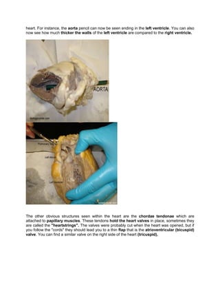
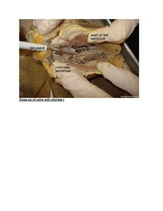
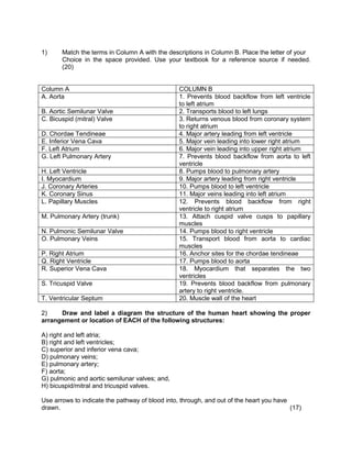
This document shows demonstration of a heart dissection, if the teacher wishes to this practical he/she should moderate the learners so that they don't hurt themselves
9953330565 Low Rate Call Girls In Rohini Delhi NCR
Grade11 life sciences practical task
1. Grade11 Life sciences Practical task
Instructions
1. Read the given reader below and understand it so that you may answer the questions
that will follow bellow.
2. Answer each question’s; don’t leave any question unanswered if you do so you will be
penalised.
3. If you didn’t write the work you will get zero and you won’t do the practical in class.
4. After the practical you will submit your work in class.
5. This practical will also prepare you for the exam that is about to come in November
Practical 1: Dissection of a sheep heart
Heart Dissection Walk Through :
The heart dissection is probably one of the most difficult dissections you will do. Part of the
reason it is so difficult to learn is that the heart is not perfectly symmetrical, but it is so close that
it becomes difficult to discern which side you are looking at (dorsal, ventral, left or right). Finding
the vessels is directly related to being able to orient the heart correctly and figuring out which
side you are looking at.
The heart is also difficult because the fatty tissue that surrounds the heart can obscure the
openings to the vessels. This means that you really must experience the heart with your hands
and feel your way to find the openings. Many people will be squeamish about this, and because
the heart is slippery, it is easy to drop. Don't be shy with the heart; use your fingers to feel your
way through the dissection.
1. Step One: Orientation
When you first remove your heart from the bag, you will see a lot of fatty tissue surrounding it. It
is usually a waste of time to try to remove this tissue. You may also want to arm yourself with
some kind of markers for the parts you find, colour pencils work great to identify a vessel and
you will see many used to mark openings on the heart.
There are a few clues to help you figure out the left and the right side, but often the packaging
and preserving process can cause the heart to be misshapen. If you are lucky, the heart will be
nicely preserved and you will see that the front (ventral) side of the heart has a couple of key
features:
1) a large pulmonary trunk(artery) that extends off the top of it
2) the flaps of the auricles covering the top of the atria.
3) thecurve of the entire front side, whereas the backside is much flatter.
2. Side of the heart Back of the heart
Auricle flap of the heart
Step 2: Locate the Aorta
Use your fingers to probe around the top of the heart. Four major vessels can be found entering
the heart:
the pulmonary trunk (artery),
aorta,
superior vena cava, and the
pulmonary vein.
Remember that if you are looking at the back of the heart, then the right and left sides are the
same as your right and left hand.
If you find the pulmonary artery, the aorta should be situated a little bit behind it. It may be
covered by fat, so use your fingers to poke around until you find the opening. Push your finger
all the way in and you will feel inside of the left ventricle. The left ventricle has a very thick
wall, unlike the right ventricle. Insert your finger through the pulmonary artery to feel the
right ventricle and you will notice and feel that it is much thinner than the left side of the heart.
3. With your fingers or probes in the aorta and the pulmonary trunk/artery you should notice that
they criss-cross each other, with the pulmonary trunk in the front.
At this point, you may want to use your colored pencils to mark these vessels so that you don't
get them confused when you are searching for the other two openings that top of the heart.
Step 3: Locate the Veins
The two major veins that enter the heart can be found on the backside, as both enter the
atria. On the left side, you should be able to find the opening of the pulmonary vein as it
enters the left atrium. The superior vena cava enters the right atrium. In many preserved
hearts, the heart was cut at these points, so you won't see the vessels themselves, you will just
find the openings. Again, use your fingers to feel around the heart to find the openings. If you've
marked the aorta and pulmonary artery then you won't mistake them for the veins you are
looking for. This picture shows all of the vessels labeled.
Sometimes, the aorta still has its branches attached to it. There are three vessels that branch
from the aorta:
4. the brachiocephalic,
left common carotid and the
leftsubclavian.
The majority of the time, these vessels are not visible because the aorta was cut too close to the
main part of the heart when the heart was removed from the animal.
Step 4: Make the Incisions
Now that you have all of the vessels located and marked, you can now open the heart to view
the inner chambers. Use the superior vena cava and pulmonary vein as guides for where to
cut. You are basically going to be cutting each side of the heart so that you can look inside. The
heart below is marked to show you where the two incisions should be made.
Step 5: Viewing the Chambers
At this point it is helpful to have two hands, one to hold the heart apart so you can take a peak
inside of it and another to use a probe to locate the specific parts. Your colour pencils you used
to mark the heart in step 2 can also now be used to see where those vessels connect within the
5. heart. For instance, the aorta pencil can now be seen ending in the left ventricle. You can also
now see how much thicker the walls of the left ventricle are compared to the right ventricle.
The other obvious structures seen within the heart are the chordae tendonae which are
attached to papillary muscles. These tendons hold the heart valves in place, sometimes they
are called the "heartstrings". The valves were probably cut when the heart was opened, but if
you follow the "cords" they should lead you to a thin flap that is the atrioventricular (bicuspid)
valve. You can find a similar valve on the right side of the heart (tricuspid).
7. 1) Match the terms in Column A with the descriptions in Column B. Place the letter of your
Choice in the space provided. Use your textbook for a reference source if needed.
(20)
Column A COLUMN B
A. Aorta 1. Prevents blood backflow from left ventricle
to left atrium
B. Aortic Semilunar Valve 2. Transports blood to left lungs
C. Bicuspid (mitral) Valve 3. Returns venous blood from coronary system
to right atrium
D. Chordae Tendineae 4. Major artery leading from left ventricle
E. Inferior Vena Cava 5. Major vein leading into lower right atrium
F. Left Atrium 6. Major vein leading into upper right atrium
G. Left Pulmonary Artery 7. Prevents blood backflow from aorta to left
ventricle
H. Left Ventricle 8. Pumps blood to pulmonary artery
I. Myocardium 9. Major artery leading from right ventricle
J. Coronary Arteries 10. Pumps blood to left ventricle
K. Coronary Sinus 11. Major veins leading into left atrium
L. Papillary Muscles 12. Prevents blood backflow from right
ventricle to right atrium
M. Pulmonary Artery (trunk) 13. Attach cuspid valve cusps to papillary
muscles
N. Pulmonic Semilunar Valve 14. Pumps blood to right ventricle
O. Pulmonary Veins 15. Transport blood from aorta to cardiac
muscles
P. Right Atrium 16. Anchor sites for the chordae tendineae
Q. Right Ventricle 17. Pumps blood to aorta
R. Superior Vena Cava 18. Myocardium that separates the two
ventricles
S. Tricuspid Valve 19. Prevents blood backflow from pulmonary
artery to right ventricle.
T. Ventricular Septum 20. Muscle wall of the heart
2) Draw and label a diagram the structure of the human heart showing the proper
arrangement or location of EACH of the following structures:
A) right and left atria;
B) right and left ventricles;
C) superior and inferior vena cava;
D) pulmonary veins;
E) pulmonary artery;
F) aorta;
G) pulmonic and aortic semilunar valves; and,
H) bicuspid/mitral and tricuspid valves.
Use arrows to indicate the pathway of blood into, through, and out of the heart you have
drawn. (17)
8. 3) Respectively, are the pulmonary arteries and pulmonary veins oxygen rich OR oxygen
poor?
(2)
4) Which chamber of the heart functions as the receiving chamber for pulmonic blood?
(1)
5) Which chamber of the heart functions as the muscular pump for pulmonic circulation?
(1)
6) Respectively, are the systemic arteries and systemic veins oxygen rich OR oxygen
poor? (2)
7) Which chamber of the heart functions as the receiving chamber for systemic blood?
(1)
8) Which chamber of the heart functions as the muscular pump for systemic circulation?
(1)
AUTHENTIC
1. opportunity to collaborate
2. provide opportunity for the learners to reflect
3. it provide the real world relevance
4. it ill-defined, requiring learners to define the task and subtask needed to complete their
activity
5. seamlessly integrated with assessment