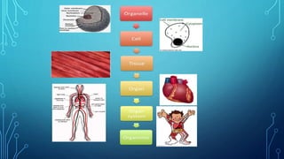
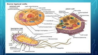
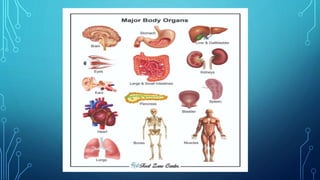
PROPER
HANDLING OF
MICROSCOPE
•Tip 1: Handle with care
•Most microscope problems occur
as a result of improper handling.
When carrying your microscope,
hold it by the base and the metal
support arm. Do not pick it up by
the stage, as this can cause
•Tip 2: Keep lenses clear of slides
•When using your microscope and
adjusting the focus you will need
to lower the objective lens down
as far as it will go. However, you
should never allow the lens to
touch the slide you are looking
•Tip3: Clean after using
•Always ensure the objectives are
cleaned immediately after use.
Objective, eyepieces, and
condenser may be removed for
cleaning. Use only lens paper and
lens cleaner. Do not use solvents.
•Tip 4: Cover when not in use
•All microscopes are sold with dust
covers. Always keep your microscope
covered when not in use even if the
microscope is stored in a cabinet. Eye
tubes also need to be kept free of dust
so do not store a microscope without the
eyepieces. If the microscope eyepieces
must be removed, cover the tubes with
•Tip 5: Look after the bulb
•After using the microscope, turn off
the illuminator and wait for it to
cool for several minutes before
putting it away. By allowing the bulb
to cool you will extend its life. When
turning the microscope on and off,
•Tip 6: Store in a clean, dry place
•Make sure you do not store your
microscope in an area that has
corrosive chemical fumes that can
destroy lenses or metal parts or
beside solutions that may leak. Salt
air and pervasive damp can also
cause damage over time. Make sure
•Tip 7: Only use special lens paper or
wipes for cleaning the lenses
•Microscope lenses can easily be scratched
and should be treated with great care. Use
an aspirator to remove dust. Sticky
residue can be removed with lens paper
moistened with distilled water or lens
cleaning solution and rubbed gently using
a circular motion. Never use sharp
•Tip 8: Keep your User's Manual and
wrenches in a safe place
•Each microscope should come with a user's
manual and specialist wrenches as required.
Always consult the User's Manual before
making any adjustments to your microscope
and use the wrenches provided. Never over-
tighten or use force when performing any
maintenance on your microscope, or use
•Tip 9: Perform an annual maintenance
check
•On an annual basis moving parts on the
microscope should be cleaned and
lubricated. Clean grease and dirt from
sliding surfaces using a clean cloth. Do
not grease the teeth of the rack and
pinion gears. Inspect the power cords
and plugs for safety and stock up on a
•Tip 10: Have your
microscope serviced
professionally
•A rule of thumb for
frequency of servicing is
every 200 hours of use or
MAGNIFICATIO
N
•Magnification is the
ability of a microscope
to produce an image of
an object at a scale
larger (or even smaller)
•The two lenses that
determine the magnification
of the microscope are the
eyepiece lens and the
objective lens. Each has a
number on it that signifies its
•OCULAR LENS (eyepiece) – Your
microscope will have either one
(monocular) or two (binocular) ocular
lenses.
•These are the lenses you will look
through when examining a specimen
with the microscope. Take a look at the
side of your ocular lens and you will
notice a label of “10X”. This indicates that
•OBJECTIVE LENSES – Notice the set of objective
lenses on the revolving nosepiece. These lenses
allow you to change the degree of magnification.
Some of our microscopes have four objective
lenses while others have only three. The degree
of magnification for each objective lens is
indicated on its side. Let’s take a look at each
progressing from the shortest to longest
objective lenses, being sure to rotate the
revolving nosepiece to click each objective lens
•4X – This objective magnifies the image by a
factor of 4. It is referred to as the “scanning
objective” since it is used to scan the slide
to locate the specimen before viewing it at
higher magnification.
•10X – This objective magnifies the image by
a factor of 10 and is referred to as the “low
power” objective.
•Total Magnification
The total magnification of an image is quite simple – it is the
product of the ocular lens magnification times the
magnification of the objective lens you are using:
•Magnification of Ocular x Magnification of Objective = Total
Magnification
For example, if the ocular lens magnifies the image by a
factor of 10 (10X), and the objective lens magnifies the
image by a factor of 50 (50X), the total magnification of the
image is 500X:
10X x 50X = 500X
LEVELS OF
BIOLOGICAL
ORGANIZATION
•Organelle
•The term ‘organelle’ means little
organs and it is a specialized part of a
cell. Just like the human body which
has organs such as heart, liver, etc., a
cell also has them in the form of
organelle. Examples are the nucleus,
•Cell
•All living organisms in the biological
organization consist of structural and
functional units called cells. For example,
bacteria, amoeba, archaebacteria, etc, are
all single-celled organisms. Humans and
many other organisms are multicellular,
which may have around
CELL
•Tissue
•A group of cells which may or may not have
similar characteristics is termed as a tissue.
Each tissue is attributed with a particular set
of functions in the body of the living
organism. Connective tissue, muscle tissue,
nervous tissue and epithelial tissue are the
tissues in animals, while meristematic
•Organ
•Now when the different types of
tissues are organized or grouped
together, they form what is known
as an organ. These tissues enable a
particular organ to carry out its own
specific task. Examples are heart,
•Organ System
•An organ system consists of a groups of
organs which are meant for performing a
broad set of functions, particularly in
multi-cellular organisms. A number of
organs in a system work together as a
single unit. For example, the circulatory
system works with the help of heart,
blood and blood vessels.
DIGESTI
VE
SYSTEM
CIRCULATO
RY SYSTEM
NERVOU
S
SYSTEM
REPRODUCTIVE SYSTEM
DIGESTIVE ORGAN
SYSTEM
•The digestive system
is the group of organs
that break down food
in order to absorb its
•Mouth
•It is the initial part of the
Digestive system. With the
help of teeth, the food that
we eat is broken down into
•Pharynx
•It is a muscular organ mostly called
a fibromuscular organ which is
located at the terminal of the
mouth. It works as a bridge between
the mouth and esophagus; it
transfers food molecules to the
•Esophagus
•The esophagus is located
near the windpipe or trachea.
It is a muscular tube
measuring about 5m long. It
is responsible for the transfer
•Stomach
•It is the main organ of the digestive
system where important parts of
digestion take place. It is a hollow
U-shaped muscular organ that is
situated at the left side of the
abdominal cavity and below the
diaphragm. It wraps over the edges
•Small Intestine
•Major separation of food molecules takes
place at this stage. It is a thin long tube-
like structure that is about 9-10 feet
long located just behind the stomach. It
is the lower part of the gastrointestinal
tract. It is completely coiled and again
this small intestine is divided into 3, the
Duodenum Jejunum and Ileum
•Pancreas
•Digestive Enzymes from the pancreas are
released to the duodenum. The pancreas
is the producer of insulin in our body.
The enzymes released by the pancreas
are responsible for the digestion of fats,
proteins, and carbohydrates in the food
molecules. The insulin produced is
responsible for maintaining the sugar
•Liver
•The liver comprises an important
part of the digestive process, it
releases bile juice which is
responsible for the digestion of
vitamins in food molecules. It is a
large reddish browned triangular-
•Gallbladder
•It is a pear-shaped organ
located under the liver. It is
again divided into 3 parts:
Fundus, Body, and Neck. It
stores the bile juice produced by
•Large Intestine
•It is responsible for transferring waste
materials outside the body. It is a long
tube-like structure that is 10-15 feet
long situated below the stomach
wrapped by a small intestine. It clears the
bowel by evacuating the waste particles
with the help of the rectum and anus.
•Rectum
•It is a long tube that connects
the large intestine and anus
when the large intestine is
full the waste is temporarily
placed in the rectum until it is
•Anus
•When the rectum is full
it starts an urge by
contracting muscles and
it finally relaxes and
•The circulatory
system (cardiovascular
system) pumps blood
from the heart to the
lungs to get oxygen.
CIRCULATO
RY SYSTEM
•Blood vessels
•A tube through which the
blood circulates in the body.
Blood vessels include a
network of arteries,
arterioles, capillaries,
•Blood is a body fluid in the
circulatory system of humans
and other vertebrates that
delivers necessary substances
such as nutrients and oxygen to
the cells, and transports
NERVOUS SYSTEM
•The nervous system is
made up of the brain,
spinal cord and nerves.
It controls much of what
you think and feel and
•The nervous system has two
main parts: The central nervous
system is made up of the brain
and spinal cord. The peripheral
nervous system is made up of
nerves that branch off from the
spinal cord and extend to all
parts of the body.
•The brain is the control
centre for all the body's
functions, such as
walking, talking,
swallowing, breathing,
•spinal cord both sends
and receives important
information about the
body and the
surrounding
•Nerves of the human body
work like the wires in an
electric circuit. Each and
every nerve is responsible for
carrying a set of signals, that
convey many messages like
REPRODUCTIVE ORGAN
SYSTEM
•human reproductive
system, organ
system by which
humans reproduce
Magnification Grade 7.pptx

Magnification Grade 7.pptx

  • 1.
  • 2.
    •Tip 1: Handlewith care •Most microscope problems occur as a result of improper handling. When carrying your microscope, hold it by the base and the metal support arm. Do not pick it up by the stage, as this can cause
  • 3.
    •Tip 2: Keeplenses clear of slides •When using your microscope and adjusting the focus you will need to lower the objective lens down as far as it will go. However, you should never allow the lens to touch the slide you are looking
  • 4.
    •Tip3: Clean afterusing •Always ensure the objectives are cleaned immediately after use. Objective, eyepieces, and condenser may be removed for cleaning. Use only lens paper and lens cleaner. Do not use solvents.
  • 5.
    •Tip 4: Coverwhen not in use •All microscopes are sold with dust covers. Always keep your microscope covered when not in use even if the microscope is stored in a cabinet. Eye tubes also need to be kept free of dust so do not store a microscope without the eyepieces. If the microscope eyepieces must be removed, cover the tubes with
  • 6.
    •Tip 5: Lookafter the bulb •After using the microscope, turn off the illuminator and wait for it to cool for several minutes before putting it away. By allowing the bulb to cool you will extend its life. When turning the microscope on and off,
  • 7.
    •Tip 6: Storein a clean, dry place •Make sure you do not store your microscope in an area that has corrosive chemical fumes that can destroy lenses or metal parts or beside solutions that may leak. Salt air and pervasive damp can also cause damage over time. Make sure
  • 8.
    •Tip 7: Onlyuse special lens paper or wipes for cleaning the lenses •Microscope lenses can easily be scratched and should be treated with great care. Use an aspirator to remove dust. Sticky residue can be removed with lens paper moistened with distilled water or lens cleaning solution and rubbed gently using a circular motion. Never use sharp
  • 9.
    •Tip 8: Keepyour User's Manual and wrenches in a safe place •Each microscope should come with a user's manual and specialist wrenches as required. Always consult the User's Manual before making any adjustments to your microscope and use the wrenches provided. Never over- tighten or use force when performing any maintenance on your microscope, or use
  • 10.
    •Tip 9: Performan annual maintenance check •On an annual basis moving parts on the microscope should be cleaned and lubricated. Clean grease and dirt from sliding surfaces using a clean cloth. Do not grease the teeth of the rack and pinion gears. Inspect the power cords and plugs for safety and stock up on a
  • 11.
    •Tip 10: Haveyour microscope serviced professionally •A rule of thumb for frequency of servicing is every 200 hours of use or
  • 12.
  • 13.
    •Magnification is the abilityof a microscope to produce an image of an object at a scale larger (or even smaller)
  • 14.
    •The two lensesthat determine the magnification of the microscope are the eyepiece lens and the objective lens. Each has a number on it that signifies its
  • 15.
    •OCULAR LENS (eyepiece)– Your microscope will have either one (monocular) or two (binocular) ocular lenses. •These are the lenses you will look through when examining a specimen with the microscope. Take a look at the side of your ocular lens and you will notice a label of “10X”. This indicates that
  • 16.
    •OBJECTIVE LENSES –Notice the set of objective lenses on the revolving nosepiece. These lenses allow you to change the degree of magnification. Some of our microscopes have four objective lenses while others have only three. The degree of magnification for each objective lens is indicated on its side. Let’s take a look at each progressing from the shortest to longest objective lenses, being sure to rotate the revolving nosepiece to click each objective lens
  • 17.
    •4X – Thisobjective magnifies the image by a factor of 4. It is referred to as the “scanning objective” since it is used to scan the slide to locate the specimen before viewing it at higher magnification. •10X – This objective magnifies the image by a factor of 10 and is referred to as the “low power” objective.
  • 18.
    •Total Magnification The totalmagnification of an image is quite simple – it is the product of the ocular lens magnification times the magnification of the objective lens you are using: •Magnification of Ocular x Magnification of Objective = Total Magnification For example, if the ocular lens magnifies the image by a factor of 10 (10X), and the objective lens magnifies the image by a factor of 50 (50X), the total magnification of the image is 500X: 10X x 50X = 500X
  • 19.
  • 21.
    •Organelle •The term ‘organelle’means little organs and it is a specialized part of a cell. Just like the human body which has organs such as heart, liver, etc., a cell also has them in the form of organelle. Examples are the nucleus,
  • 23.
    •Cell •All living organismsin the biological organization consist of structural and functional units called cells. For example, bacteria, amoeba, archaebacteria, etc, are all single-celled organisms. Humans and many other organisms are multicellular, which may have around
  • 24.
  • 25.
    •Tissue •A group ofcells which may or may not have similar characteristics is termed as a tissue. Each tissue is attributed with a particular set of functions in the body of the living organism. Connective tissue, muscle tissue, nervous tissue and epithelial tissue are the tissues in animals, while meristematic
  • 27.
    •Organ •Now when thedifferent types of tissues are organized or grouped together, they form what is known as an organ. These tissues enable a particular organ to carry out its own specific task. Examples are heart,
  • 29.
    •Organ System •An organsystem consists of a groups of organs which are meant for performing a broad set of functions, particularly in multi-cellular organisms. A number of organs in a system work together as a single unit. For example, the circulatory system works with the help of heart, blood and blood vessels.
  • 30.
  • 31.
  • 32.
  • 33.
  • 34.
  • 35.
    •The digestive system isthe group of organs that break down food in order to absorb its
  • 37.
    •Mouth •It is theinitial part of the Digestive system. With the help of teeth, the food that we eat is broken down into
  • 38.
    •Pharynx •It is amuscular organ mostly called a fibromuscular organ which is located at the terminal of the mouth. It works as a bridge between the mouth and esophagus; it transfers food molecules to the
  • 40.
    •Esophagus •The esophagus islocated near the windpipe or trachea. It is a muscular tube measuring about 5m long. It is responsible for the transfer
  • 42.
    •Stomach •It is themain organ of the digestive system where important parts of digestion take place. It is a hollow U-shaped muscular organ that is situated at the left side of the abdominal cavity and below the diaphragm. It wraps over the edges
  • 44.
    •Small Intestine •Major separationof food molecules takes place at this stage. It is a thin long tube- like structure that is about 9-10 feet long located just behind the stomach. It is the lower part of the gastrointestinal tract. It is completely coiled and again this small intestine is divided into 3, the Duodenum Jejunum and Ileum
  • 46.
    •Pancreas •Digestive Enzymes fromthe pancreas are released to the duodenum. The pancreas is the producer of insulin in our body. The enzymes released by the pancreas are responsible for the digestion of fats, proteins, and carbohydrates in the food molecules. The insulin produced is responsible for maintaining the sugar
  • 48.
    •Liver •The liver comprisesan important part of the digestive process, it releases bile juice which is responsible for the digestion of vitamins in food molecules. It is a large reddish browned triangular-
  • 50.
    •Gallbladder •It is apear-shaped organ located under the liver. It is again divided into 3 parts: Fundus, Body, and Neck. It stores the bile juice produced by
  • 52.
    •Large Intestine •It isresponsible for transferring waste materials outside the body. It is a long tube-like structure that is 10-15 feet long situated below the stomach wrapped by a small intestine. It clears the bowel by evacuating the waste particles with the help of the rectum and anus.
  • 54.
    •Rectum •It is along tube that connects the large intestine and anus when the large intestine is full the waste is temporarily placed in the rectum until it is
  • 56.
    •Anus •When the rectumis full it starts an urge by contracting muscles and it finally relaxes and
  • 57.
    •The circulatory system (cardiovascular system)pumps blood from the heart to the lungs to get oxygen.
  • 58.
  • 60.
    •Blood vessels •A tubethrough which the blood circulates in the body. Blood vessels include a network of arteries, arterioles, capillaries,
  • 62.
    •Blood is abody fluid in the circulatory system of humans and other vertebrates that delivers necessary substances such as nutrients and oxygen to the cells, and transports
  • 64.
  • 65.
    •The nervous systemis made up of the brain, spinal cord and nerves. It controls much of what you think and feel and
  • 66.
    •The nervous systemhas two main parts: The central nervous system is made up of the brain and spinal cord. The peripheral nervous system is made up of nerves that branch off from the spinal cord and extend to all parts of the body.
  • 68.
    •The brain isthe control centre for all the body's functions, such as walking, talking, swallowing, breathing,
  • 70.
    •spinal cord bothsends and receives important information about the body and the surrounding
  • 72.
    •Nerves of thehuman body work like the wires in an electric circuit. Each and every nerve is responsible for carrying a set of signals, that convey many messages like
  • 73.
  • 74.