PE 459 LECTURE 2- natural gas basic concepts and properties
WHO - The microscope - A practical guide.pdf
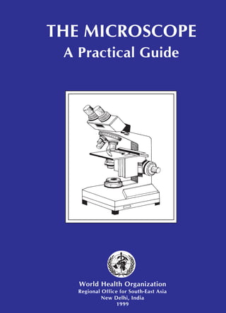
1. THE MICROSCOPE
A Practical Guide
World Health Organization
Regional Office for South-East Asia
New Delhi, India
1999
2. THE MICROSCOPE
A Practical Guide
World Health Organization
Regional Office for South-East Asia
New Delhi, India
1999
SME/BME/2
3.
4. THE MICROSCOPE
A Practical Guide
World Health Organization
Regional Office for South-East Asia
New Delhi, India
1999
WHO Project: ICP TUB 001
5. The issue of this document does not constitute formal publication.
It should not be reviewed, abstracted or quoted without the agreement
of the World Health Organization.
ACKNOWLEDGEMENTS
The World Health Organization wishes to acknowledge the assistance of
Mr K.K. Khanna in the preparation of this document and The Research
Institute of Tuberculosis (RIT), Japan for permission to use illustrations
originally prepared by them. This document is also based in part on
information given in Function, Use and Maintenance of Routine
Microscope, 1986 (Zeiss, West Germany) and Chapter 6 has been adapted
from TB Microscopy by A. Fujiki (RIT, Japan). The document was edited
and designed by Byword Editorial Consultants. Dr Armand Van Deun
provided helpful suggestions. Dr Thomas R. Frieden, Medical Officer
(Tuberculosis), was responsible for project design and implementation.
6. Reliable microscopy is a mainstay of primary health
care, including programmes to diagnose and cure
malaria and tuberculosis. For effective diagnosis to
occur, the entire health care team must function
effectively. The doctor must request the appropriate
test and must motivate the patient to have the test
done. The administrative authorities must ensure that
equipment, supplies and trained staff are present. The
microscopist must perform the examination and report
the results to the doctor promptly and accurately.
And, the doctor must make the appropriate treatment
decisions. If even a single step in this process fails,
the patient will not be accurately diagnosed and
treated, and may develop disability, may spread the
disease to others, or may die.
Laboratory technicians are thus on the forefront
of primary health care and of efforts to control
emerging and re-emerging infections. Our ability to
detect, cure, and hence control serious epidemics
such as tuberculosis and malaria depends on reliable
laboratory technicians. This practical booklet is
intended to help laboratory technicians to perform
their work, both accurately and for a long time, by
ensuring the proper use and maintenance of the
microscope.
Dr Uton Muchtar Rafei
Regional Director
Foreword
7.
8. 1 Introduction 1
2 Types of Microscopy 2
3 Parts of the Microscope 4
4 Routine Operation of the Microscope 13
5 Maintenance of the Microscope 18
6 Care of the Microscope 23
7 Materials for Care and Maintenance 24
8 Fungal Growth on the Microscope 26
9 Dos for Good Microscopy 28
10 Don’ts for Good Microscopy 32
11 Repair/Service 34
Annexures
I Maintenance Record Form 36
II Troubleshooting Guide 37
Contents
v
v
v
v
v
9. 1
1
1
1
1
1 Introduction
The microscope is a valuable instrument. There are many small objects or
details of objects which cannot be seen by the unaided human eye. The
microscope magnifies the image of such objects thus making them visible to
the human eye. Microscopes are used to observe the shape of bacteria,
fungi, parasites and host cells in various stained and unstained preparations.
There are many different microscopes available. This guide provides:
❍ A brief background on microscopes and microscopy (Chapters 2–4).
❍ How to maintain a microscope in good condition. Chapters 5–10
describe routine maintenance procedures as well as Dos and Don’ts for
proper use of the microscope.
❍ When and how minor repairs should be undertaken at the local level.
Chapter 11 includes brief guidelines regarding minor repairs at the local
level.
❍ A troubleshooting guide for common problems.
This guide is intended for peripheral health staff who use,
maintain, and repair microscopes.
10. 2
2
2
2
2
2 Types of Microscopy
Microscopes used in clinical practice are light microscopes. They are called
light microscopes because they use a beam of light to view specimens.
A compound light microscope is the most common microscope used in
microbiology. It consists of two lens systems (combination of lenses) to
magnify the image. Each lens has a different magnifying power. A
compound light microscope with a single eye-piece is called monocular; one
with two eye-pieces is said to be binocular.
Microscopes that use a beam of electrons (instead of a beam of light) and
electromagnets (instead of glass lenses) for focusing are called electron
microscopes. These microscopes provide a higher magnification and are
used for observing extremely small microorganisms such as viruses.
Light microscopy
Brightfield microscopy
This is the commonly used type of microscope. In brightfield microscopy
the field of view is brightly lit so that organisms and other structures are
visible against it because of their different densities. It is mainly used with
stained preparations. Differential staining may be used depending on the
properties of different structures and organisms.
Darkfield microscopy
In darkfield microscopy the field of view is dark and the organisms are
illuminated. A special condenser is used which causes light to reflect from
the specimen at an angle. It is used for observing bacteria such as
treponemes (which cause syphilis) and leptospires (which cause
leptospirosis).
11. 3
3
3
3
3
Types of Microscopy
Phase-contrast microscopy
Phase-contrast microscopy allows the examination of live unstained
organisms. For phase-contrast microscopy, special condensers and objectives
are used. These alter the phase relationships of the light passing through the
object and that passing around it.
Fluorescence microscopy
In fluorescence microscopy specimens are stained with fluorochromes/
fluorochrome complexes. Light of high energy or short wavelengths (from
halogen lamps or mercury vapour lamps) is then used to excite molecules
within the specimen or dye molecules attached to it. These excited
molecules emit light of different wavelengths, often of brilliant colours.
Auramine differential staining for acid-fast bacilli is one application of the
technique; rapid diagnostic kits have been developed using fluorescent
antibodies for identifying many pathogens.
12. 4
4
4
4
4
3 Parts of the Microscope
The main parts of the microscope are the eye-pieces, microscope tube, nose-
piece, objective, mechanical stage, condenser, coarse and fine focusing
knobs, and light source.
Fig. 3.1
EYE-PIECE
MICROSCOPE TUBE
OBJECTIVE
MECHANICAL STAGE
NOSE-PIECE
CONDENSER
ILLUMINATOR
COARSE FOCUSING KNOB
FINE FOCUSING KNOB
STAND
SLIDE HOLDER
ARM
IRIS DIAPHRAGM
13. 5
5
5
5
5
Eye-pieces
❍ The specimen is viewed through the
eye-piece (Fig. 3.2). It has a lens
which magnifies the image formed
by the objective. The magnifying
power of the eye-piece is in the
range 5x–20x. A movable pointer
may be attached to the inside of the
eye-piece.
❍ In binocular microscopes, the two eye-pieces can be moved closer or
farther apart to adjust for the distance between the eyes by pulling–
pushing motion or by moving a knurled ring.
Microscope tube
❍ The microscope tube is attached on top of the arm. It can be of the
monocular or binocular type. It supports the eye-piece on the upper end.
Mechanical tube length
❍ Mechanical tube length is the distance between the place where the
objective is inserted and the top of the draw-tube into which the eye-
pieces fit.
Parts of the Microscope
Fig. 3.2
10x
14. 6
6
6
6
6
❍ In modern microscopes it is not
tubular; it contains prisms that
bend the light coming up, thus
providing a comfortable viewing
angle (Fig. 3.3). In a binocular
tube, the light is also split and sent
to both eye-pieces.
Parts of the Microscope
Fig. 3.4
PATH OF LIGHT IN A BINOCULAR TUBE
PRISMS
EYE-PIECE
Fig. 3.3
MICROSCOPE TUBE
Do not interchange the objectives of two microscopes if the
specified mechanical tube length is different.
Nose-piece
❍ The nose-piece is attached under
the arm of the microscope tube.
The nose-piece (Fig. 3.4) houses
the objectives and rotates them.
The objectives are arranged in
sequential order of their
magnifying power, from lower to
higher. This helps to prevent the
immersion oil from getting onto
the intermediate objectives.
15. 7
7
7
7
7
Objectives
❍ The image of the specimen first
passes through the objective (Fig.
3.5). Objectives with magnifying
powers 4x, 10x, 40x and 100x are
commonly used. The magnifying
power is marked on the lens and is
usually colour-coded for easy
identification.
The 100x objective is for oil immersion.
Parts of the Microscopee
Fig. 3.5
The numerical aperture (NA) is the measure of light-gathering power of a
lens. The NA corresponding to the various magnifying powers of the
objective is:
Magnification Numerical aperture
10x 0.25
40x 0.65
100x 1.25
A high NA indicates a high resolving power and thus useful magnification
(see page 10).
To provide the best image at high magnification, immersion oil is placed
between the slide and the oil immersion objective (100x). Unlike air,
immersion oil has the same refractive index as glass. Therefore, it improves
the quality of the image. If immersion oil is not used, the image appears
blurred or hazy.
16. 8
8
8
8
8
Mechanical stage
❍ The mechanical stage holds the slide and allows it to be moved to the
left, right, forward or backward by rotating the knobs.
❍ It is fitted with fine vernier graduations as on a ruler. This helps in
relocating a specific field of examination.
Parts of the Microscope
Fig. 3.6
IRIS DIAPHRAGM LEVER
Condenser
❍ The condenser (Fig. 3.6) illuminates
the specimen and controls the
amount of light and contrast. There
are different types of condensers.
Some condensers have a rack-and
pinion mechanism for up-and-down
adjustment.
❍ The NA of a condenser should be equal to or greater than that of the
objective with maximum NA.
❍ An iris diaphragm is provided below the condenser. This adjusts the NA
of the condenser when using objectives having low magnifying power.
❍ A swing-out type filter holder may be fitted above or under the
condenser. In some microscopes the filter holder may not be swing-out
type. The filter holder holds detachable filters when required.
❍ Condenser centring screws, when present, are used to align the
condenser with the objective.
❍ A condenser raising knob may be present (if centring screws are not
there), or the distance may be fixed.
17. 9
9
9
9
9
Two-sided mirror
❍ A mirror (Fig. 3.7) is the simplest
illuminator. The two-sided mirror
provides necessary illumination
through reflection of natural or
artificial light. It has two surfaces,
one plain for artificial light and
other concave for natural light. It is
supported on two sides by a fork
fixed on a mount in a way that
permits free rotation.
Parts of the Microscope
Fig. 3.7
A mirror is usually fitted on a mount or at the base of the
microscope.
Built-in light sources
An illuminator is built into the base of the microscope. A halogen bulb
provides the best illumination. On top of the illuminator is an in-built filter
holder to fit the filter of desired quality.
Filters
❍ Blue filters are used to change the light from ordinary electric bulbs into
a more natural white light.
❍ Neutral density filters are used to reduce brightness without changing
the colour of the background.
❍ Green filters may be useful in some situations.
18. 10
10
10
10
10
The object of AFB (Ziehl–Neelsen) microscopy is to find AFB, which are
stained red by carbol fuchsin. The intensity of the red colour decreases
when blue/green filters are used. Blue/green filters are, therefore, not
recommended for Ziehl–Neelsen microscopy.
Parts of the Microscope
Cedar wood oil should not be used as it leaves a sticky residue on the
objective. If cedar wood oil is used, particular care then needs to be taken to
ensure that the objective is thoroughly and promptly cleaned with xylene
after each session of use. Petrol can be used in place of xylene for cleaning
if xylene is not available.
Liquid paraffin should not be used as it has a low refractive index which
produces an inferior image. It is also unsuitable for scanning specimens for
long periods, as is required for accurate microscopy.
GLASS ROD SEEMS TO DISAPPEAR IN
IMMERSION OIL WITH REFRACTIVE
INDEX OF 1.5
Fig. 3.8
Immersion oil
❍ Immersion oil must be used with
objectives having NA more than
1.0. This increases the resolving
power of the objective.
❍ An immersion oil of medium
viscosity and refractive index of
1.5 is adequate. Any synthetic
non-drying oil with a refractive
index of 1.5 and/or as
recommended by the
manufacturer should be used.
19. 11
11
11
11
11
Coarse and fine focusing knobs
The coarse and fine focusing knobs are used to change the distance between
the specimen slide and the objective. The coarse focusing knob alters this
distance rapidly and is used to bring the specimen into the field of view
using an objective having low magnification power. The fine focusing knob
changes the distance very slowly and permits better viewing of the object.
One revolution of the fine focusing knob should generally move the
mechanical stage by 100 µm. The movement should be smooth and free
from jerks.
Halogen lamp
Halogen lamps are low wattage, high intensity lamps and are the preferred
light source. Though costlier, these have the following advantages over
tungsten lamps:
❍ emit white light
❍ have higher luminosity (brighter)
❍ have compact filament
❍ have longer life.
Functioning of the microscope
There are three main optical pieces in the compound light microscope. All
three are essential for a sharp and clear image. These are:
❍ Condenser
❍ Objectives
❍ Eye-pieces.
Parts of the Microscope
20. 12
12
12
12
12
Parts of the Microscope
The condenser illuminates the object by converging a parallel beam of light
on it from a built-in or natural source. The objective forms a magnified
inverted (upside down) image of the object. The eye-piece magnifies the
image formed by the objective. This image is formed below the plane of the
slide.
The total magnification of the microscope is the product of the magnifying
powers of the objective and the eye-piece.
For example, if the magnifying power of the eye-piece is 10x and that of the
objective is 100x, then the total magnification of the compound light
microscope is: 10x X 100x = 1000-fold magnification.
21. 13
13
13
13
13
4 Routine Operation of the Microscope
❍ Ensure that the voltage supply in the laboratory corresponds to that
permitted for the microscope; use a voltage protection device, if
necessary.
Fig. 4.1
Fig. 4.2
❍ Turn on the light source of the
microscope (Fig. 4.1).
❍ With the light intensity knob,
decrease the light while using the
low magnification objective.
❍ Place a specimen slide on the stage.
Make sure the slide is not placed
upside down. Secure the slide to the
slide holder of the mechanical stage
(Fig. 4.2).
❍ Rotate the nose-piece to the 10x objective, and raise the stage to its
maximum.
❍ Move the stage with the adjustment knobs to bring the desired section of
the slide into the field of view.
22. 14
14
14
14
14
❍ Focus the specimen under 10x
objective using the coarse
focusing knob and lowering the
stage (Fig. 4.3).
❍ Make sure the condenser is
almost at its top position. Centre
the condenser using condenser
centring screws if these are
provided in the microscope. For
this take out one eye-piece and
while looking down the tube,
close the iris diaphragm till only a
pin-hole remains. Check if this is
located in the centre of the tube.
❍ Open the condenser iris
diaphragm to 70%–80% to adjust
the contrast so that the field is
evenly lighted (Fig. 4.4) .
Routine Operation of the Microscope
Fig. 4.4
Many modern microscopes have pre-centred and fixed
condensers. In these no adjustments are required. To reduce
glare adjust the opening of the iris diaphragm.
Exit pupil of objective
Iris diaphragm
Fig. 4.3
23. 15
15
15
15
15
❍ Adjust the interpupillary distance
till the right and left images become
one (Fig. 4.5).
❍ Focus the image with the right eye
by looking into the right eye-piece
and turning the focusing knob
(Fig. 4.6).
❍ Focus the image with the left eye by
looking into the left eye-piece by
turning the diopter ring (Fig. 4.7).
Routine Operation of the Microscope
Fig. 4.7
Fig. 4.5
Fig. 4.6
24. 16
16
16
16
16
❍ Put one drop of immersion oil on
the specimen (Fig. 4.8).
❍ Change to 100x objective
(Fig. 4.9).
Routine Operation of the Microscope
❍ Increase the light by turning the intensity knob until a bright but
comfortable illumination is achieved.
❍ Focus the specimen by turning the fine focusing knob.
❍ When the reading/observation has been recorded, rotate the objective
away from the slide.
❍ Release the tension of the slide holder, and remove the slide.
❍ If immersion oil was used, wipe it from the objective with lens paper or
muslin cloth at the end of each session of use. In general, avoid wiping
the objective except when it seems to be dirty.
❍ Turn off the light.
Fig. 4.8
Fig. 4.9
25. 17
17
17
17
17
❍ Cover the microscope when not in use and take necessary precautions
against fungus.
Eye strain should not develop if the microscope is used
properly.
Never adjust the stage upward while looking through the eye-
piece. It will cause the objective to push against the slide and
may damage it.
Only the 100x objective can be used for viewing under
immersion oil. All other lenses are to be used without
immersion oil; keep them dry and avoid applying oil or any
liquid to these lenses.
Routine Operation of the Microscope
26. 18
18
18
18
18
Dust is the worst enemy of the microscope. Always keep the
microscope properly covered. Fungus is also a major problem.
Always keep the microscope in dry surroundings.
5 Maintenance of the Microscope
(NOTE: In all cases, the manufacturer’s manual should be consulted for
specific instructions.)
Installation and storage
❍ Install the microscope on a sturdy, level table. Equipment and
instruments which generate vibrations, such as centrifuges and
refrigerators, should not be placed on or near this table.
❍ The height of the table should be convenient for the user. As an
alternative or in addition, an adjustable stool should be made available
to make microscopy comfortable.
❍ The table should be away from water, sinks, and racks containing
chemicals, to prevent damage to the microscope from splashes or spills.
❍ If the microscope does not have a built-in light source then the table
should be placed near a window away from direct sunlight and
arrangements made for the provision of a lamp.
❍ In so far as is possible, the microscopy room should be free from dust
and should not be damp.
❍ If the microscope is to be used every day, do not remove it from the site
of installation, provided security is assured.
❍ When the microscope is not in use, keep it covered with a polythene or
plastic cover and take necessary precautions against fungus.
27. 19
19
19
19
19
Maintenance of the Microscope
❍ In humid areas, store the microscope every night in a cabinet fitted with
an electric bulb (5 W or 40 W). This is switched on at night to reduce
humidity.
❍ If the microscope is used intermittently and requires storage for
prolonged periods, keep it in an air-tight plastic bag with about 100g of
drying agent. Remember to regenerate/replace drying agents (silica gel
or dry rice) fortnightly or as needed.
❍ If only a wooden box is available, keep the microscope in it with some
dry silica gel or dry rice (see page 25).
Maintenance of lenses
Avoid collection of dust and immersion oil on the objectives and eye-pieces
by keeping the microscope covered. Do not allow immersion oil to touch
any of the objectives other than the oil immersion objective. Always keep
the eye-pieces in place to protect the inner surface of the objective. Close the
holes of missing objectives in the nose-piece by using special caps that are
provided, or by sealing with adhesive tape.
Removal of dust from lenses
Check for dust or dirt on the lenses (eye-pieces, objective, condenser and
illuminator lenses) if the image appears hazy or with black dots.
❍ If the black dot moves when the eye-piece is rotated, this means that the
dust is on the eye-piece.
❍ If the black dot moves when the slide moves then the dust is present on
the slide.
❍ If these two are ruled out, presume that the dust is on the objective.
Dust on objectives shows as dots if it is inside. If the dust is outside the
objective it shows as a hazy image.
28. 20
20
20
20
20
Do not remove the dust from the lenses by wiping these with a
cloth as this can scratch the lens and damage it permanently.
Use an airbrush or a camel-hair/artist’s brush.
Maintenance of the Microscope
Dust can be removed with a camel-hair/artist’s brush or by blowing air over
the lens with an airbrush. Dust on the inner surface of the objective can be
removed by using a soft camel-hair brush (artist’s brush).
Removal of oil from lenses
The presence of oil on the lenses produces a hazy image. The localization
of oil can be done by the same method as has been described above for
localization of dust.
Oil should be removed with the help of lens paper using lens
cleaning fluid as recommended by the manufacturer. This can
be applied gently with lens paper. Do not use force to remove
oil as this might result in scratches on the lens.
If the field of view is not clear despite cleaning, and the microscope works
well with another lens, then the lens has been permanently damaged and
must be repaired or replaced.
If the field of view is not clear even after changing the lenses (objective and
eye-piece) there is probably dirt or fungus on the tube prisms. These can be
checked by removing the eye-pieces, and examining the upper part of the
microscope tube with the light fully open. Fungus is seen as threads, dots or
a woolly layer.
29. 21
21
21
21
21
Maintenance of the Microscope
Inspection of the objective
❍ Carefully unscrew the objective from the nose-piece.
❍ Gently remove one eye-piece to use as a magnifier (or use a magnifying
glass).
❍ Grasp the objective in one hand with the front lens face up.
❍ Hold the eye-piece in the other hand with the top lens facing down.
❍ Bring the eye-piece very close to your eye and focus on the objective.
Change the angle of the objective so that light can reflect off its surface.
The two lens surfaces will be about 2.5 cm apart. Try to avoid letting
them touch each other.
❍ Inspect the objective for scratches, nicks, cracks, deterioration of seal
around the lens, or oil seepage into the lens.
Maintenance of mechanical moving parts
Mechanical moving parts of the microscope may become too stiff or too
loose.
Stiffness is due to accumulation of dust or because the sliding channel has
become rough. This problem can be overcome by cleaning, polishing and
lubricating the sliding channel and the rack and pinion. First remove the
dust with a camel-hair/artist’s brush or by blowing air; clean it with a
solvent such as petrol, polish with metal polish and apply high quality
silicone grease to lubricate the moving parts.
Stiff movements may also be due to mechanical bending of some part.
Rectify the fault or call the service engineer.
30. 22
22
22
22
22
Maintenance of the Microscope
With prolonged use, the up and down movement of the mechanical stage
becomes loose. The stage, therefore, slides down during examination
resulting in loss of focus. Adjust the tension with the tension adjustment
device as recommended by the manufacturer.
Maintenance of light source
The supply of voltage (110 V or 220 V) must always conform to that
specified for the microscope. Adequate number of spare bulbs and fuses
should be available. Do not touch the bulbs with bare hands. Provide
adequate ventilation to take care of heat generated by light. Provide voltage
protection, if necessary. Before switching the lamp on, adjust the variable
voltage regulator to minimum. Switch on the lamp and slowly increase the
voltage until the desired intensity is achieved.
31. 23
23
23
23
23
6 Care of the Microscope
After daily use
❍ Bring the variable voltage regulator setting to the minimum before
turning off the lamp. Turn off the light source of the microscope.
❍ Gently wipe the immersion oil off the objective, condenser and
mechanical stage with lens paper or muslin cloth.
❍ Replace the cover of the microscope and take necessary precautions
against fungus.
Each month
❍ Use an air brush to blow away dust. Clean the objectives, eye-pieces, and
condenser with lens cleaning fluid. Do not put fluid directly on the
lenses; instead, apply it to the lens paper and then clean.
❍ Remove the slide holder from the mechanical stage and clean.
❍ With a tissue moistened with water, wipe the dust off the body of the
microscope and the window of the illuminator in the base of the unit.
Every six months
Thoroughly inspect, clean, and lubricate the microscope after consulting the
manufacturer’s manual. This should preferably be done by professional
service personnel.
32. 24
24
24
24
24
7 Materials for Care and Maintenance
(NOTE: In all cases, the manufacturer’s manual should be consulted for
specific instructions.)
Lens cleaning fluid
Lens cleaning fluid is used to clean optical surfaces. It does not harm the
coatings of the lens and does not soften the sealers and cements around the
lens.
Consult the manufacturer’s manual for specifications regarding
lens cleaning fluids as requirements are different depending on
the microscope.
Ethyl ether and xylene are the commonly used lens cleaning fluids. Petrol
can be used if xylene is not available. Ethyl ether is extremely flammable
and xylene is toxic. These must, therefore, be stored safely to avoid any
accident. Alcohol, acetones or any other ketones should not be used, unless
recommended by the manufacturer, since these may dissolve the sealants
around the lens.
Lens paper
Lens paper is specially prepared paper free from abrasive particles. If lens
paper is not available, muslin cloth or soft silk cloth may be used.
Light bulbs and fuses
Maintain a sufficient supply of bulbs and fuses for every microscope.
33. 25
25
25
25
25
Air brush
Use air to blow away particles from the
surface of the microscope. Be careful
when cleaning the mechanical stage as
tiny pieces of broken glass may be
present. A simple air brush (Fig. 7.1) can
be made in the laboratory by attaching a
Pasteur pipette to a rubber bulb.
Microscope cover
After use, the microscope should be covered with a polythene or a plastic
bag and necessary precautions against fungus should be taken (see
Chapter 8).
Drying agents
Keep dry silica gel or any other drying agent in the microscope cabinet to
reduce moisture. Regenerate the drying agent when necessary. Dry silica gel
(blue in colour) absorbs moisture inside the box. Its colour changes to pink
when it is unable to absorb more moisture. When this occurs, it should be
dried by keeping in a hot air oven or heating in a saucepan. When
completely dry it regains its original blue colour and can be reused.
If silica gel is not available, disposable and cheap drying agents like salt and
rice can be used. Rice is convenient and inexpensive. As soon as it is no
longer dry and crisp, it must be replaced.
This method will work only if the cabinet or box closes tightly. If no good
closed space is available, a plastic bag may be used provided it is made of
thick polythene and sealed each time. If a lamp for heating is used at night,
then simultaneous ventilation is an advantage, and the space does not have
to be closed tightly.
Materials for Care and Maintenance
Fig. 7.1
34. 26
26
26
26
26
8 Fungal Growth on the Microscope
Fungus is common in hot and humid climates. These conditions prevail for
most of the year in South-East Asia, and therefore precautions are
necessary. Fungal growth should be suspected when part or all of the image
becomes unclear or hazy. If fungal growth is advanced, the image becomes
dim and hardly anything can be seen.
Fungus can attack all microscopes within few years if no
precautions are taken, even if “anti-fungal treated” lenses are
used.
The lenses, the eye-piece tube and prisms of the microscope are often the
first places for fungal growth. The eye-piece tube can be checked by taking
out the eye-pieces and inspecting the inner part of the tube with the light on.
Cleaning of the eye-piece tube is difficult and should be done only by
authorized personnel.
Factors facilitating fungal growth
❍ Hot and humid environment
❍ Storage cabinets made of wood, leather or plastic without a desiccant
❍ Storage in cupboards or drawers
❍ Storage in small, dark unventilated rooms.
How to prevent fungal growth
❍ Store the microscope every night in a cabinet fitted with an electric bulb
(5 W or 40 W). The bulb should be preferably fitted on the top of the
cabinet so that it is near the tube (head of the microscope). Keep the
bulb switched on overnight. If this technique is used, the cabinet should
have holes for ventilation so that air flows freely.
35. 27
27
27
27
27
or alternatively,
❍ Use a drying agent, such as silica gel or rice, continuously. When using a
drying agent be sure the microscope is confined to a wooden box or air-
tight plastic bag. Be sure to regenerate/change the drying agent as
described on page 25.
❍ Clean the microscope regularly. Wear thin cloth/latex gloves when
handling microscope lenses. Otherwise, fungus may grow where
fingerprints were left.
❍ If none of the above are feasible, keep the microscope in a place with
good circulation of air. When not in use, the microscope can be kept in
direct sunlight for a few hours to reduce moisture.
❍ Although generally not feasible in peripheral centres, continuous air
conditioning is very effective in preventing fungal growth. Keeping
microscopes in AC stores is only recommended for prolonged storage,
not if they have to be taken out daily.
How to remove a film of fungus
Remove fungal growth as soon as it appears and frequently thereafter.
Moisten a wad of cotton wool with a fungus cleaner which is recommended
by the manufacturer. Use lens cleaner if fungus cleaner is not available.
Clean the lens by moving the cotton wool in circles or back and forth under
moderate pressure. If necessary, repeat the same procedure with a fresh wad
of cotton wool. Wipe the lens with a fresh dry wad of cotton. Contact the
service engineer if this does not remove fungal growth.
Do not attempt to clean parts of the microscope which are not
accessible (such as prisms) and which may require
disassembling the instrument.
Fungal Growth on the Microscope
36. 28
28
28
28
28
9 Dos for Good Microscopy
❍ Place the microscope on a level
vibration-free surface. Never keep it
on the surface where a centrifuge is
placed. Also, keep it away from
refrigerators and air conditioners
(Fig. 9.1).
❍ Store the microscope in a cabinet
fitted with an electric bulb (5 W or
40 W) which is switched on in order
to reduce humidity (Fig. 9.2).
❍ Always carry the microscope with
one hand supporting the base and
the other hand around the arm
(Fig. 9.3).
Fig. 9.1
Fig. 9.2
Fig. 9.3
37. 29
29
29
29
29
❍ Place the microscope in a
location from which it need
not be moved frequently
(Fig. 9.4).
❍ Turn the nose-piece to the objective
with lowest magnifying power before
removing the slide and when the
microscope is not in use (Fig. 9.5).
❍ Cover the microscope when not in
use, taking all precautions to prevent
growth of fungus (Fig. 9.6).
Dos for Good Microscopy
Fig. 9.5
Fig. 9.6
Fig. 9.4
38. 30
30
30
30
30
Dos for Good Microscopy
❍ Adjust the variable voltage regulator setting to minimum before
switching on the lamp and increase the voltage slowly until the desired
intensity of light is achieved.
❍ Always keep the condenser up, adjusting the light intensity by using the
illuminator regulator. Remember to adjust the iris diaphragm opening to
about 80% of its maximum when using the immersion objective, or to
slightly less for lower power objectives.
Fig. 9.8
Fig. 9.7
❍ Always place the slide with the
specimen side up (Fig. 9.7).
❍ For focusing, always turn the stage up toward the objectives while
looking from the side and not through the eye-pieces, so as to avoid
turning it up too far and damaging the objective. Only thereafter do the
actual focusing, looking through the eye-pieces, by lowering the stage
away from the objectives.
❍ Always keep the immersion oil
bottle capped and free from dust
and debris (Fig. 9.8).
39. 31
31
31
31
31
Fig. 9.9
Fig. 9.10
❍ Use a dropper and not a glass rod
to put immersion oil on the slides
without touching it (Fig. 9.9).
❍ Gently wipe off immersion oil from
the lens after each session of use
with lens paper or muslin cloth. This
is sufficient if good quality oil is
used (use synthetic oil recommended
by the manufacturer) (Fig. 9.10).
❍ The cover slip should conform to the specifications for the objective of
the microscope. Most oil immersion objectives are corrected for cover
slip of 0.17 mm thickness.
Dos for Good Microscopy
40. 32
32
32
32
32
10 Don’ts for Good Microscopy
❍ Do not increase the intensity of the light source beyond the maximum
permitted value (Fig. 10.1).
Fig. 10.1
Fig. 10.2
Fig. 10.3
SAFE
UNSAFE
❍ Do not use bad quality facial tissue
or coarse cloth to clean the lens as
the coarse fibres can scratch the
surface of the lens (Fig. 10.2).
❍ Never touch electric bulbs with bare
fingers. Natural oil from the skin
may burn and darken its surface
causing premature decrease in light
intensity. Use lens paper to hold the
bulb when inserting it (Fig. 10.3).
FACIAL TISSUE
41. 33
33
33
33
33
Fig. 10.4
❍ Do not introduce bubbles into the immersion oil by stirring it, or
sucking or expelling the oil violently. A bubble under the objective will
cause glare and lower contrast, thus reducing the quality of the image.
❍ Do not use xylene (or petrol) excessively to clean the lens. Excess oil can
be usually wiped off with lens paper or muslin cloth. If good quality
immersion oil is used xylene is usually not needed. Avoid using
cedarwood oil.
❍ Do not clean lenses frequently. This may cause scratching and chipping
of lenses.
Don’ts for Good Microscopy
❍ Do not exchange objectives of two
microscopes unless you are certain
that their mechanical tube length
specifications are identical
(Fig. 10.4).
❍ Do not keep the microscope in a closed space or under a cover in a
humid climate without taking precautions against fungal growth. If
nothing in this regard can be done, then the microscope should be kept
without a cover in a well-ventilated space, preferably under a working
fan.
42. 34
34
34
34
34
11 Repair/Service
Repairs and service that can be undertaken in the laboratory
The microscope is a high precision instrument and care must be taken to
preserve its accuracy. In modern day microscopes, there are not many parts
that can be serviced by the user. Proper maintenance of the microscope to
avoid damage to its lenses is of prime importance. This chapter summarizes
some of the important repairs that can be undertaken in the laboratory.
Electric systems
❍ Replace blown out fuse.
❍ Replace burnt out lamp.
❍ Replace power cord or three-pin plug.
Focus adjustment mechanism
❍ Tighten the screws controlling the movement of the mechanical stage.
❍ Adjust the focusing tension as recommended by the manufacturer.
Optical system
❍ Gently remove the oil which has stuck to or dried on the objectives with
lens paper soaked in lens cleaning fluid recommended by the
manufacturer. Be careful not to scratch the surface of the lens. If the oil
film is hard, repeated applications may be necessary.
❍ Remove fungus as described in Chapter 8.
43. 35
35
35
35
35
Remove dust
❍ Before cleaning the lens by wiping or rubbing, remove dust with an
artist’s brush or air brush, otherwise wiping the lenses may cause
scratches.
❍ To remove dust from the outside surface, use a soft camel-hair/artist’s
brush or an air brush. In case the problem persists, use the lens cleaning
fluid recommended by the manufacturer. If this is not available, clean
the surface with a soft silk cloth/muslin cloth which has been washed
well.
❍ For dust inside the objective, unscrew it out of the nose-piece and clean
with a camel-hair/artist’s brush or air brush.
❍ For dust inside the eye-piece (this happens only if it has been tampered
with), unscrew the top-most lens of the eye-piece and remove the dust
with the help of camel-hair/artist’s brush or air brush. Lenses may also
be cleaned with a swab of cotton wool moistened with lens cleaning
fluid or distilled water.
❍ For dust on the surface of the prism, remove the observation tube and
clean the surface of the prisms with soft tissue moistened with lens
cleaning fluid or distilled water. Never remove the prisms. Clean them in
their original position only. Clean the observation tube by blowing air
through it.
Never open the prism case or remove the prism. This will
completely alter the alignment and the microscope will have to
be sent to the manufacturer for repair.
Repair/Service
44. 36
36
36
36
36
Annex I
Maintenance Record Form
Identification Location Sl. No.
Year
Month Date Routine maintenance by
January
February
March
April
May
June
July
August
September
October
November
December
Problem/corrective action
Name/address/phone no. of Service Engineer/Dealer/Manufacturer
45. 37
37
37
37
37
Trouble Cause Remedy
Loose plug connection at the wall socket, Secure the loose connections
transformer or power supply to the microscope
Improperly installed light bulb Reinstall the bulb
Dirty bulb contacts Gently file away the crusty deposits
at the contacts
Erratic voltage supply Use a voltage stabilizer
Damaged wiring Fix the faulty wiring
Faulty on–off switch Replace the switch
If there are dark spots on the bulb, the filament of Replace the bulb
the bulb is likely to burn out
Lead of the light source is not plugged in Plug in the lead
Light bulb has burned out Replace the bulb
Faulty switch Replace the switch
Fuse blown out Replace the fuse
Light source is not centred Adjust the centring of the condenser
Objective is not aligned with the path of light Gently rotate the nose-piece until it
clicks into position
Light source does not
turn on
Specimen unevenly
illuminated
Light from the source
flickers
Annex II
Troubleshooting Guide
46. 38
38
38
38
38
Troubleshooting
Guide
Trouble Cause Remedy
Iris diaphragm is almost closed/not centred Adjust the opening of the iris diaphragm
Dirt, or fungal growth Gently wipe the condenser lens with lens paper/
soft cloth. If the trouble persists clean
with lens paper soaked in xylene, or lens
cleaning fluid, or fungus cleaner
Condenser is too low Raise the condenser
Heavy fungal growth somewhere on lenses Clean the lens using lens cleaning fluid as
recommended by the manufacturer
Iris diaphragm is almost closed Adjust diaphragm opening
Voltage supply is too high or too low Ensure proper voltage supply
Install voltage protection device
Bulb is not of standard quality Use bulb of standard quality as
recommended by the manufacturer
Iris diaphragm too far open Close the iris diaphragm to make the opening
narrower
Specimen slide is placed upside down Place the slide with the side on which
the specimen has been placed
facing upward
Coverslip and mounting fluid too thick Use coverslip of right thickness and
mount properly
Specimen poorly
illuminated even at
maximum voltage
Excessive image contrast
Illuminator too bright
or too dark
Unclear image with glare
Specimen gets focused
at 10x but not at higher
magnifications
47. 39
39
39
39
39
Troubleshooting
Guide
Trouble Cause Remedy
Lens has been accidently smeared with oil Gently remove the oil with lens paper
or muslin cloth
Damaged lens Examine the objective. If it has scratches,
nicks or cracks, get it serviced
professionally or replace it
Fungal growth Clean the lens using fungus cleaning
fluid as recommended by the manufacturer
Slide is not placed correctly on the stage Remove the slide, clean the stage of dust
and broken glass pieces. Place the slide
and clamp gently
It is being used without oil Apply immersion oil
Immersion oil is of poor quality Use good quality immersion oil
(low refractive index)
Surface of the lens is dirty or oil is inside the Clean lens with lens paper, if required with
objective lens cleaning fluid; replace lens if
necessary
Bubbles in immersion oil Remove air bubbles
Dust on the collector lens of the light source Remove the dust particles with a camel-
Dust on the top-most lens of the condenser hair/artist’s brush
Dust on the eye-piece
Poor quality of image
with 40x objective
Specimen goes out of
focus more than usual at
high magnification
Oil immersion objective
does not give a clear
image
Dust/dirt visible in the
field of view
48. 40
40
40
40
40
Trouble Cause Remedy
Tension adjustment on the mechanical stage Adjust tension with tension adjustment
is loose or tight device
Mechanical stage is locked too low Unlock the pre-focus locking lock,
adjust to proper height and lock
Eye-pieces are not matched Use matched eye-pieces
Improper adjustment of interpupillary distance Adjust the interpupillary distance
Diopter adjustment was not done Make diopter adjustment
Faulty (lower rating) fuse Use proper fuse
High line voltage Use voltage protection device
Defect in the electrical circuit Get the help of a qualified service
engineer
Troubleshooting
Guide
Mechanical stage drops
and specimen goes out
of focus or stiff up-and-
down movement of the
stage
Mechanical stage cannot
be raised to its upper
limit
Incomplete binocular
vision
Fuse blows out
frequently