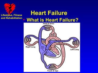
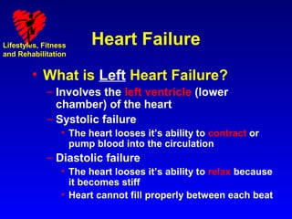
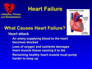
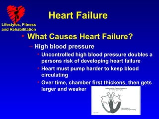
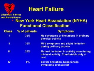
The document discusses heart failure, including its causes, types, signs and symptoms. It defines heart failure as when the heart cannot pump enough blood to meet the body's needs. Left heart failure involves the left ventricle, and can be systolic or diastolic. Right heart failure usually occurs secondary to left heart failure. Common causes include coronary artery disease, heart attack, high blood pressure, and diabetes. Signs and symptoms include shortness of breath, cough, edema, fatigue, lack of appetite, and confusion.
This document provides information about myocardial infarction (MI) or heart attack. It defines MI as death of heart muscle cells due to lack of oxygen, usually caused by a blockage in the coronary arteries. It lists risk factors for MI such as smoking, diabetes, hypertension, and family history. It describes the signs and symptoms of MI, diagnostic tests including ECG and cardiac enzymes, types of MI, and treatments including thrombolytics, angioplasty, medications, and lifestyle changes to prevent future heart attacks. The nursing management of MI focuses on reducing pain, improving perfusion, preventing complications, health education, and calling for help if symptoms worsen.
This document provides information on heart failure (CHF), including:
1) CHF is a clinical syndrome that results from any structural or functional cardiac disorder that impairs the ventricle's ability to fill or eject blood.
2) The main mechanisms of CHF are increased blood volume, increased resistance to blood flow, decreased contractility, and decreased filling.
3) CHF can be compensated initially through mechanisms like the Frank-Starling mechanism and neurohormonal activation, but these can eventually worsen heart failure.
Cardiomyopathy refers to diseases of the heart muscle that weaken the heart's ability to pump blood effectively. The three main types are dilated, hypertrophic, and restrictive cardiomyopathy. Dilated cardiomyopathy causes the left ventricle to enlarge and weaken, impairing its ability to pump blood. Causes include viral infections, toxins, genetic factors, and hypertension. Symptoms include fatigue, shortness of breath, and fluid retention. Diagnosis involves echocardiograms, electrocardiograms, and cardiac catheterization. Treatment focuses on managing symptoms through medications, lifestyle changes, and potentially surgery or transplantation.
Coronary artery disease is caused by atherosclerosis and is a leading cause of death worldwide. As atherosclerosis progresses, fatty deposits build up in the arteries and restrict blood flow. Over many years, risk factors such as high cholesterol, hypertension, smoking, and obesity can cause plaque to build up and rupture, restricting blood flow. Managing modifiable risk factors through lifestyle changes, medication, and medical treatment can help prevent complications from coronary artery disease.
This document discusses the management of hypertensive heart disease. It notes that hypertension accounts for about 25% of heart failure cases and uncontrolled hypertension can lead to left ventricular hypertrophy and dysfunction. For the case of the 78-year-old woman admitted with congestive heart failure, the key points are that she is elderly with signs of heart failure, accelerated hypertension, and normal left ventricular function. Her treatment should focus on controlling heart failure, hypertension, and any ischemia through medications like diuretics, nitrates, and antihypertensives.
ECG- Atrial Fibrillation, CXR-P/A view-Cardiomegaly,
Echocardiogram-severe mitral stenosis with severe MR with
moderate pulmonary hypertension. Patient underwent MVR and
she is doing well.
This document defines heart failure and discusses its key characteristics. It describes how heart failure occurs when the heart is unable to pump enough blood to meet the body's needs due to problems like abnormal heart muscle function or excessive loads on the heart. The document outlines the pathophysiology and progression of heart failure, including ventricular dilation and hypertrophy as compensatory mechanisms that ultimately fail. It also covers the clinical features, diagnostic tests, medical management, and nursing care considerations for patients with heart failure.
Coronary artery disease involves the buildup of plaque in the heart's arteries, reducing blood flow. It is the most common cardiovascular disease and can cause stable or unstable angina, heart attack, and heart failure. Risk factors include high cholesterol, smoking, hypertension, diabetes, and obesity. Diagnosis involves ECG, stress tests, imaging, and angiography. Treatment includes medications like nitrates, statins, and ACE inhibitors, as well as surgical procedures like angioplasty, stenting, and bypass surgery. Lifestyle changes such as quitting smoking, diet, exercise, and weight control also help manage the disease.
This document provides an overview of ischemic heart disease (IHD). IHD is caused by reduced blood flow to the heart muscle and includes conditions like angina and myocardial infarction. The main causes are atherosclerotic lesions in the coronary arteries leading to plaque buildup and blockages. Over time, plaques can rupture, causing blood clots that fully or partially block blood flow to the heart. This leads to insufficient oxygen delivery and cell death. The document outlines the pathogenesis and morphological changes that occur during angina and myocardial infarction as well as risk factors, diagnosis, and complications of IHD.
The document provides guidelines for the diagnosis and management of chronic stable angina, defining it as chest discomfort caused by myocardial ischemia that is typically triggered by exertion or stress. It discusses the pathophysiology, risk factors, diagnostic testing options including ECG, stress testing, and imaging, and recommendations for invasive coronary angiography. The guidelines are intended to help clinicians properly evaluate and treat patients experiencing chronic stable angina.
ISCHEMIA HEART DISEASE AND MYOCARDIAL INFARETIONfikri asyura
This document discusses ischemic heart disease and myocardial infarction. It covers the pathophysiology of coronary ischemia, including how myocardial oxygen demand and supply are determined. When demand exceeds supply, ischemia occurs. The document details the physiology of coronary blood flow, autoregulation, and flow reserve. It then covers the clinical syndromes of stable angina, unstable angina, and acute myocardial infarction. Key concepts include the progression of atherosclerotic plaque, the vulnerable plaque that can rupture in acute coronary syndromes, and the treatment approaches for stable and unstable ischemia.
Mitral stenosis is characterized by obstruction of blood flow from the left atrium to the left ventricle due to thickening and immobility of the mitral valve leaflets. The most common cause is rheumatic heart disease. As the stenosis progresses, the left atrial pressure rises, leading to pulmonary congestion and right-sided heart failure over time. On examination, findings may include an accentuated S1, opening snap, and mid-diastolic murmur with presystolic accentuation. Chest x-ray may show an enlarged left atrium and signs of pulmonary congestion. Treatment involves rate control for atrial fibrillation, diuretics, and potentially balloon valvuloplasty or
Myocarditis is an inflammatory disease of the heart muscle that can be caused by infectious or non-infectious triggers. It has a variable clinical presentation ranging from mild symptoms to life-threatening conditions. The diagnosis is challenging due to the heterogeneity of symptoms but can involve electrocardiogram, cardiac biomarkers, echocardiogram, cardiac MRI and endomyocardial biopsy. About half of acute cases resolve in 2-4 weeks but some develop heart failure or arrhythmias. Treatment focuses on supporting heart function and managing symptoms while the disease runs its course.
This document discusses the classification, symptoms, and treatment of heart failure. It begins by classifying heart failure based on its onset (acute or chronic), which side of the heart is affected (left or right), and its severity according to the New York Heart Association stages. The main symptoms of heart failure are then described using the mnemonic "FACES" (fatigue, activities limited, chest congestion, edema, shortness of breath). The document goes on to outline pharmacological treatments including ACE inhibitors, beta blockers, spironolactone, diuretics, and digoxin. Non-pharmacological interventions like diet, exercise, and cardiac rehabilitation are also mentioned.
Heart failure is defined as a structural or functional impairment of the heart that prevents it from maintaining an adequate blood supply to meet the body's needs. It can be caused by conditions that overload or damage the heart such as hypertension, heart attacks, and cardiomyopathy. Heart failure is classified as acute or chronic and as forward or backward based on its location and symptoms. While compensatory mechanisms initially help the heart function, long term they cause further damage and worsening of heart failure over time. Diagnosis involves tests like echocardiograms, EKGs and blood tests, and treatment focuses on diuretics, ACE inhibitors and beta blockers to manage symptoms and slow progression of the disease.
This document discusses the echocardiographic evaluation of mitral valve prolapse (MVP). It describes the use of M-mode, 2D, and 3D echocardiography to diagnose MVP and assess mitral regurgitation severity. Measurement of the vena contracta and use of the proximal isovelocity surface area method are emphasized for accurate regurgitant quantification. Surgical indications and repair techniques are also summarized.
This document discusses aortic stenosis, including its causes, symptoms, diagnosis, and treatment. It notes that aortic stenosis can be valvular, subvalvular, or supravalvular and can be caused by rheumatic fever, congenital defects, or age-related calcification. Common symptoms include chest pain, shortness of breath, fatigue, and murmurs. Diagnosis involves echocardiogram, EKG, chest X-ray and cardiac catheterization. Treatment options range from medication to manage symptoms, balloon valvuloplasty, or aortic valve replacement surgery.
The document outlines mitral regurgitation (MR), including its definition, causes, progression, signs and symptoms, and diagnostic tests. MR occurs when the mitral valve does not close properly, allowing blood to flow back into the left atrium. It begins as an acute condition but can progress to chronic compensated and decompensated phases. In acute and decompensated phases, symptoms include heart failure signs. Chronic compensated MR often presents without symptoms. Diagnosis involves physical exam, echocardiogram, and other cardiac tests.
This document summarizes the echocardiographic assessment of mitral stenosis (MS). It describes the anatomy of the mitral valve and causes of MS. Methods for assessing MS severity include measuring the pressure gradient, mitral valve area using planimetry and pressure half-time, and pulmonary artery pressure. Suitability for percutaneous transvenous mitral commissurotomy is evaluated. Concomitant valve lesions are also identified. Stress echocardiography may be used when symptoms are equivocal. Transesophageal echocardiography is recommended in some cases.
Hypertensive heart disease is caused by high blood pressure that places chronic pressure on the heart. It results in thickening of the heart muscle and reduced compliance of the left ventricle. Over time, this can lead to dilation and dysfunction of the left ventricle. The right side of the heart can also be affected by pulmonary hypertension, resulting in right ventricular hypertrophy and dilation known as cor pulmonale. Management involves controlling blood pressure to reduce the hypertrophy and restore heart size.
The aortic valve has three cusps that open and close to regulate blood flow from the heart to the aorta. Aortic stenosis occurs when the valve opening narrows due to calcium buildup on the cusps. In the elderly, aortic stenosis is usually caused by age-related degeneration and calcification of the valve. Symptoms include chest pain, shortness of breath, and fainting. Diagnosis involves echocardiogram, Doppler ultrasound and cardiac catheterization. Treatment options include medications, balloon valvuloplasty, open-heart surgery to replace the valve, and newer transcatheter aortic valve replacement procedures for high-risk elderly patients.
This document summarizes the evaluation of aortic valve stenosis using echocardiography. It describes the normal aortic valve anatomy and various types of aortic valve stenosis including calcific, bicuspid, rheumatic, and supravalvular or subvalvular stenosis. Doppler echocardiography is used to evaluate aortic valve stenosis severity based on valve area, mean gradient, and peak jet velocity. Stress echocardiography with dobutamine can help distinguish true severe from pseudo-severe low-flow, low-gradient aortic stenosis.
Cardiomyopathy slideshare Dilated cardiomyopathy Hypertrophic cardiomyopathy ...Adarsh SA
Cardiomyopathy is a disease of the heart muscle that makes it harder for the heart to pump blood. There are several types classified by structural and functional abnormalities. The main types are dilated, hypertrophic, restrictive, and arrhythmogenic right ventricular cardiomyopathy. Dilated cardiomyopathy is the most common type and causes the heart chambers to enlarge and the heart muscle to thin and weaken over time, reducing its ability to pump blood. Management involves identifying underlying causes, treating heart failure symptoms, and controlling arrhythmias.
The document discusses mitral stenosis, including its anatomy, causes, pathogenesis, clinical presentation, investigations and treatments. Some key points:
- Mitral stenosis is a narrowing of the mitral valve opening caused by conditions like rheumatic fever. It reduces blood flow from the left atrium to the left ventricle.
- Symptoms include shortness of breath, cough, fatigue and leg swelling due to increased pulmonary pressures. Examination may reveal an irregular heartbeat and diastolic murmur.
- Echocardiogram can measure the valve area and assess complications. Treatment options range from medical management to surgical procedures like balloon valvuloplasty or valve replacement.
Myocarditis is an inflammatory disease of the heart muscle that is usually caused by viral infections. It can lead to dilated cardiomyopathy and heart failure. Viruses are the most common cause, with adenovirus now more prevalent than coxsackievirus. Myocarditis presents with symptoms of heart failure, chest pain, or arrhythmias. Diagnosis involves EKG, cardiac biomarkers, echocardiogram, cardiac MRI, and endomyocardial biopsy. Treatment focuses on managing arrhythmias and heart failure with medications, while immunosuppression may benefit some forms of myocarditis but not others.
This document discusses acute heart failure, including its definition, causes, clinical presentations, and pathophysiology. Acute heart failure is rapid onset heart failure that can occur with or without previous cardiac disease. It is often life-threatening and requires urgent treatment. The patient may present with acute decompensated heart failure, hypertensive acute heart failure, pulmonary edema, cardiogenic shock, or high output failure. The pathophysiology involves a vicious cycle where the heart cannot maintain sufficient cardiac output to meet demands, leading to further worsening if not treated.
Heart failure is a condition where the heart cannot pump enough blood to meet the body's needs. It has many potential causes, but is often due to problems with the heart muscle itself or valves. Treatment focuses on managing symptoms with diuretics, and slowing progression with ACE inhibitors, beta-blockers, and aldosterone antagonists. Other therapies aim to improve heart function or treat underlying causes. Prognosis depends on severity but ranges from 5-50% annual mortality.
Acute left ventricular failure is sudden worsening of dyspnea or congestive heart failure caused by conditions that increase left ventricular pressure like aortic stenosis or acute valve regurgitation. It leads to pulmonary edema as hydrostatic pressure in lungs exceeds plasma oncotic pressure, causing fluid to exude into alveoli. Patients experience terrifying breathing difficulties and pink frothy sputum. On exam, they are pale, sweaty and tachycardic with rales and S3 gallop, indicating severe dysfunction. Treatment focuses on oxygen, diuretics, morphine and positioning to relieve anxiety and dyspnea while reducing preload on the heart.
Left ventricular failure occurs when the left ventricle fails to pump blood successfully, causing backpressure in the pulmonary circulation and pooling of blood. This can lead to acute pulmonary edema if left untreated. The patient presented with shortness of breath, tachycardia, crackles in both lungs, and edema. Treatment focused on improving oxygenation, decreasing venous return, reducing myocardial oxygen demand and off-loading the ventricles using oxygen, nitrates like nitroglycerin, and furosemide. Timely diagnosis and treatment of congestive cardiac failure remains important.
The document summarizes key information about acute heart failure, including epidemiology, pathophysiology, treatment approaches, and trial data. It describes the ASCEND-HF trial which investigated the effects of nesiritide vs placebo on outcomes in over 7,000 patients hospitalized for acute decompensated heart failure. The trial found no significant differences between nesiritide and placebo for its co-primary endpoints of 30-day mortality or heart failure rehospitalization and dyspnea relief at 6 and 24 hours.
Left-sided heart failure occurs when the left side of the heart, which pumps oxygenated blood to the body, cannot pump sufficiently. This causes a back-up of blood in the lungs, resulting in respiratory symptoms like shortness of breath and cough. It is usually due to conditions that damage or weaken the heart muscle over time such as heart attacks or high blood pressure. Treatment involves lifestyle changes and medications to help the heart function more efficiently and reduce symptoms.
Acute heart failure: diagnosing and managing acute heart failure in adultsEmergency Live
The need for this guideline was identified as the NICE guidelines on chronic heart failure were being updated. We recognised at this time that there were important aspects of the diagnosis and management of acute heart failure that were not being addressed by the chronic heart failure guideline, which focussed on long term management rather than the immediate care of someone who is acutely unwell as a result of heart failure. The aim of this guideline is to provide guidance to the NHS on the diagnosis and management of acute heart failure.
Heart failure is a condition in which the heart does not pump enough blood to meet all the needs of the body. It is caused by heart muscle damage or dysfunction, valve problems, heart rhythm disturbances and other rarer causes. Acute heart failure can present as new-onset heart failure in people without known cardiac dysfunction, or as acute decompensation of chronic heart failure.
Acute heart failure is a common cause of admission to hospital (over 67,000 admissions in England and Wales per year) and is the leading cause of hospital admission in people 65 years or older in the UK.
This guideline includes important aspects of the diagnosis and management of acute heart failure that are not addressed by the NICE guideline on chronic heart failure (NICE clinical guideline 108). The guideline on chronic heart failure focused on long-term management rather than the immediate care of someone who is acutely unwell as a result of heart failure.
This guideline covers the care of adults (aged 18 years or older) who have a diagnosis of acute heart failure, have possible acute heart failure, or are being investigated for acute heart failure. It includes the following key clinical areas.
This document discusses heart failure, including its definitions, types, causes, clinical manifestations, and management from a dental perspective. It defines heart failure as the heart's inability to supply sufficient oxygenated blood to the body's needs. The types include left ventricle, right ventricle, and combined failure. Causes include congenital heart defects and conditions like cardiomyopathy. Clinical manifestations vary depending on which ventricle is affected but may include edema, dyspnea, fatigue, and cyanosis. Dental management involves recognizing signs of failure, terminating procedures if needed, positioning the patient comfortably, and providing post-procedure care and follow-up.
This document discusses congestive heart failure (CHF). It provides epidemiological data on CHF, showing it affects millions of people worldwide and costs billions of dollars annually. It defines CHF as the heart's inability to meet circulatory demands and classifies it based on location (left vs right heart) and time course (acute vs chronic). Causes of acute and chronic CHF include myocardial infarction, hypertension, valvular diseases, and cardiomyopathies. The pathophysiology of CHF involves systolic and diastolic dysfunction that can lead to ventricular hypertrophy, dilation, and neurohormonal activation causing further organ damage.
Pathophysiology of congestive heart failurethunderrajesh
This document provides an overview of congestive heart failure, including its definition, types, causes, symptoms, complications, diagnosis, and treatment. Congestive heart failure occurs when the heart muscle is weakened and cannot pump blood effectively, leading to fluid buildup in tissues and organs. The main types are systolic and diastolic dysfunction. Common causes include hypertension, coronary artery disease, and valvular issues. Symptoms involve fatigue, shortness of breath, and swelling. Treatment focuses on medications like ACE inhibitors, diuretics, beta blockers, and lifestyle changes such as diet, exercise, and stress reduction.
This document provides an overview of the management of acute left ventricular failure (ALVF). It begins with definitions and causes of acute heart failure and ALVF. The pathophysiology, signs and symptoms, classifications, diagnosis and differential diagnosis of ALVF are described. The goals and algorithms for treatment in the emergency department and pharmacological therapies including diuretics, vasodilators, inotropes, vasopressors and other drugs are summarized. Non-pharmacological therapies, mechanical circulatory support, post-stabilization care, and long-term management are also outlined. The document concludes with a discussion of potential new therapies, prognosis, and underlying diseases/comorbidities in acute heart failure.
The document discusses the inpatient management of acute decompensated heart failure. Key points include identifying signs and symptoms of ADHF, obtaining pertinent labs and imaging to evaluate the patient, and treating with intravenous diuretics followed by chronic medications like ACE inhibitors, beta blockers, and spironolactone prior to discharge with close follow up.
This document summarizes the treatment of acute decompensated heart failure (ADHF). It describes ADHF as rapid accumulation of fluid in the lungs due to left ventricular dysfunction, often from conditions like coronary artery disease or hypertension. Initial treatment involves stabilization, monitoring, diuresis, and supplemental oxygen. Diuretics are the primary treatment but vasodilators may also be used, and choices depend on the patient's blood pressure and volume status. Additional treatments discussed include inotropes, vasopressors, beta blockers, and mechanical cardiac support.
A brief synopsis of acute decompensated heart failureDr Emad efat
This document provides an overview of acute decompensated heart failure (ADHF). It defines ADHF as a clinical syndrome characterized by the development of respiratory distress due to rapidly accumulated fluid in the lungs. The document categorizes heart failure based on systolic vs diastolic function, left vs right sided, acute vs chronic onset, and NYHA functional classification. Common symptoms, physical exam findings, causes, risk factors, differential diagnoses, and initial investigations are described. Imaging findings on chest x-ray indicative of different stages of heart failure are also summarized.
This document discusses the management of bradycardia. It defines bradycardia as a heart rate under 50 beats per minute and distinguishes between symptomatic, unstable bradycardia requiring immediate intervention and asymptomatic cases not in imminent danger. It outlines treatment for different types of heart block including first, second-degree Mobitz I and II, and third-degree AV block. The document was presented by Eder Augusto Hernández Ruiz from the Universidad del Norte.
Bradyarrhythmias are caused by problems with impulse formation in the sinus node or impulse conduction through the AV node. Sinus node dysfunction can cause sinus bradycardia, sinus pause/arrest, or chronotropic incompetence. Atrioventricular block is classified as first, second, or third degree and may be caused by conditions like CAD, drugs, or infiltrative diseases. Second degree AV block is further classified as Mobitz type I or II based on PR interval characteristics. Third degree AV block causes complete dissociation between atrial and ventricular rhythms.
- Right heart failure is characterized by low cardiac output, hypotension, hepatic enlargement and raised jugular venous pressure. It has a high mortality rate comparable to left heart failure.
- Failure occurs when the right ventricle can no longer compensate for increased volume. Determining preload is difficult but high right atrial pressures indicate elevated right ventricular pressures and volume.
- Treatment aims to reduce afterload and optimize preload. Afterload reduction can be achieved through selective pulmonary vasodilation using inhaled nitric oxide, prostacyclins or phosphodiesterase inhibitors.
This document discusses acute heart failure (AHF), including its definition, initial signs of end organ dysfunction, potential end organ dysfunction, and causes. It notes that AHF often arises from a deterioration of chronic heart failure and may be a first presentation. It also discusses biomarkers like BUN and hyponatremia as predictors of outcomes. The document covers diuretic use and its association with adverse events, as well as approaches to decongestion. It defines cardiorenal syndrome in AHF and notes its association with worse outcomes. Various inotropic agents are also discussed.
Heart failure occurs when the heart cannot pump enough blood to meet the body's needs. It is usually caused by conditions that damage or overwork the heart over time. Symptoms include shortness of breath, swelling, and fatigue. Treatment focuses on lifestyle changes, medications to improve pumping ability and reduce workload, and occasionally surgery. The goal is to control symptoms and slow the progression of the disease.
Congestive cardiac failure occurs when the heart cannot pump enough blood to meet the body's demands. It can be caused by conditions that damage the heart or make it work harder over time. Symptoms include shortness of breath, swelling, fatigue, and confusion. Treatment focuses on lifestyle changes, medications, and sometimes surgery. Medications aim to reduce the heart's workload by dilating blood vessels, removing fluid buildup, or increasing pumping ability. The goal is to manage symptoms and slow the progression of heart failure.
Heart failure occurs when the heart cannot pump enough blood to meet the body's needs. Fluid backs up in the lungs and body, causing congestion. Common causes include coronary artery disease, heart attacks, high blood pressure, and congenital or valvular heart defects. Symptoms include shortness of breath, swelling, fatigue, and reduced exercise capacity. Treatment involves medications, lifestyle changes, and potentially surgery or devices. Nursing focuses on managing symptoms like fatigue and anxiety while monitoring for fluid retention.
The document calculates the average lifetime total heartbeats for a person based on average heart rate, lifespan, and conversions between heartbeats per minute/hour/day/year. It finds that the average lifetime total heartbeats is approximately 2.8 billion beats.
A brief presentation on anatomy of heart, different types of cardiovascular disease, their symptoms, causes, prevention methods and medical treatments available to cure heart disease.
Heart disease refers to conditions that affect the heart and impair its function. The main types of heart disease are coronary artery disease, angina, heart attack, heart failure, and arrhythmia. Risk factors for heart disease include smoking, high cholesterol, high blood pressure, diabetes, being overweight, physical inactivity, and an unhealthy diet. Lifestyle changes like quitting smoking, eating healthy, exercising regularly, losing excess weight, and reducing stress can help reduce the risk of heart disease.
Heart disease and its risk factors 17103053ahique1234
Heart disease refers to conditions that affect the heart and impair its function. The main types of heart disease are coronary artery disease, angina, heart attack, heart failure, and arrhythmia. Risk factors for heart disease include smoking, high cholesterol, high blood pressure, diabetes, being overweight, physical inactivity, and an unhealthy diet. Lifestyle changes like quitting smoking, eating healthy, exercising regularly, losing excess weight, and reducing stress can help reduce the risk of heart disease.
These slides are for Yoga Teachers or students of Yoga for understanding the disease and what Yoga program we can offer to our client when they reach you for help. Although every individual is unique and Yoga Therapy should also be made considering what level of disease they are going through.
Disclaimer: We dont take any responsibility if someone starts to follow the program as mentioned in the PPT for any harm or injury.
Heart failure is a condition where the heart cannot pump enough blood to meet the body's needs. It affects nearly 5 million Americans and is common, though not the same as a heart attack. The main symptoms are fatigue, limited activity, chest congestion, swelling, and shortness of breath. While there is no cure, early diagnosis and treatment with medications like diuretics, ACE inhibitors, and beta blockers can slow the progression, prevent hospitalization, and save lives for those living with heart failure.
Hypertension (high blood pressure) is when the pressure in your blood vessels is too high (140/90 mmHg or higher). It is common but can be serious if not treated. People with high blood pressure may not feel symptoms. The only way to know is to get your blood pressure checked.
The document discusses hypertension (high blood pressure) including its causes, symptoms, risks, diagnosis, treatment, and self-management. It defines normal and high blood pressure, explains how it is measured, and lists lifestyle factors and medications that can control it. The ten commandments of blood pressure control are also outlined, emphasizing weight, diet, exercise, medication adherence, and regular monitoring by a healthcare provider.
The document discusses hypertension (high blood pressure) including definitions of normal and high blood pressure, symptoms, causes, health risks, and treatment options. It defines blood pressure and the normal range as below 140/90 mmHg. High blood pressure usually has no symptoms and is called the "silent killer". Risk factors include age, genetics, obesity, lack of exercise, alcohol, stress and salt intake. Treatment involves lifestyle changes like diet, exercise, weight loss and medication if needed.
This document discusses hypertension (high blood pressure) including:
1. It defines hypertension as persistent high blood pressure that can damage vessels and organs like the heart, brain, kidneys, and eyes.
2. It identifies risk factors for primary (essential) hypertension such as age, heredity, sex, race, obesity, sodium intake, alcohol, stress, and other conditions.
3. It describes potential complications of uncontrolled hypertension like heart disease, heart failure, stroke, kidney damage, and retinal damage.
4. It provides an overview of hypertension diagnosis, treatment including lifestyle changes and medications, management, and nursing considerations.
This document discusses heart disease and dietary recommendations for prevention. It defines different types of cardiovascular diseases and risk factors like atherosclerosis, hypertension, and diabetes. Lifestyle factors that can increase risk are discussed such as unhealthy diet, physical inactivity, obesity, smoking, and excessive alcohol. The document provides dietary recommendations from reducing saturated fats, sodium, and sugar intake to eating more fruits/vegetables and whole grains. Physical activity guidelines and maintaining a healthy weight are also summarized as important prevention strategies.
Congestive heart failure is a condition where the heart cannot pump enough blood to meet the body's needs. It occurs when the heart muscle is damaged, such as from a heart attack or conditions that overwork the heart like high blood pressure. Symptoms include shortness of breath, swelling in the legs or abdomen, and fatigue. Treatment focuses on lifestyle changes, medications to manage symptoms and improve heart function, and sometimes surgery or implanted devices. Ongoing monitoring is needed to watch for changes in symptoms or physical signs that may indicate worsening of the condition.
Congestive heart failure occurs when the heart muscle is unable to pump enough blood to meet the body's needs. There are two main types - left-sided heart failure which prevents the body from getting enough oxygen-rich blood, and right-sided heart failure which occurs when the right side of the heart cannot perform effectively. Symptoms range from fatigue at first to shortness of breath, swelling, and fainting at more severe stages. Risk factors include high blood pressure, coronary artery disease, diabetes, and certain medications. Treatment involves lifestyle changes and medications, with the condition managed to varying degrees depending on its stage and severity.
Congestive heart failure occurs when the heart cannot adequately pump blood to meet the body's needs. It can be caused by conditions like coronary artery disease, heart attacks, or viruses. Common symptoms include shortness of breath, fatigue, swelling, and coughing. Medications help decrease cardiac workload and fluid retention. Community paramedics help with medication compliance by identifying reasons for non-compliance and providing education. They monitor for signs of acute heart failure like sudden fluid buildup or rapid heartbeat. Proper medical care of CHF can improve symptoms, though lifestyle changes and lifelong treatment are usually needed.
The document discusses the heart, including its anatomy, function, common diseases, symptoms, treatment, and prevention. It describes how the heart works by pumping blood through two circuits to oxygenate blood and supply the body. Common heart diseases include coronary artery disease, congenital defects, arrhythmias, and heart failure. Symptoms vary but can include chest pain and fatigue. Treatment depends on the condition but may involve lifestyle changes, medications, or surgery. Controling risk factors like high blood pressure and smoking can help prevent heart disease. The prognosis of heart failure is generally poor without treatment.
Rasamanikya is a excellent preparation in the field of Rasashastra, it is used in various Kushtha Roga, Shwasa, Vicharchika, Bhagandara, Vatarakta, and Phiranga Roga. In this article Preparation& Comparative analytical profile for both Formulationon i.e Rasamanikya prepared by Kushmanda swarasa & Churnodhaka Shodita Haratala. The study aims to provide insights into the comparative efficacy and analytical aspects of these formulations for enhanced therapeutic outcomes.
Here is the updated list of Top Best Ayurvedic medicine for Gas and Indigestion and those are Gas-O-Go Syp for Dyspepsia | Lavizyme Syrup for Acidity | Yumzyme Hepatoprotective Capsules etc
8 Surprising Reasons To Meditate 40 Minutes A Day That Can Change Your Life.pptxHolistified Wellness
We’re talking about Vedic Meditation, a form of meditation that has been around for at least 5,000 years. Back then, the people who lived in the Indus Valley, now known as India and Pakistan, practised meditation as a fundamental part of daily life. This knowledge that has given us yoga and Ayurveda, was known as Veda, hence the name Vedic. And though there are some written records, the practice has been passed down verbally from generation to generation.
These lecture slides, by Dr Sidra Arshad, offer a quick overview of the physiological basis of a normal electrocardiogram.
Learning objectives:
1. Define an electrocardiogram (ECG) and electrocardiography
2. Describe how dipoles generated by the heart produce the waveforms of the ECG
3. Describe the components of a normal electrocardiogram of a typical bipolar lead (limb II)
4. Differentiate between intervals and segments
5. Enlist some common indications for obtaining an ECG
6. Describe the flow of current around the heart during the cardiac cycle
7. Discuss the placement and polarity of the leads of electrocardiograph
8. Describe the normal electrocardiograms recorded from the limb leads and explain the physiological basis of the different records that are obtained
9. Define mean electrical vector (axis) of the heart and give the normal range
10. Define the mean QRS vector
11. Describe the axes of leads (hexagonal reference system)
12. Comprehend the vectorial analysis of the normal ECG
13. Determine the mean electrical axis of the ventricular QRS and appreciate the mean axis deviation
14. Explain the concepts of current of injury, J point, and their significance
Study Resources:
1. Chapter 11, Guyton and Hall Textbook of Medical Physiology, 14th edition
2. Chapter 9, Human Physiology - From Cells to Systems, Lauralee Sherwood, 9th edition
3. Chapter 29, Ganong’s Review of Medical Physiology, 26th edition
4. Electrocardiogram, StatPearls - https://www.ncbi.nlm.nih.gov/books/NBK549803/
5. ECG in Medical Practice by ABM Abdullah, 4th edition
6. Chapter 3, Cardiology Explained, https://www.ncbi.nlm.nih.gov/books/NBK2214/
7. ECG Basics, http://www.nataliescasebook.com/tag/e-c-g-basics
TEST BANK For An Introduction to Brain and Behavior, 7th Edition by Bryan Kol...rightmanforbloodline
TEST BANK For An Introduction to Brain and Behavior, 7th Edition by Bryan Kolb, Ian Q. Whishaw, Verified Chapters 1 - 16, Complete Newest Versio
TEST BANK For An Introduction to Brain and Behavior, 7th Edition by Bryan Kolb, Ian Q. Whishaw, Verified Chapters 1 - 16, Complete Newest Version
Basavarajeeyam is a Sreshta Sangraha grantha (Compiled book ), written by Neelkanta kotturu Basavaraja Virachita. It contains 25 Prakaranas, First 24 Chapters related to Rogas& 25th to Rasadravyas.
Muktapishti is a traditional Ayurvedic preparation made from Shoditha Mukta (Purified Pearl), is believed to help regulate thyroid function and reduce symptoms of hyperthyroidism due to its cooling and balancing properties. Clinical evidence on its efficacy remains limited, necessitating further research to validate its therapeutic benefits.
1. Lifestyles, Fitness
and Rehabilitation
Heart Failure
CCF/CHF
LVF 1st + RVF 2nd = CCF/CHF
By :
Mr Murdin b Amit
Bsc in Health Science (Hons), Ba in Buissness Administration (Hrm)(Hons)
Head of Medical Science Subject
College of Assistant Medical Officer,
Seremban, Negeri Sembilan
murdin_1966@yahoo.com
HP: 012 - 6044385
2. Lifestyles, Fitness Heart Failure
and Rehabilitation
• What is Heart Failure?
– The heart is not pumping as well as it
should
– Usually, the heart has been weakened
by an underlying condition
• Blocked arteries
• Heart attack
• High blood pressure
• Infections
• Heart valve abnormalities
3. Lifestyles, Fitness Heart Failure
and Rehabilitation
• What is Heart Failure?
– Heart failure can involve the left or right
side of the heart or both
– Usually the left side is affected first
– Heart failure occurs when either side of
the heart cannot keep up with the flow
of blood
4. Lifestyles, Fitness Heart Failure
and Rehabilitation
• What is Heart Failure?
5. Lifestyles, Fitness Heart Failure
and Rehabilitation
• What is Left Heart Failure?
– Involves the left ventricle (lower
chamber) of the heart
– Systolic failure
• The heart looses it’s ability to contract or
pump blood into the circulation
– Diastolic failure
• The heart looses it’s ability to relax because
it becomes stiff
• Heart cannot fill properly between each beat
6. Lifestyles, Fitness Heart Failure
and Rehabilitation
• What is Left Heart Failure?
– Systolic and diastolic heart failure are
treated with different types of
medications
– In both types, blood may “back up” in
the lungs causing fluid to leak into the
lungs (pulmonary edema)
– Fluid may also build up in tissues
throughout the body (edema)
7. Lifestyles, Fitness Heart Failure
and Rehabilitation
• What is Right Heart Failure?
– Usually occurs as a result of left heart
failure
– The right ventricle pumps blood to the
lungs for oxygen
– Occasionally isolated right heart failure
can occur due to lung disease or blood
clots to the lung (pulmonary embolism)
8. Lifestyles, Fitness Heart Failure
and Rehabilitation
• How fast does heart failure
develop?
– Usually a chronic disease
– The heart tries to compensate for the
loss in pumping function by:
• Developing more muscle mass
• Enlarging
• Pumping faster
9. Lifestyles, Fitness Heart Failure
and Rehabilitation
• What Causes Heart Failure?
– Health conditions that either damage
the heart or make it work too hard
• Coronary artery disease
• Heart attack
• High blood pressure
• Abnormal heart valves
• Heart muscle diseases (cardiomyopathy)
• Heart inflammation (myocarditis)
10. Lifestyles, Fitness Heart Failure
and Rehabilitation
• What Causes Heart Failure?
• Congenital heart defects
• Severe lung disease
• Diabetes
• Severe anemia
• Overactive thyroid gland (hyperthyroidism)
• Abnormal heart rhythms
11. Lifestyles, Fitness Heart Failure
and Rehabilitation
• What Causes Heart Failure?
– Coronary artery disease
• Cholesterol and fatty deposits build up in
the heart’s arteries
• Less blood and oxygen reach the heart
muscle
• This causes the heart to work harder and
occasionally damages the heart muscle
12. Heart Failure
Lifestyles, Fitness
and Rehabilitation
• What Causes Heart Failure?
– Heart attack
• An artery supplying blood to the heart
becomes blocked
• Loss of oxygen and nutrients damages
heart muscle tissue causing it to die
• Remaining healthy heart muscle must pump
harder to keep up
13. Heart Failure
Lifestyles, Fitness
and Rehabilitation
• What Causes Heart Failure?
– High blood pressure
• Uncontrolled high blood pressure doubles a
persons risk of developing heart failure
• Heart must pump harder to keep blood
circulating
• Over time, chamber first thickens, then gets
larger and weaker
14. Heart Failure
Lifestyles, Fitness
and Rehabilitation
• What Causes Heart Failure?
– Abnormal heart valves
– Heart muscle disease
• Damage to heart muscle due to drugs,
alcohol or infections
– Congenital heart disease
– Severe lung disease
15. Heart Failure
Lifestyles, Fitness
and Rehabilitation
• What Causes Heart Failure?
– Diabetes
• Tend to have other conditions that make the
heart work harder
• Obesity
• Hypertension
• High cholesterol
16. Heart Failure
Lifestyles, Fitness
and Rehabilitation
• What Causes Heart Failure?
– Severe anemia
• Not enough red blood cells to carry oxygen
• Heart beats faster and can become overtaxed with
the effort
– Hyperthyroidism
• Body metabolism is increased and overworks the
heart
– Abnormal Heart Rhythm
• If the heart beats too fast, too slow or irregular it may
not be able to pump enough blood to the body
17. Heart Failure
Lifestyles, Fitness
and Rehabilitation
• Signs and Symptoms of Heart Failure
– Shortness of Breath (dyspnea)
• WHY?
– Blood “backs up” in the pulmonary veins
because the heart can’t keep up with the supply
an fluid leaks into the lungs
• SYMPTOMS
– Dyspnea on exertion or at rest
– Difficulty breathing when lying flat
– Waking up short of breath
18. Heart Failure
Lifestyles, Fitness
and Rehabilitation
• Signs and Symptoms of Heart Failure
– Persistent Cough or Wheezing
• WHY?
– Fluid “backs up” in the lungs
• SYMPTOMS
– Coughing that produces white or pink blood-
tinged sputum (Froty Sputum)
19. Heart Failure
Lifestyles, Fitness
and Rehabilitation
• Signs and Symptoms of Heart Failure
– Edema
• WHY?
– Decreased blood flow out of the weak heart
– Blood returning to the heart from the veins
“backs up” causing fluid to build up in tissues
• SYMPTOMS
– Swelling in feet, ankles, legs or abdomen
– Weight gain
20. Heart Failure
Lifestyles, Fitness
and Rehabilitation
• Signs and Symptoms of Heart Failure
– Tiredness, fatigue
• WHY?
– Heart can’t pump enough blood to meet needs of
bodies tissues
– Body diverts blood away from less vital organs
(muscles in limbs) and sends it to the heart and
brain
• SYMPTOMS
– Constant tired feeling
– Difficulty with everyday activities
21. Heart Failure
Lifestyles, Fitness
and Rehabilitation
• Signs and Symptoms of Heart Failure
– Lack of appetite/ Nausea
• WHY?
– The digestive system receives less blood
causing problems with digestion
– Physiological disturbance to Vagus nerve
• SYMPTOMS
– Feeling of being full or sick to your stomach
22. Heart Failure
Lifestyles, Fitness
and Rehabilitation
• Signs and Symptoms of Heart Failure
– Confusion/ Impaired thinking
• WHY?
– Changing levels of substances in the blood
( sodium) can cause confusion
– Lack of O2 reach to the brain
• SYMPTOMS
– Memory loss or feeling of disorientation
– Relative or caregiver may notice this first
23. Heart Failure
Lifestyles, Fitness
and Rehabilitation
• Signs and Symptoms of Heart Failure
– Increased heart rate
• WHY?
– The heart beats faster to “make up for” the loss
in pumping function
• SYMPTOMS
– Heart palpitations
– May feel like the heart is racing or throbbing
24. Heart Failure
Lifestyles, Fitness
and Rehabilitation
• New York Heart Association (NYHA)
Functional Classification
Class % of patients Symptoms
I 35% No symptoms or limitations in ordinary
physical activity
II 35% Mild symptoms and slight limitation
during ordinary activity
III 25% Marked limitation in activity even during
minimal activity. Comfortable only at
rest
IV 5% Severe limitation. Experiences
symptoms even at rest
25. Heart Failure
Lifestyles, Fitness
and Rehabilitation
• Treatment Options
– The more common forms of heart failure
cannot be cured, but can be treated
• Lifestyle changes
• Medications
• Surgery
27. Heart Failure
Lifestyles, Fitness
and Rehabilitation
• Lifestyle changes
– Reduce stress
– Keep track of symptoms and weight and
report any changes or concern to the
doctor
– Limit fluid intake
– See the doctor more frequently
28. Heart Failure
Lifestyles, Fitness
and Rehabilitation
• Medications used to treat Heart
Failure
– ACE Inhibitors (Enalapril 5-10mg bd/tds)
• Cornerstone of heart failure therapy
• Proven to slow the progression of heart
failure
• Vasodilator – cause blood vessels to
expand lowering blood pressure and the
hearts work load
29. Heart Failure
Lifestyles, Fitness
and Rehabilitation
• Medications used to treat Heart Failure
– Diuretics (water pills) – Frusemide 40-80mg
• Prescribed for fluid build up, swelling or edema
• Cause kidneys to remove more sodium and
water from the bloodstream
• Decreases workload of the heart and edema
• Fine balance – removing too much fluid can
strain kidneys or cause low blood pressure
30. Heart Failure
Lifestyles, Fitness
and Rehabilitation
• Medications used to treat Heart
Failure
– Potassium
• Most diuretics remove potassium from the
body
• Potassium pills compensate for the amount
lost in the urine
• Potassium helps control heart rhythm and
is essential for the normal work of the
nervous system and muscles
31. Heart Failure
Lifestyles, Fitness
and Rehabilitation
• Medications used to treat Heart Failure
– Vasodilators
• Hydrallazine 100mg stat or Prazosin 1-2mg bd/tds
• Cause blood vessel walls to relax
• Occasionally used if patient cannot tolerate ACE
• Decrease workload of the heart
32. Heart Failure
Lifestyles, Fitness
and Rehabilitation
• Medications used to treat Heart Failure
– Digitalis preparations
• Digoxin 0.25mg-0.5mg bd
• Increases the force of the hearts contractions,
increases the blood stroke volume
• Relieves symptoms
• Slows heart rate and certain irregular heart beats
33. Heart Failure
Lifestyles, Fitness
and Rehabilitation
• Medications used to treat Heart Failure
– Beta-blockers (Tab Metoprolol 50-200mg)
• Lower the heart rate and blood pressure
• Decrease the workload of the heart
– Blood-thinners (coumadin)
• Used in patients at risk for developing blood clots
in the blood vessels, legs, lung and heart
• Used in irregular heart rhythms due to risk of
stroke
34. Heart Failure
Lifestyles, Fitness
and Rehabilitation
• Treatment options
– Surgery and other Medical Procedures
• Not often used in heart failure unless there
is a correctable problem
• Coronary artery bypass
• Angioplasty
• Valve replacement
• Defibrillator implantation
• Heart transplantation
• Left ventricular assist device (LVAD)