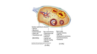
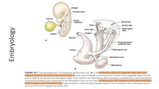
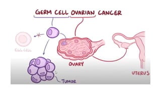
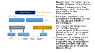
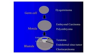
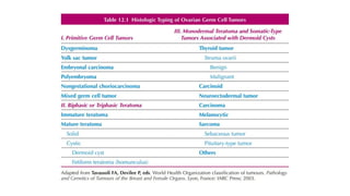
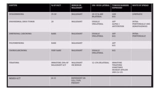
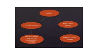
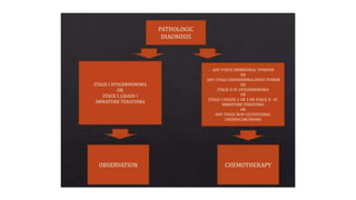
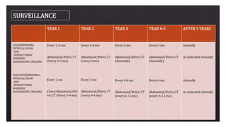
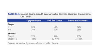
This document discusses germ cell tumors of the ovary. It begins by explaining that germ cell tumors originate from primordial germ cells and make up about 90-95% of ovarian malignancies in young women. It then covers the various subtypes of germ cell tumors, including their incidence rates, typical patient demographics, clinical presentations, diagnostic markers, pathological classifications, treatment approaches, and prognosis. Dysgerminoma is discussed as the most common subtype, while immature teratoma, endodermal sinus tumor, embryonal carcinoma, and choriocarcinoma are also described in terms of their defining characteristics and management. Throughout, the focus remains on applying knowledge from testicular germ cell tumor research
The document discusses breast-conserving treatment for early-stage breast cancer. Breast-conserving treatment, including wide local excision of the tumor, axillary lymph node dissection, and breast irradiation, is now the standard of care for most women with early-stage invasive breast cancer. Ideal candidates for breast-conserving treatment have unicentric primary tumors less than 4-5 cm in diameter. Contraindications include positive margins, advanced or multicentric disease, pregnancy, and prior radiation. The addition of a radiation boost to the tumor bed after whole breast irradiation reduces the risk of local recurrence. Hypofractionated regimens have been shown to be as effective as conventional fractionation with shorter treatment times.
This document discusses various radiotherapy techniques used for breast cancer treatment planning and delivery. It covers topics like positioning, immobilization, target volume delineation, treatment planning, dose prescription, and techniques to minimize doses to organs at risk. Some key points include the use of breast boards and immobilization devices for reproducible patient setup, delineating whole breast or chest wall as well as nodal clinical target volumes, employing tangential fields with wedges or compensators for uniform dose distribution, and using additional fields like supraclavicular or internal mammary nodes when indicated. The goal is to adequately cover targets while minimizing doses to lungs, heart, and contralateral breast.
Radiotherapy is used as primary treatment for early-stage Hodgkin lymphoma or as part of combined modality treatment with chemotherapy. Historically, large mantle fields covering lymph node regions from the skull to the pelvis were used. More modern approaches use smaller involved field radiotherapy targeting only initially involved lymph node regions after chemotherapy based on imaging. Proper delineation of clinical target volumes requires pre-chemotherapy imaging ideally with PET/CT to define original disease extent.
This document discusses hemi body irradiation (HBI) technique used to treat metastatic cancer. HBI involves irradiating only the upper or lower half of the body using parallel opposed radiation fields. It has advantages over total body irradiation like smaller field size and less side effects. HBI is used to palliate widely metastatic disease and as adjuvant therapy for certain cancers. Potential complications include nausea, diarrhea, pneumonitis and hematological effects. The document also provides an overview of cancer registries in India, which systematically collect cancer data to help understand cancer patterns and guide control programs. Population-based and hospital-based registries use active and passive methods to collect data on cancer incidence, stages and survival.
This is a made easy summary of ICRU 89 guidelines for gynecological brachytherapy. Extra practical questions for MD/DNB Radiotherapy exams are also attached.
The document discusses key concepts and parameters for prescribing, recording, and reporting photon beam therapy as defined by ICRU Report 50. It outlines volumes such as the gross tumor volume (GTV), clinical target volume (CTV), planning target volume (PTV), treated volume, and irradiated volume. It also discusses doses like the prescribed dose, maximum dose, and minimum dose. Parameters like the internal margin, set-up margin, and ICRU reference point and dose are introduced to help standardize reporting across centers for effective information exchange.
1) The document discusses various radiation techniques for treating cancer of the esophagus including 2D, 3D conformal radiation therapy, IMRT, and IGRT.
2) It covers topics like target volume delineation, field design considerations for different esophageal subsites, and evolution from 2D to 3D treatment planning.
3) While there is no consensus, most contemporary trials use margins of 3-5cm cranially and caudally on the gross tumor with approximately a 2cm radial margin.
This is a presentation on total body irradiation. This presentation explains about various techniques. positions used for TBI. Advantages and disadvantages of TBI.
It also gives an idea on Dosage and side effects.
This document discusses the management of urinary bladder carcinomas. It begins with epidemiology and risk factors, then covers diagnosis and staging. For non-muscle invasive bladder cancer (NMIBC), it describes transurethral resection of bladder tumor (TURBT) followed by adjuvant BCG or chemotherapy. For muscle invasive bladder cancer (MIBC), options discussed are radical cystectomy or bladder preservation protocols using trimodality therapy. Radiotherapy plays a role in bladder preservation or post-operatively in certain high risk cases.
The combined use of radiation therapy and chemotherapy in cancer treatment is a logical and reasonable approach that has already proven beneficial for several malignancies.
Prophylactic cranial irradiation (PCI) is used to prevent brain metastases in cancers with a high risk of spreading to the brain. It is indicated for small cell lung cancer and certain leukemias. PCI significantly reduces the rate of brain metastases in small cell lung cancer, especially when administered early at higher doses. For extensive stage small cell lung cancer, MRI surveillance may be an alternative to PCI. While PCI reduces brain metastases in leukemia, the risk of brain involvement is low for some types such as AML. The standard dose for PCI is 1200-1800 cGy in fractions, with timing and volumes depending on the cancer type. Potential toxicities include neurocognitive effects, endocrine disorders, and secondary cancers.
This document discusses dose-volume histograms (DVHs) which are used to analyze and compare radiation dose distributions in radiotherapy treatment planning. It describes how DVHs are generated by counting the number of voxels receiving different dose levels. DVHs can be displayed cumulatively or differentially and show the volume of structures receiving particular doses. The document outlines some limitations of DVHs including their insensitivity to small hot or cold spots and lack of spatial information. It emphasizes that DVHs should be used along with visual analysis of dose distributions and dose-volume statistics when evaluating treatment plans.
Early stage colorectal cancer is treated with surgery, while more advanced stages receive surgery plus chemotherapy or radiation and chemotherapy. Metastatic or recurrent disease is treated with chemotherapy, targeted therapy, and sometimes radiation or surgery. Radiation is commonly used to treat rectal cancer before or after surgery to reduce the risk of local recurrence. It can safely expand the surgical resection area and increase the chance of sphincter preservation. Radiation techniques use imaging like CT and PET scans to precisely target the radiation dose to areas at risk while minimizing side effects. Radiation can also effectively palliate symptoms from recurrent or metastatic colorectal cancer.
This slide explains the radiotherapy contouring guidelines for carcinoma esophagus. It has detailed explanations in a quite simple way, so that you need not go anywhere else for esophageal contouring guidelines.
This document discusses radiation therapy options for prostate cancer. It notes that treatment depends on risk level: low risk may receive external beam radiation or seeds alone, intermediate risk should receive some external beam, and high risk should receive hormone therapy plus radiation. Newer techniques like IMRT and IGRT reduce side effects by more precisely targeting the prostate. Side effects of radiation include short term issues like urinary frequency and diarrhea as well as long term risks like radiation cystitis and impotence in some cases.
This document discusses radiotherapy planning for vulvar cancer. It begins with an introduction that notes vulvar cancer is rare but usually presents as early stage squamous cell carcinoma in elderly women. It then covers anatomy, lymphatic spread, investigations, staging, indications for radiotherapy, patient positioning and immobilization, target volumes, field arrangements, doses, and toxicities. The target volumes include the vulvar tumor bed, inguinal lymph nodes, and sometimes pelvic lymph nodes. Doses depend on whether radiotherapy is adjuvant or definitive and if there is gross disease or positive margins. Toxicities are a concern especially for organs at risk like the bowels and bladder.
The document provides guidelines for contouring the clinical target volume (CTV) and organs at risk for carcinoma of the cervix treated with 3D conformal radiation therapy or intensity-modulated radiation therapy. The CTV includes the involved lymph nodes (GTV N) and relevant draining nodal groups. The CTV for the primary tumor (CTV-P) includes the gross tumor, uterus, cervix, parametrium, vagina, and ovaries. Detailed guidelines are provided for contouring the lymph node regions, uterus, vagina, and parametrium. A planning target volume (PTV) is created by adding a 10 mm margin to the total CTV. Additional margins are used to create an
This document discusses radiotherapy techniques for the treatment of esophageal cancer. It covers indications for radiotherapy including resectable, unresectable, and advanced/metastatic cancer. Techniques described include external beam radiotherapy, brachytherapy, dose schedules, treatment planning, and recent improvements in radiation delivery methods. Side effects of treatment are also outlined. Overall survival rates are provided for different treatment approaches including chemotherapy, surgery, and combined modality therapy.
1. Radiation therapy plays an important role in the treatment of Wilms tumor, especially for advanced or high-risk cases.
2. It is used preoperatively, postoperatively, and for metastatic disease to reduce the risk of recurrence.
3. The indications and techniques for radiation therapy depend on factors like tumor stage, histology, response to chemotherapy, and whether metastases are present. Precise radiation treatment planning is required to effectively target tumors while sparing healthy tissues.
This document discusses the clinical implementation of volumetric modulated arc therapy (VMAT) at UT M.D. Anderson Cancer Center. It provides an overview of VMAT, the advantages it offers over other radiation therapy techniques, and the steps taken to configure the accelerator, treatment planning system, and quality assurance processes for VMAT delivery. Key aspects covered include accelerator prerequisites, TPS commissioning, patient-specific quality assurance using films and ion chambers, monthly constancy checks, and tips for rapid arc treatment planning for prostate cases.
This document summarizes recent developments in breast cancer radiotherapy. It discusses a recent meta-analysis of breast cancer radiotherapy trials involving 42,000 women which found that radiotherapy reduces 15-year breast cancer mortality by 5-10% and overall mortality by 4.4%, compared to no radiotherapy. Newer targeted radiotherapy techniques like partial breast irradiation and intensity-modulated radiation therapy (IMRT) are explored which may improve outcomes by more conformally targeting radiation doses to the breast tissue and reducing doses to nearby organs. However, long-term data on these newer techniques is still pending and questions remain around patient selection and appropriate treatment margins.
Conventional Brachytherapy in carcinoma cervixIsha Jaiswal
Brachytherapy plays a vital role in treating cervical cancer. It allows a high dose of radiation to be delivered to the tumor while sparing surrounding normal tissues. Historically, different brachytherapy systems such as Stockholm, Paris, and Manchester systems were used to prescribe dose based on empirical rules and measurements at reference points. More recently, the ICRU recommends a standardized approach for prescribing, recording, and reporting brachytherapy treatments based on dose distributions and volumes rather than single points to allow better comparison between treatments.
Radiation therapy is an important treatment for esophageal cancer. It can be used preoperatively to downstage tumors and improve resection rates, definitively for inoperable locally advanced cancers, or palliatively to relieve symptoms like difficulty swallowing. The document discusses optimal radiation targets, doses, and limits to nearby organs. Combined modality approaches using chemotherapy with radiation have significantly improved survival compared to radiation alone.
Overview about evolution of the term Oligometastases,the paradigm and various states of oligometastases,treat options ,clinical trials and relevance in current clinical practice
- Reirradiation or retreatments after initial radiotherapy is possible for 10% of cancer patients who experience a second cancer. However, if the radiation tolerance of a normal organ or tissue was exceeded in the initial treatment, reirradiation cannot be done safely.
- Early-responding tissues like skin generally recover better than late-responding tissues like fibrosis and can tolerate reirradiation with reduced doses. Spinal cord and lung data from rodent and monkey studies show some reirradiation is possible. Kidney and bladder do not recover from late damage.
- Clinical studies on reirradiation are limited but show it can provide local control and possibly survival for head and neck cancers, though with high risks of toxicity and functional
This document discusses the importance of treatment verification in radiotherapy and outlines the process. It notes that even small errors can have negative consequences so treatment verification is essential to ensure the right dose is delivered to the right area. The key aspects of treatment verification are machine setup, monitor units, patient positioning and imaging by comparing images to references. Errors can be systematic from planning or random from daily variations; various methods are described to reduce errors and ensure treatments are accurately delivered.
- Ovarian germ cell tumors (OGCTs) are derived from primordial germ cells and can be benign or malignant, comprising 20-25% of ovarian neoplasms. Common types include teratomas, dysgerminomas, yolk sac tumors, and embryonal carcinomas.
- Teratomas are the most common OGCT and can be mature/benign, immature/malignant, or contain malignant transformations. Diagnosis involves tumor markers and imaging. Staging follows FIGO guidelines.
- Treatment prioritizes fertility-sparing surgery like unilateral salpingo-oophorectomy. The role of lymphadenectomy is debated but adjuvant chemotherapy is often used given high
This document provides an overview of the management of testicular tumors. It discusses the epidemiology, relevant anatomy, classification, risk factors, pathogenesis, and treatment of germ cell tumors, which account for 90-95% of primary testicular cancers. It describes the various histologic types including seminoma, embryonal carcinoma, teratoma, choriocarcinoma, and yolk sac tumor. Patterns of spread are also reviewed. Serum tumor markers such as AFP, HCG, and LDH are important in diagnosis and monitoring.
Ovarian cancer is a malignant proliferation of ovarian cells. The most common types are serous, endometrioid, mucinous, clear cell, and undifferentiated epithelial ovarian cancers. Risk factors include increasing age, family history, BRCA gene mutations, nulliparity, infertility, and obesity. Symptoms include abdominal pain, bloating, and changes in bowel or bladder habits. Diagnosis involves imaging tests and blood markers like CA-125. Staging determines prognosis and treatment, which may include surgery, chemotherapy, and radiation. Prognosis depends on cancer type and stage, with 5-year survival rates over 90% for localized disease but only 30% for advanced stages.
- Borderline ovarian tumors, also known as tumors of low malignant potential, are noninvasive epithelial ovarian neoplasms with an intermediate behavior between benign cystadenomas and invasive carcinomas.
- They account for 14-15% of primary ovarian neoplasms and are most commonly serous histology. The majority are diagnosed at stage I and have an excellent prognosis with five-year survival rates of 99% for stage I disease.
- Treatment involves surgical staging and tumor debulking. For apparent unilateral stage I disease, conservative surgery such as salpingo-oophorectomy may be considered. Bilateral salpingo-oophorectomy is recommended for bilateral tumors. Chemotherapy is not routinely used except in
This document provides a summary of testicular carcinoma and germ cell tumors. It discusses the classification, spread, clinical features, investigations, and staging of testicular tumors. The majority are germ cell tumors, with seminomas and non-seminomatous germ cell tumors being the most common. Metastatic sites vary between seminomas and non-seminomas. Prognostic factors are important for determining treatment. Screening is recommended for high risk groups to detect tumors early.
This document provides an overview of malignant ovarian tumors. It discusses the epidemiology, risk factors, pathology, diagnosis, screening, staging, prognosis, and management of ovarian cancer. Some key points include:
- Ovarian cancer has a high mortality rate and is often diagnosed at an advanced stage. The most common type is epithelial ovarian cancer.
- Risk factors include age, nulliparity, family history, and hereditary conditions like BRCA mutations. Protective factors include pregnancy and oral contraceptive use.
- Theories on pathogenesis involve repeated ovulation and inflammation damaging the ovarian surface.
- Staging involves determining if the cancer is confined to ovaries or has spread within the pelvis or abdomen.
The document provides information on normal ovaries and ovarian masses. It discusses:
1. The typical size of normal ovaries and factors that can affect size.
2. Risks of ovarian neoplasms - a woman has a 5-10% lifetime risk of surgery for a suspected ovarian mass, of which 13-21% will be malignant.
3. Differential diagnosis of adnexal masses varies with age, with masses in pre-menarchal/post-menopausal women considered highly abnormal.
Dr. Aisha Nazeer presented on malignant ovarian tumors. Key points include:
- Ovarian cancer is the second most common gynecological cancer and a major cause of death.
- 75% of cases are diagnosed at an advanced stage when survival is only 10-20%.
- Risk factors include age, reproductive history, family history, and genetic mutations.
- Symptoms are often vague in early stages leading to it being called the "silent killer".
- Staging and treatment involves surgical staging and chemotherapy or radiation depending on the type and stage of cancer.
1. The document discusses ovarian cancer, including its classification, risk factors, staging, symptoms, diagnosis, and treatment.
2. Ovarian cancer is classified into epithelial tumors (90% of cases) and non-epithelial tumors. Epithelial tumors are further divided into serous, mucinous, endometrioid, clear cell, and other subtypes.
3. Treatment involves surgery to optimally debulk the tumor, followed by chemotherapy for most patients. Chemotherapy is also used for palliative purposes in advanced or recurrent disease.
This document discusses ovarian cancer, including its presentation, types, staging, and management. It notes that ovarian cancer is the second most common gynecological cancer and a major cause of death. Epithelial ovarian cancer accounts for about 90% of cases and often presents at an advanced stage with vague symptoms. Treatment typically involves surgery to remove as much of the tumor as possible followed by chemotherapy. The document reviews the different histological types of ovarian cancer and sex cord-stromal tumors and germ cell tumors, which each have distinct characteristics and management approaches focused on fertility preservation when possible.
Epithelial ovarian cancer is the fifth most common cause of cancer death in women. The peak incidence is around age 60. Serous carcinomas are the most common type and often originate from the fallopian tubes. Symptoms are often vague until late stages, making early detection challenging. Screening methods have not proven effective at reducing mortality from ovarian cancer. Surgical staging and optimal debulking surgery along with chemotherapy are the mainstay of treatment.
- Neuroblastoma and Wilms tumor are the two most common childhood solid tumors that arise from neural crest cells and kidney respectively.
- Neuroblastoma has a peak incidence between ages 1-2 years and can occur anywhere along the sympathetic nervous system. Wilms tumor has a peak incidence below 5 years of age and presents as an abdominal mass.
- Both tumors are diagnosed through imaging such as CT/MRI and biopsy. Treatment involves surgery, chemotherapy and radiation depending on disease stage and risk factors. Prognosis is generally good, especially for early stage disease. Long term follow-up is important to monitor recurrence.
This document discusses cervical cancer, including its incidence, risk factors, diagnosis, staging, treatment, and prognosis in Bangladesh. It notes that cervical cancer rates are high in Bangladesh due to lack of screening and various social risk factors. Diagnosis involves examination, biopsy, and imaging. Staging follows the FIGO system and considers tumor size and spread. Treatment options include surgery, radiation, chemotherapy, or combinations. Prognosis depends on stage, tumor size and type, age, lymph node involvement, and HPV status.
ca uterus cancer in uterus, common female problemSasiSoman3
Endometrial cancer arises from the lining of the uterus. The major risk factors include increased estrogen exposure unopposed by progesterone, obesity, and hereditary nonpolyposis colorectal cancer. Symptoms include abnormal uterine bleeding, especially in postmenopausal women. Diagnosis is made by endometrial biopsy, which is recommended for women with risk factors or abnormal bleeding. Most cases are diagnosed early and treated with hysterectomy and radiation, resulting in high survival rates.
Sacrococcygeal teratomas are benign or malignant tumors composed of germ cells that most commonly occur in the sacrococcygeal region. They can be classified as mature teratomas containing fully differentiated tissues, immature teratomas with incompletely differentiated tissues, or malignant teratomas containing malignant elements. Diagnosis involves prenatal ultrasound, postnatal radiological imaging and tumor marker testing. Treatment is complete surgical excision of the tumor and coccyx to prevent recurrence, with chemotherapy potentially used for malignant components. Long term follow up monitors for recurrence or complications.
This document discusses neoplasms of the testis, specifically germ cell tumors. It covers the main types of germ cell tumors - seminomas and non-seminomatous germ cell tumors (NSGCTs). Seminomas arise from a precursor lesion called germ cell neoplasia in situ (GCNIS). NSGCTs include embryonal carcinoma, choriocarcinoma, teratoma, and mixed forms. Risk factors, pathogenesis, signs and symptoms, diagnostic testing, staging, and prognosis of germ cell tumors are described. Serum markers AFP, HCG, and LDH are important for diagnosis and management.
This document provides an overview of malignant ovarian tumors. It discusses the epidemiology, risk factors, pathogenesis, classification, and management of ovarian cancers. Some key points include:
- Ovarian cancer has a high mortality rate and accounts for over 50% of gynecological cancer deaths.
- Risk factors include nulliparity, family history, and hereditary conditions like BRCA mutations.
- Theories for pathogenesis include incessant ovulation and retrograde menstruation.
- The majority are epithelial tumors, most commonly serous carcinomas. Other types include mucinous, endometrioid, clear cell, and germ cell tumors.
- Early stages are often asymptomatic, contributing to late
- Thyroid tumors can be benign or malignant. Benign tumors are not cancerous and do not spread, while malignant tumors are cancerous and can invade nearby tissues and spread via the bloodstream or lymphatic system.
- The major types of thyroid cancer are papillary, follicular, medullary, and anaplastic carcinoma. Papillary and follicular cancers together account for 80-90% of cases and generally have a good prognosis. Medullary cancer arises from C-cells and can be hereditary. Anaplastic carcinoma is very aggressive.
- Risk factors for thyroid cancer include radiation exposure, family history, being female, older age, and iodine deficiency. Symptoms may
This document provides guidelines for contouring the clinical target volumes (CTVs) and planning target volumes (PTVs) for cervical cancer radiotherapy treatment planning. It describes how to delineate the gross tumor volume (GTV), CTVs for the primary tumor (CTV2) and lymph nodes (CTV1), and the internal target volume (ITV) and PTV margins. CTV2 includes the cervix, uterus, vagina and parametrium. CTV1 covers the pelvic lymph nodes. The ITV expands CTV2 for uterine motion. The PTV adds setup error margins to the total CTV (CTV1 + CTV2) and incorporates the ITV expansion around CTV2.
1. Locally advanced rectal cancers are defined as T4 or node-positive lesions that cannot be completely resected without a high risk of residual disease. Management involves pre-operative chemotherapy with or without radiation therapy followed by surgery and adjuvant chemotherapy.
2. For resectable stage II/III cancers, pre-operative chemoradiation or radiation followed by surgery and adjuvant chemotherapy improves local control and survival compared to surgery alone.
3. For unresectable T4 cancers, induction chemotherapy and long-course chemoradiation may enable resection. Adjuvant chemotherapy is recommended in all cases.
The document discusses craniospinal irradiation (CSI), which delivers radiation to the entire cranial-spinal axis to treat intracranial tumors. It was pioneered in the 1950s and is commonly used to treat tumors that may spread through the cerebrospinal fluid such as medulloblastoma. The document outlines the techniques, challenges, indications, and evolving approaches for CSI such as reduced dose protocols and hyperfractionated regimens. It discusses topics like patient positioning, target volumes, critical structures, field arrangements, and the use of newer technologies like virtual simulation.
1) Electron beam therapy uses high-energy electrons between 6-20MeV to treat superficial tumors less than 5cm deep. It is useful for cancers of the skin, eye, breast, head and neck, and gastrointestinal tract.
2) Electron beams have distinct advantages over x-rays and brachytherapy in minimizing dose to deeper tissues and providing dose uniformity.
3) The depth that receives 90% of the maximum dose, called R90, is typically one-third to one-fourth of the electron energy in MeV. This determines the maximum treatment depth.
VEDANTA AIR AMBULANCE SERVICES IN REWA AT A COST-EFFECTIVE PRICE.pdfVedanta A
Air Ambulance Services In Rewa works in close coordination with ground-based emergency services, including local Emergency Medical Services, fire departments, and law enforcement agencies.
More@: https://tinyurl.com/2shrryhx
More@: https://tinyurl.com/5n8h3wp8
Research, Monitoring and Evaluation, in Public Healthaghedogodday
This is a presentation on the overview of the role of monitoring and evaluation in public health. It describes the various components and how a robust M&E system can possitively impact the results or effectiveness of a public health intervention.
Test bank clinical nursing skills a concept based approach 4e pearson educati...rightmanforbloodline
Test bank clinical nursing skills a concept based approach 4e pearson education
At Malayali Kerala Spa Ajman, Full Service includes individualized care for every client. We specifically design each massage session for the individual needs of the client. Our therapists are always willing to adjust the treatments based on the client's instruction and feedback. This guarantees that every client receives the treatment they expect.
By offering a variety of massage services, our Ajman Spa Massage Center can tackle physical, mental, and emotional illnesses. In addition, efficient identification of specific health conditions and designing treatment plans accordingly can significantly enhance the quality of massaging.
At Malayali Kerala Spa Ajman, we firmly believe that everyone should have the option to experience top-quality massage services regularly. To achieve that goal we offer cheap massage services in Ajman.
If you are interested in experiencing transformative massage treatment at Malayali Kerala Spa Ajman, you can use our Ajman Massage Center WhatsApp Number to schedule your next massage session.
Contact @ +971 529818279
Visit @ https://malayalikeralaspaajman.com/
India Home Healthcare Market: Driving Forces and Disruptive Trends [2029]Kumar Satyam
According to the TechSci Research report titled "India Home Healthcare Market - By Region, Competition, Forecast and Opportunities, 2029," the India home healthcare market is anticipated to grow at an impressive rate during the forecast period. This growth can be attributed to several factors, including the rising demand for managing health issues such as chronic diseases, post-operative care, elderly care, palliative care, and mental health. The growing preference for personalized healthcare among people is also a significant driver. Additionally, rapid advancements in science and technology, increasing healthcare costs, changes in food laws affecting label and product claims, a burgeoning aging population, and a rising interest in attaining wellness through diet are expected to escalate the growth of the India home healthcare market in the coming years.
Browse over XX market data Figures spread through 70 Pages and an in-depth TOC on "India Home Healthcare Market”
https://www.techsciresearch.com/report/india-home-healthcare-market/15508.html
Health Tech Market Intelligence Prelim Questions -Gokul Rangarajan
The Ultimate Guide to Setting up Market Research in Health Tech part -1
How to effectively start market research in the health tech industry by defining objectives, crafting problem statements, selecting methods, identifying data collection sources, and setting clear timelines. This guide covers all the preliminary steps needed to lay a strong foundation for your research.
This lays foundation of scoping research project what are the
Before embarking on a research project, especially one aimed at scoping and defining parameters like the one described for health tech IT, several crucial considerations should be addressed. Here’s a comprehensive guide covering key aspects to ensure a well-structured and successful research initiative:
1. Define Research Objectives and Scope
Clear Objectives: Define specific goals such as understanding market needs, identifying new opportunities, assessing risks, or refining pricing strategies.
Scope Definition: Clearly outline the boundaries of the research in terms of geographical focus, target demographics (e.g., age, socio-economic status), and industry sectors (e.g., healthcare IT).
3. Review Existing Literature and Resources
Literature Review: Conduct a thorough review of existing research, market reports, and relevant literature to build foundational knowledge.
Gap Analysis: Identify gaps in existing knowledge or areas where further exploration is needed.
4. Select Research Methodology and Tools
Methodological Approach: Choose appropriate research methods such as surveys, interviews, focus groups, or data analytics.
Tools and Resources: Select tools like Google Forms for surveys, analytics platforms (e.g., SimilarWeb, Statista), and expert consultations.
5. Ethical Considerations and Compliance
Ethical Approval: Ensure compliance with ethical guidelines for research involving human subjects.
Data Privacy: Implement measures to protect participant confidentiality and adhere to data protection regulations (e.g., GDPR, HIPAA).
6. Budget and Resource Allocation
Resource Planning: Allocate resources including time, budget, and personnel required for each phase of the research.
Contingency Planning: Anticipate and plan for unforeseen challenges or adjustments to the research plan.
7. Develop Research Instruments
Survey Design: Create well-structured surveys using tools like Google Forms to gather quantitative data.
Interview and Focus Group Guides: Prepare detailed scripts and discussion points for qualitative data collection.
8. Sampling Strategy
Sampling Design: Define the sampling frame, size, and method (e.g., random sampling, stratified sampling) to ensure representation of target demographics.
Participant Recruitment: Plan recruitment strategies to reach and engage the intended participant groups effectively.
9. Data Collection and Analysis Plan
Data Collection: Implement methods for data gathering, ensuring consistency and validity.
Analysis Techniques: Decide on analytical approaches (e.g., statistical
English Drug and Alcohol Commissioners June 2024.pptxMatSouthwell1
Presentation made by Mat Southwell to the Harm Reduction Working Group of the English Drug and Alcohol Commissioners. Discuss stimulants, OAMT, NSP coverage and community-led approach to DCRs. Focussing on active drug user perspectives and interests
Joker Wigs has been a one-stop-shop for hair products for over 26 years. We provide high-quality hair wigs, hair extensions, hair toppers, hair patch, and more for both men and women.
2024 Media Preferences of Older Adults: Consumer Survey and Marketing Implica...Media Logic
When it comes to creating marketing strategies that target older adults, it is crucial to have insight into their media habits and preferences. Understanding how older adults consume and use media is key to creating acquisition and retention strategies. We recently conducted our seventh annual survey to gain insight into the media preferences of older adults in 2024. Here are the survey responses and marketing implications that stood out to us.
The Ultimate Guide in Setting Up Market Research System in Health-TechGokul Rangarajan
How to effectively start market research in the health tech industry by defining objectives, crafting problem statements, selecting methods, identifying data collection sources, and setting clear timelines. This guide covers all the preliminary steps needed to lay a strong foundation for your research.
"Market Research it too text-booky, I am in the market for a decade, I am living research book" this is what the founder I met on the event claimed, few of my colleagues rolled their eyes. Its true that one cannot over look the real life experience, but one cannot out beat structured gold mine of market research.
Many 0 to 1 startup founders often overlook market research, but this critical step can make or break a venture, especially in health tech.
But Why do they skip it?
Limited resources—time, money, and manpower—are common culprits.
"In fact, a survey by CB Insights found that 42% of startups fail due to no market need, which is like building a spaceship to Mars only to realise you forgot the fuel."
Sudharsan Srinivasan
Operational Partner Pitchworks VC Studio
Overconfidence in their product’s success leads founders to assume it will naturally find its market, especially in health tech where patient needs, entire system issues and regulatory requirements are as complex as trying to perform brain surgery with a butter knife. Additionally, the pressure to launch quickly and the belief in their own intuition further contribute to this oversight. Yet, thorough market research in health tech could be the key to transforming a startup's vision into a life-saving reality, instead of a medical mishap waiting to happen.
Example of Market Research working
Innovaccer, founded by Abhinav Shashank in 2014, focuses on improving healthcare delivery through data-driven insights and interoperability solutions. Before launching their platform, Innovaccer conducted extensive market research to understand the challenges faced by healthcare organizations and the potential for innovation in healthcare IT.
Identifying Pain Points: Innovaccer surveyed healthcare providers to understand their difficulties with data integration, care coordination, and patient engagement. They found widespread frustration with siloed systems and inefficient workflows.
Competitive Analysis: Analyzed competitors offering similar solutions in healthcare analytics and interoperability. Identified gaps in comprehensive data aggregation, real-time analytics, and actionable insights.
Regulatory Compliance: Ensured their platform complied with HIPAA and other healthcare data privacy regulations. This compliance was crucial to gaining trust from healthcare providers wary of data security issues.
Customer Validation: Conducted pilot programs with several healthcare organizations to validate the platform's effectiveness in improving care outcomes and operational efficiency. Gathered feedback to refine features and user interface.
Mental Health and well-being Presentation. Exploring innovative approaches and strategies for enhancing mental well-being. Discover cutting-edge research, effective strategies, and practical methods for fostering mental well-being.
3. • Germ cell tumors are derived from the primordial germ cells of the
ovary.
• Occur with only about one-tenth the incidence of malignant germ cell
tumors of the testis.
• Although they can arise in extragonadal sites such as the
mediastinum and the retroperitoneum, the majority of germ cell
tumors arise in the gonad from undifferentiated germ cells.
• The variation in the site of these cancers is explained by the
embryonic migration of the germ cells from the caudal part of the
yolk sac to the dorsal mesentery before their incorporation into the
sex cords of the developing gonads
4. • Germ cell tumors are a model of a curable cancer.
• The management of patients with ovarian germ cell tumors has largely been
extrapolated from the much greater experience of treating males with the more
common testicular germ cell tumors.
• There have been many randomized trials for testicular germ cell tumors, which
have provided a strong evidence base for treatment decision making.
• The outcome of patients with testicular germ cell tumors is better in experienced
centers, and it is reasonable to suggest the same will be true for the less common
ovarian counterparts.
• The cure rate is high, and attention is now being directed at reducing toxicity
without compromising survival.
• There are still a small number of patients who die from the disease, and studies
are in progress to try to improve the outcome for this high-risk, poor-prognostic
subset
5. • Murugaesu et al. reported that stage and elevated tumor markers
were independent poor prognostic indicators.
• These findings are important because they identify similar prognostic
factors for ovarian and testicular germ cell tumors, and are in
accordance with the clinical observation that testicular and ovarian
germ cell tumors behave similarly.
12. • Ovarian germ cell tumors have a
variable pattern of differentiation.
• Dysgerminomas are primitive
neoplasms that do not have the
potential for further
differentiation.
• Embryonal carcinomas are
composed of multipotential cells
that are capable of further
differentiation.
• This lesion is the precursor of
several other types of
extraembryonic (yolk sac tumor,
choriocarcinoma) or embryonic
(teratoma) germ cell tumors.
• The process of differentiation is
dynamic, and the resulting
neoplasms may be composed of
different elements that show
various stages of development
(Teilum, 1965).
13.
14.
15. Serum markers
• Both α fetoprotein (AFP) and human chorionic gonadotropin (hCG) are
secreted by some germ cell malignancies.
• An elevated AFP and β-hCG can be clinically useful in the differential
diagnosis of patients with a pelvic mass, and in monitoring patients after
surgery.
• Placental alkaline phosphatase (PLAP) and lactate dehydrogenase (LDH) are
elevated in up to 95% of patients with dysgerminomas, and serial
monitoring of serum LDH levels may be useful for monitoring the disease.
• PLAP is more useful as an immunohistochemical marker than as a serum
marker.
• The classification of germ cell tumors is based both on histologic features
and immunohistochemical expression of tumor markers.
16. • In this scheme, embryonal carcinoma, which is composed of
undifferentiated cells that synthesize both hCG and AFP, is the progenitor
of several other germ cell tumors.
• More differentiated germ cell tumors—such as the endodermal sinus
tumor (EST) (Yolk sac tumor), which secretes AFP, and choriocarcinoma,
which secretes hCG—are derived from the extraembryonic tissues;
immature teratomas are derived from the embryonic cells and do not
secrete hCG, but may be associated with an elevated AFP.
• Elevated hCG levels are seen in 3% of dysgerminomas and the level is
typically less than 100 International Unit.
• AFP is never elevated in pure dysgerminomas.
17.
18.
19. Symptoms
• In contrast to the relatively slow-growing epithelial ovarian tumors,
germ cell malignancies grow rapidly, and often are characterized by
subacute pelvic pain related to capsular distention, hemorrhage, or
necrosis.
• The rapidly enlarging pelvic mass may produce pressure symptoms on
the bladder or rectum, and menstrual irregularities also may occur in
menarchal patients.
• Acute symptoms associated with torsion or rupture can develop.
• In more advanced cases, ascites may develop, and the patient may
present with abdominal distention.
20. Diagnosis and treatment
• Adnexal masses measuring 2 cm or more in premenarchal girls or complex masses 8 cm
or more in premenopausal patients will usually require surgical exploration.
• In young patients, preoperative blood tests should include serum hCG, AFP, LDH and
CA125 levels, a complete blood count, and liver function tests.
• A radiograph of the chest is important because germ cell tumors can metastasize to the
lungs or mediastinum.
• A karyotype should ideally be obtained preoperatively on all premenarchal girls because
of the propensity of these tumors to arise in dysgenetic gonads, but this may not be
practical.
• A preoperative computed tomographic (CT) scan or magnetic resonance imaging (MRI)
may document the presence and extent of retroperitoneal lymphadenopathy or liver
metastases, but unless there is very extensive metastatic disease, is unlikely to influence
the decision to operate on the patient initially.
• If postmenarchal patients have predominantly cystic lesions up to 8 cm in diameter, they
may undergo observation or a trial of hormonal suppression for two cycles.
30. • Dysgerminomas are the most common malignant germ cell tumor,
accounting for approximately 30–40% of all ovarian cancers of germ
cell origin.
• They represent only 1–3% of all ovarian cancers, but represent as
many as 5–10% of ovarian cancers in patients younger than 20 years
of age.
• Seventy-five percent of dysgerminomas occur between the ages of 10
and 30 years, 5% occur before the age of 10 years and they rarely
occur after age 50.
• They typically occur in young women and 20–30% of ovarian
malignancies associated with pregnancy are dysgerminomas.
31. • Approximately 5% of dysgerminomas occur in phenotypic females
with abnormal gonads.
• Dysgerminomas can be associated with patients who have pure
gonadal dysgenesis (46XY, bilateral streak gonads), mixed gonadal
dysgenesis (45X/46XY, unilateral streak gonad, contralateral testis),
and the androgen insensitivity syndrome (46XY, testicular
feminization).
• Therefore, in premenarchal patients with a pelvic mass, the karyotype
should be determined, particularly if a dysgerminoma is considered as
the likely diagnosis.
32. • Approximately 65% of dysgerminomas are stage I at diagnosis.
• 85-90% of stage I tumors are confined to one ovary, while 10–15% are
bilateral.
• All other germ cell tumors are rarely bilateral.
• In patients whose contralateral ovary has been preserved, a
dysgerminoma can develop in 5–10% of them over the next 2 years.
• This figure includes patients who have not received systemic
chemotherapy, as well as patients with gonadal dysgenesis
33. • In the 25% of patients who present with metastatic disease, the
tumor most commonly spreads via the lymphatics, particularly to the
higher para-aortic nodes.
• Metastases to the contralateral ovary may be present
• An uncommon site of metastatic disease is bone, and when
metastasis to this site occurs, the metastases are seen typically in the
lower vertebrae.
• Metastases to the lungs, liver, brain, mediastinum and supraclavicular
lymph nodes are rare.
34. Treatment
• The treatment of patients with early dysgerminoma is primarily
surgical, including
• resection of the primary lesion and
• limited surgical staging–washings, omental biopsy, careful palpation of all
peritoneal surfaces and retroperitoneal nodes, and biopsy of anything
suspicious.
• Chemotherapy is administered to patients with metastatic disease.
• Because the disease principally affects young women, special
consideration must be given to the preservation of fertility.
35.
36. • The role of surgery to resect residual masses following chemotherapy for
dysgerminomas is not clear, as the vast majority of these patients will only
have necrotic tissue and nonviable tumor.
• In general, these patients should be closely monitored with scans and
tumor markers.
• A positron emission tomography (PET–CT) scan should be considered in
patients who have bulky residual masses larger than 3 cm more than 4
weeks after chemotherapy.
• If the PET–CT is positive or if there is a suggestion of progressive disease on
scans, ideally there should be histologic confirmation of residual disease
before embarking on salvage therapy.
37. • In patients with stage IA dysgerminoma, unilateral oophorectomy
alone results in a 5-year disease-free survival rate of greater than
95%.
• The 5-year survival for patients with negative nodes was 95.7%
compared to 82.8% for patients with positive nodes.
39. • Immature teratomas typically contain immature neuroepithelium and
may be pure immature teratomas or occur in combination with other
germ cell tumors as mixed germ cell tumors.
• The pure immature teratoma accounts for fewer than 1% of all
ovarian cancers, but it is the second most common germ cell
malignancy and represents 10–20% of all ovarian malignancies seen
in women younger than 20 years of age.
• Approximately 50% of pure immature teratomas of the ovary occur
between the ages of 10 and 20 years, and they rarely occur in
postmenopausal women.
40. Grading
• Those with less than one lower-power field of immature neuroepithelium
on the slide with the greatest amount of immature neuroepithelium
(grade 1) have a survival of at least 95%, whereas greater amounts of
immature neuroepithelium (grades 2 and 3) appear to have a lower
overall survival (approximately 85%).
• This may not apply to immature teratomas of the ovary in children,
because they appear to have a very good outcome with surgery alone,
regardless of the degree of immaturity.
41. Treatment
• Surgery-
• In a premenopausal patient where the tumor appears confined to a
single ovary, unilateral oophorectomy and limited surgical staging
should be performed.
• Contralateral involvement is rare, and routine resection or wedge
biopsy of the contralateral ovary is unnecessary.
• Any suspicious lesions on the peritoneal surfaces should be sampled
and submitted for histologic evaluation.
42. • The most frequent site of dissemination is the peritoneum and, much
less commonly, the retroperitoneal lymph nodes.
• Blood-borne metastases to organ parenchyma such as the lungs, liver,
or brain are uncommon.
• Cure ultimately depends on the ability to deliver chemotherapy
promptly.
• Any surgical resection that may be potentially morbid and therefore
delay chemotherapy should be resisted.
• Surgical resection of any residual disease should be considered at the
completion of chemotherapy.
43. • Patients with stage IA, grade 1 tumors have an excellent prognosis, and no
adjuvant therapy is required. In patients with high-grade, stage IA
immature teratomas, adjuvant chemotherapy has commonly been given.
• The inferiority of carboplatin was confirmed in a larger randomized trial
reported by Horwich et al..
• In view of these results, BEP is the preferred treatment regimen.
• The 3-day schedule has been found to be equivalent to a 5-day schedule
for BEP chemotherapy.
• A cycle of BEP consisted of etoposide 500 mg/m2 , administered at either
100 mg/m2 days 1 through 5 or 165 mg/m2 days 1 through 3, cisplatin 100
mg/m2 , administered at either 20 mg/m2 days 1 through 5 or 50 mg/m2
days 1 and 2. Bleomycin 30,000 International Unit is administered on days
1, 8, and 15 during cycles 1 through 3.
44. Mature cystic teratoma
• Malignant transformation of a mature teratoma is a rare event.
• Squamous cell carcinoma is the most frequent subtype of malignancy, but
adenocarcinomas, primary melanomas, and carcinoids may also rarely
occur.
• The risk is reported to be between 0.5% and 2% of teratomas, and usually
occurs in postmenopausal patients.
• Platinum-based chemotherapy with or without pelvic radiation is most
often used for adjuvant treatment of early-stage disease (DosSantos,
2007).
• However, regardless of treatment received. patients with advanced disease
do poorly
46. • yolk sac carcinomas because they are derived from the primitive yolk sac.
• They are the third most frequent malignant germ cell tumor of the ovary.
• median age of 18 years at diagnosis.
• Approximately one-third of the patients are premenarchal at presentation.
• Abdominal or pelvic pain occurs in approximately 75% of patients, whereas
an asymptomatic pelvic mass is documented in 10% of patients.
• Most ESTs secrete AFP and rarely may also elaborate detectable alpha-1-
antitrypsin (AAT).
• There is a good correlation between the extent of disease and the level of
AFP, although discordance also has been observed.
• The serum level of AFP is useful in monitoring the patient’s response to
treatment, as well as in follow-up.
47. Treatment
• The treatment of an EST consists of surgical exploration, unilateral salpingo-
oophorectomy, a frozen section for diagnosis, and limited surgical staging.
• A hysterectomy and contralateral salpingo-oophorectomy should not be done.
• Conservative surgery and adjuvant chemotherapy allow fertility preservation as
with other germ cell tumors.
• In patients with metastatic disease, all gross disease should be resected if
possible.
• Bilaterality is not seen in EST, and the other ovary is involved with metastatic
disease only.
• Most patients have early-stage disease: 71% stage I, 6% stage II, and 23% stage III
48. • All patients should be treated with a cisplatin-based regimen such as BEP,
which is considered the standard of care.
• The chance of cure now approaches 100% for patients with early-stage
disease, and is at least 75% for patients with more advanced-stage disease.
• 3 cycles of BEP is considered optimal for good prognosis for low risk
patients and four cycles for patients with intermediate to high-risk tumors.
(extrapolated from GCT testis)
• An alternative approach is to use VIP (etoposide, ifosfamide, and cisplatin)
in patients with more advanced disease in whom bleomycin is
contraindicated.
• Four cycles of VIP are equivalent to four cycles of BEP.
49. • Neoadjuvant chemotherapy followed by fertility-sparing surgery may
also be a reasonable option for patients with advanced ovarian germ
cell tumors not suitable for optimal cytoreduction.
• POMB-ACE (cisplatin, vincristine, methotrexate, bleomycin,
actinomycin D, cyclophosphamide, etoposide) regimen for high-risk
germ cell tumors of any histologic type.
• This protocol introduces seven drugs into the initial management,
which is intended to reduce the chances of developing drug
resistance, which may be particularly relevant for patients with large
volume metastatic disease
50. Embryonal Carcinoma
• Embryonal carcinoma of the ovary is an extremely rare tumor that is distinguished from a
choriocarcinoma of the ovary by the absence of syncytiotrophoblastic and
cytotrophoblastic cells.
• The patients are very young, their ages ranging between 4 and 28 years (median = 14
years).
• Older patients have been reported.
• Embryonal carcinomas may secrete estrogens, with the patient exhibiting symptoms and
signs of precocious pseudopuberty or irregular bleeding.
• The presentation is otherwise similar to that of the EST.
• The primary lesions tend to be large, and approximately two-thirds are confined to one
ovary at the time of presentation.
• These lesions frequently secrete AFP and hCG, which are useful for following the
response to subsequent therapy.
• The treatment of embryonal carcinomas is the same as that for ESTs
51. Choriocarcinoma of the Ovary
• Pure nongestational choriocarcinoma of the ovary is an extremely
rare tumor.
• Histologically, it has the same appearance as gestational
choriocarcinoma metastatic to the ovaries.
• The majority of patients with this cancer are younger than 20 years.
• The presence of hCG can be useful in monitoring the patient’s
response to treatment.
• In the presence of high hCG levels, isosexual precocity has been seen,
occurring in approximately 50% of patients whose tumors appear
before menarche.
52. • There are only a few limited reports on the use of chemotherapy for
these nongestational choriocarcinomas, but complete responses have
been reported to the MAC regimen (methotrexate, actinomycin D,
and cyclophosphamide) as described for gestational trophoblastic
disease.
• These tumors are so rare that no good data are available, but the
options also include the BEP or POMB-ACE regimens.
• The prognosis for ovarian choriocarcinomas has been poor.
• The majority of patients have metastases to organ parenchyma at the
time of initial diagnosis, and they should be managed as high-risk
germ cell tumors.
53. Polyembryoma
• Polyembryoma of the ovary is another extremely rare tumor, which is
composed of “embryoid bodies.”
• This tumor replicates the structures of early embryonic differentiation (i.e.,
the three somatic layers: Endoderm, mesoderm, and ectoderm).
• They occur in very young, premenarchal girls with signs of pseudopuberty,
and AFP and hCG levels are elevated.
• Women with polyembryomas confined to one ovary may be followed with
serial tumor markers and diagnostic-imaging techniques to avoid cytotoxic
chemotherapy.
• In patients who require chemotherapy, the BEP regimen is appropriate.
54. Mixed germ cell malignancies
• Mixed germ cell malignancies of the ovary contain two or more elements of the
tumors described above.
• In one series (107), the most common component of a mixed germ cell tumor
was dysgerminoma, which occurred in 80%, followed by EST in 70%, immature
teratoma in 53%, choriocarcinoma in 20%, and embryonal carcinoma in 16%.
• The most frequent combination was a dysgerminoma and an EST.
• The mixed germ cell tumors may secrete either AFP or hCG— or both or
neither—depending on the components.
• These tumors should be managed with combination chemotherapy, preferably
BEP.
• The serum marker, if positive initially, may become negative during
chemotherapy, but this may reflect regression of only a particular component of
the mixed lesion.
55. • Therefore, a second-look laparotomy may be indicated if there is
residual disease following chemotherapy, particularly if there was an
immature teratomatous component in the original tumor.
• The most important prognostic features are the size of the primary
tumor and the relative percentage of its most malignant component.
• In stage IA lesions smaller than 10 cm, survival is 100%.
• Tumors composed of less than one-third EST, choriocarcinoma, or
grade 3 immature teratoma also have an excellent prognosis, but it is
possibly less favorable when these components comprise the
majority of the tumor.
56. Take home message
• Germ cell tumors are derived from the primordial germ cells of the
ovary.
• The primary treatment is surgery followed by chemotherapy.
• Surgical treatment of GCT ovary is resection of tumor with limited
surgical staging. Fertility is preserved if required.
• BEP is the most effective chemotherapy.
• Tumor markers are very very important for response to treatment and
follow up.