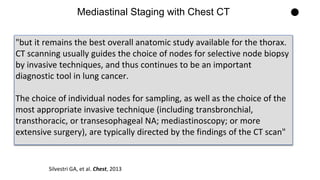
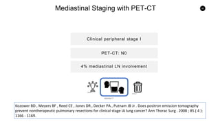
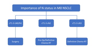
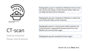
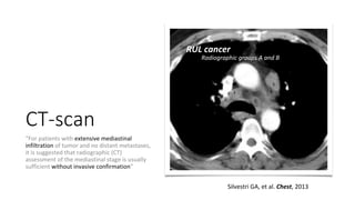
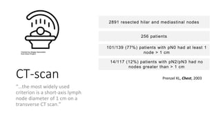
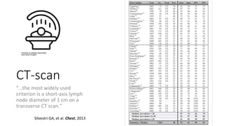
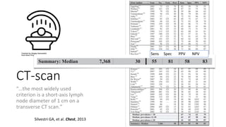
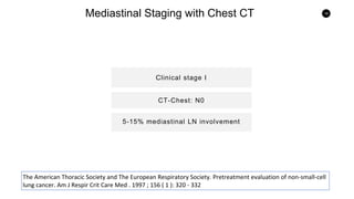
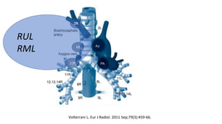
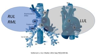
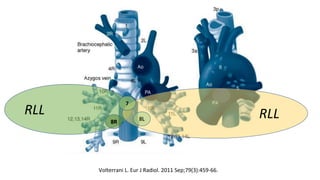
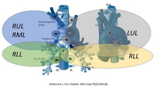
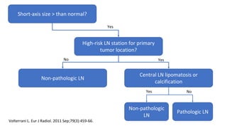
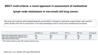
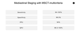
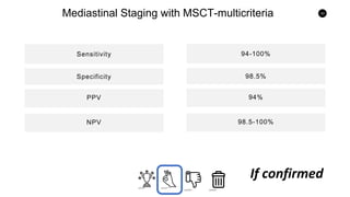
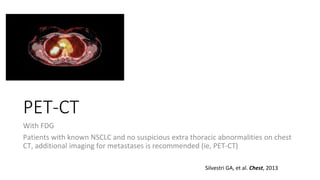
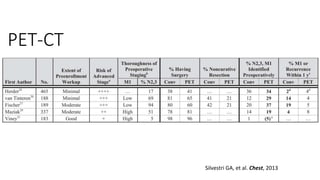
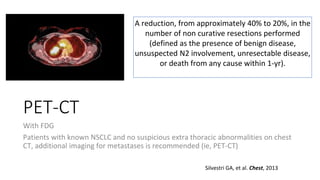
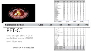
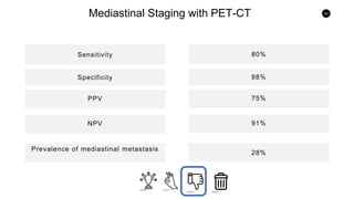
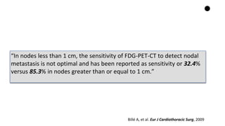
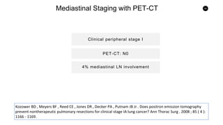
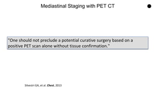
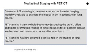
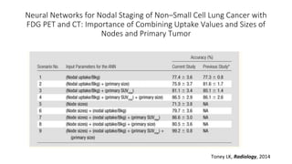
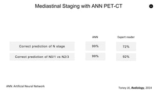
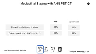
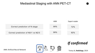
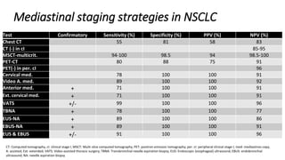
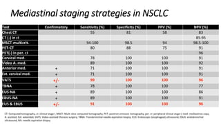
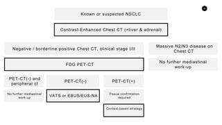
Mediastinal Staging in Clinical Stage I and II NSCLC
Mauricio Lema Medina MD
Clínica de Oncología Astorga / Clínica SOMA, Medellín, Colombia
Medellín, 21.05.2018
Conflicts of interest for this lecture
Mauricio Lema
None for this lecture
@onconerd
Mediastinal LN status MUST be as
accurate as posible BEFORE undergoing
surgery with curative intent
Importance of N status in M0 NSCLC
cT1-3 cN0/N1 cT1-3 cN3cT1-3 cN2
Surgery
Pre-Op/Definitive
Chemo-RT
Definitive Chemo-RT
CT-scan
Contrast-enhanced
Thorax, liver & adrenal
Silvestri GA, et al. Chest, 2013
Radiographic group A: mediastinal infiltration that encircles
the vessels and airways, so that discrete lymph nodes can
no longer be discerned or measured
Radiographic group B: mediastinal infiltration in whom the
size of discrete nodes can be measured
Radiographic group C: central tumor (within proximal 1/3
of the thorax) or suspected N1 disease (Level 10 ≥1 cm)
(N2/N3 risk in the 20-25% range).
Radiographic group D: peripheral clinical stage I
CT-scan
“For patients with extensive mediastinal
infiltration of tumor and no distant metastases,
it is suggested that radiographic (CT)
assessment of the mediastinal stage is usually
sufficient without invasive confirmation”
Silvestri GA, et al. Chest, 2013
RUL cancer
Radiographic groups A and B
CT-scan
“…the most widely used
criterion is a short-axis lymph
node diameter of 1 cm on a
transverse CT scan.”
Silvestri GA, et al. Chest, 2013
CT-scan
“…the most widely used
criterion is a short-axis lymph
node diameter of 1 cm on a
transverse CT scan.”
Prenzel KL, Chest, 2003
2891 resected hilar and mediastinal nodes
101/139 (77%) patients with pN0 had at least 1
node > 1 cm
256 patients
14/117 (12%) patients with pN2/pN3 had no
nodes greater than > 1 cm
CT-scan
“…the most widely used
criterion is a short-axis lymph
node diameter of 1 cm on a
transverse CT scan.”
Silvestri GA, et al. Chest, 2013
CT-scan
“…the most widely used
criterion is a short-axis lymph
node diameter of 1 cm on a
transverse CT scan.”
Silvestri GA, et al. Chest, 2013
Sens Spec PPV NPV
15
Mediastinal Staging with Chest CT
Sensitivity
PPV
Specificity
NPV
Prevalence of mediastinal metastasis
55%
58%
81%
83%
30%
Excellent Acceptable Insufficient Useless
17
Mediastinal Staging with Chest CT
Sensitivity
PPV
Specificity
NPV
Prevalence of mediastinal metastasis
55%
58%
81%
83%
30%
18
Mediastinal Staging with Chest CT
Clinical stage I
5-15% mediastinal LN involvement
CT-Chest: N0
The American Thoracic Society and The European Respiratory Society. Pretreatment evaluation of non-small-cell
lung cancer. Am J Respir Crit Care Med . 1997 ; 156 ( 1 ): 320 - 332
19
Mediastinal Staging with Chest CT
Clinical stage I
5-15% mediastinal LN involvement
CT-Chest: N0
The American Thoracic Society and The European Respiratory Society. Pretreatment evaluation of non-small-cell
lung cancer. Am J Respir Crit Care Med . 1997 ; 156 ( 1 ): 320 - 332
"but it remains the best overall anatomic study available for the thorax.
CT scanning usually guides the choice of nodes for selective node biopsy
by invasive techniques, and thus continues to be an important
diagnostic tool in lung cancer.
The choice of individual nodes for sampling, as well as the choice of the
most appropriate invasive technique (including transbronchial,
transthoracic, or transesophageal NA; mediastinoscopy; or more
extensive surgery), are typically directed by the findings of the CT scan"
Mediastinal Staging with Chest CT
Silvestri GA, et al. Chest, 2013
Can we improve on mediastinal
staging with CT-scan?
Lymph node stations Descriptor Abnormal size (short axis)
1 Highest mediastinal
2 Upper para-tracheal >7 mm
3 Pre-vascular / retro-tracheal
4 Lower para-tracheal >9 mm
5 Sub-aortic (AP window) >8 mm
6 Para-aortic >8 mm
7 Sub-carinal >12 mm
8L Para-oesophageal >7 mm
8R Para-oesophageal >10 mm
Volterrani L. Eur J Radiol. 2011 Sep;79(3):459-66.
RUL
RML
2R
4R
10R
Volterrani L. Eur J Radiol. 2011 Sep;79(3):459-66.
RUL
RML
2R
4R
10R
LUL
2L
4L
6
5
Volterrani L. Eur J Radiol. 2011 Sep;79(3):459-66.
RLL
8R
RLL8L
7
Volterrani L. Eur J Radiol. 2011 Sep;79(3):459-66.
RUL
RML
2R
4R
10R
RLL
8R
LUL
2L
4L
6
RLL8L
7
5
Volterrani L. Eur J Radiol. 2011 Sep;79(3):459-66.
High-risk LN station for primary
tumor location?
Short-axis size > than normal?
Central LN lipomatosis or
calcification
Non-pathologic LN
Pathologic LN
Non-pathologic
LN
Yes
YesNo
NoYes
Volterrani L. Eur J Radiol. 2011 Sep;79(3):459-66.
MSCT multi-criteria: a novel approach in assessment of mediastinal
lymph node metastases in non-small cell lung cancer.
Volterrani L. Eur J Radiol. 2011 Sep;79(3):459-66.
86 consecutive patients with histopathologically proven NSCLC. All patients underwent surgical lymph node resection
within 30 days from the CT examination. In all cases pathological and CT results were reviewed and correlated.
30
Mediastinal Staging with MSCT-multicriteria
Sensitivity
PPV
Specificity
NPV
94-100%
94%
98.5%
98.5-100%
?
32
Mediastinal Staging with MSCT-multicriteria
Sensitivity
PPV
Specificity
NPV
94-100%
94%
98.5%
98.5-100%
33
Mediastinal Staging with MSCT-multicriteria
Sensitivity
PPV
Specificity
NPV
94-100%
94%
98.5%
98.5-100%
If confirmed
PET-CT
With FDG
Patients with known NSCLC and no suspicious extra thoracic abnormalities on chest
CT, additional imaging for metastases is recommended (ie, PET-CT)
Silvestri GA, et al. Chest, 2013
PET-CT
Silvestri GA, et al. Chest, 2013
PET-CT
PET-CT
With FDG
Patients with known NSCLC and no suspicious extra thoracic abnormalities on chest
CT, additional imaging for metastases is recommended (ie, PET-CT)
Silvestri GA, et al. Chest, 2013
A reduction, from approximately 40% to 20%, in the
number of non curative resections performed
(defined as the presence of benign disease,
unsuspected N2 involvement, unresectable disease,
or death from any cause within 1-yr).
PET-CT
Meta-analysis of PET + CT in
mediastinal staging of NSCLC
n= 4105 patients
Silvestri GA, et al. Chest, 2013
PET-CT
Meta-analysis of PET + CT in
mediastinal staging of NSCLC
n= 4105 patients
Silvestri GA, et al. Chest, 2013
PET-CT
Meta-analysis of PET + CT in
mediastinal staging of NSCLC
n= 4105 patients
Silvestri GA, et al. Chest, 2013
Sens Spec PPV NPV
PET-CT
Meta-analysis of PET + CT in mediastinal
staging of NSCLC
Years 2004-2011
n= 2,014 patients
Silvestri GA, et al. Chest, 2013
42
Mediastinal Staging with PET-CT
Sensitivity
PPV
Specificity
NPV
Prevalence of mediastinal metastasis
80%
75%
88%
91%
28%
“In nodes less than 1 cm, the sensitivity of FDG-PET-CT to detect nodal
metastasis is not optimal and has been reported as sensitivity or 32.4%
versus 85.3% in nodes greater than or equal to 1 cm.”
Billé A, et al. Eur J Cardiothoracic Surg, 2009
44
Mediastinal Staging with PET-CT
Clinical peripheral stage I
4% mediastinal LN involvement
PET-CT: N0
Kozower BD , Meyers BF , Reed CE , Jones DR , Decker PA , Putnam JB Jr . Does positron emission tomography
prevent nontherapeutic pulmonary resections for clinical stage IA lung cancer? Ann Thorac Surg . 2008 ; 85 ( 4 ):
1166 - 1169.
45
Mediastinal Staging with PET-CT
Clinical peripheral stage I
4% mediastinal LN involvement
PET-CT: N0
Kozower BD , Meyers BF , Reed CE , Jones DR , Decker PA , Putnam JB Jr . Does positron emission tomography
prevent nontherapeutic pulmonary resections for clinical stage IA lung cancer? Ann Thorac Surg . 2008 ; 85 ( 4 ):
1166 - 1169.
"One should not preclude a potential curative surgery based on a
positive PET scan alone without tissue confirmation."
Mediastinal Staging with PET CT
Silvestri GA, et al. Chest, 2013
"However, PET scanning is the most accurate noninvasive imaging
modality available to evaluate the mediastinum in patients with lung
cancer.
PET scanning is also a whole-body study (excluding the brain), offers
additional information relating to extrathoracic sites of possible disease
involvement, and can reduce noncurative resections.
PET scanning has now assumed a central role in the staging of lung
cancer.”
Mediastinal Staging with PET CT
Silvestri GA, et al. Chest, 2013
Can we improve on mediastinal
staging with PET-CT?
Neural Networks for Nodal Staging of Non–Small Cell Lung Cancer with
FDG PET and CT: Importance of Combining Uptake Values and Sizes of
Nodes and Primary Tumor
Toney LK, Radiology, 2014
“The authors developed a back-
propagation ANN with one hidden layer
and eight processing units”.
Neural Networks for Nodal Staging of Non–Small Cell Lung Cancer with
FDG PET and CT: Importance of Combining Uptake Values and Sizes of
Nodes and Primary Tumor
Toney LK, Radiology, 2014
Neural Networks for Nodal Staging of Non–Small Cell Lung Cancer with
FDG PET and CT: Importance of Combining Uptake Values and Sizes of
Nodes and Primary Tumor
Toney LK, Radiology, 2014
52
Mediastinal Staging with ANN PET-CT
Correct prediction of N stage
Correct prediction of N0/1 vs N2/3
99%
99%
72%
92%
ANN Expert reader
ANN: Artificial Neural Network
Toney LK, Radiology, 2014
53
Mediastinal Staging with ANN PET-CT
Correct prediction of N stage
Correct prediction of N0/1 vs N2/3
99%
99%
72%
92%
ANN Expert reader
ANN: Artificial Neural Network
Toney LK, Radiology, 2014
54
Mediastinal Staging with ANN PET-CT
Correct prediction of N stage
Correct prediction of N0/1 vs N2/3
99%
99%
72%
92%
ANN Expert reader
ANN: Artificial Neural Network
Toney LK, Radiology, 2014
If confirmed
?
Pancoast syndrome is characterized by a
malignant neoplasm of the superior sulcus of the
lung with destructive lesions of the thoracic inlet
and involvement of the brachial plexus and
cervical sympathetic nerves (stellate ganglion, go
here).
This is accompanied by
(1) severe pain in the shoulder region
radiating toward the axilla and scapula along the
ulnar aspect of the muscles of the hand,
(2) atrophy of hand and arm muscles,
(3) Horner syndrome (ptosis, miosis,
hemianhidrosis, enophthalmos), and
(4) compression of the blood vessels with
edema.
Silvestri GA, et al. Chest, 2013
Chest MRI
MRI of the chest should not be performed
routinely for staging of the mediastinum.
MRI is useful in patients with NSCLC when
there is concern about involvement of the
superior sulcus or the brachial plexus.
Silvestri GA, et al. Chest, 2013
Mediastinoscopy
“…involves an incision just above the
suprasternal notch, insertion of a
mediastinoscope alongside the trachea,
and biopsy of mediastinal nodes.”
Silvestri GA, et al. Chest, 2013
Morbidity: 2%
Mortality: 0.08%
2R
4R
7
2L
4L
1
3a
Mediastinoscopy
Node groups that cannot undergo a
biopsy with this technique include:
Posterior subcarinal (station 7)
Inferior mediastinal (stations 8, 9),
Aortopulmonary window (APW) (station 5)
Anterior mediastinal (station 6) nodes.
Mediastinoscopy
“…involves an incision just above the
suprasternal notch, insertion of a
mediastinoscope alongside the trachea,
and biopsy of mediastinal nodes.”
Silvestri GA, et al. Chest, 2013
Mediastinoscopy
“…involves an incision just above the
suprasternal notch, insertion of a
mediastinoscope alongside the trachea,
and biopsy of mediastinal nodes.”
Silvestri GA, et al. Chest, 2013
Sens Spec PPV NPV
Video Assisted Mediastinoscopy
“…A videomediastinoscope allows better visualization, more extensive
sampling (including posterior station 7)…”
Silvestri GA, et al. Chest, 2013
63
Mediastinal Staging with Mediastinoscopy
Sensitivity
PPV
Specificity
NPV
78%
(100%)
(100%)
91%
89%
(100%)
(100%)
92%
Mediastinoscopy VAM
6
5
The problem of
LN stations 5 and
6
Very significant for LUL tumors
66
Mediastinal Staging with Anterior Mediastinoscopy and
Extended Cervical Mediastinoscopy
Sensitivity
PPV
Specificity
NPV
71%
(100%)
(100%)
91%
71%
(100%)
(100%)
91%
Anterior Extended Cervical
PPV
Video-Assisted Thoracic Surgery
“This is performed under general
anesthesia and, in general, is limited
to an assessment of only one side of
the mediastinum.
Access to the R-sided nodes is
straightforward, but access to the L
paratracheal nodes is more difficult”
Silvestri GA, et al. Chest, 2013
Morbidity: 2%
Mortality: 0%
68
Mediastinal Staging with VATS
Sensitivity
PPV
Specificity
NPV
71%
(100%)
(100%)
91%
71%
(100%)
(100%)
91%
PPV
69
Mediastinal Staging with VATS
Sensitivity
PPVSpecificity
NPV
PPV
99%
(100%)
(100%)
96%
Trans-
bronchial
Needle
Aspiration
Silvestri GA, et al. Chest, 2013
Trans-
bronchial
Needle
Aspiration
Silvestri GA, et al. Chest, 2013
Used mostly to corroborate
tumor involvement in enlarged
LN
Trans-
bronchial
Needle
Aspiration
Silvestri GA, et al. Chest, 2013
Used mostly to corroborate
tumor involvement in enlarged
LN
73
Mediastinal Staging with Transbronchial Needle Aspiration
Sensitivity
PPVSpecificity
NPV
PPV
78%
(100%)
(100%)
77%
74
Mediastinal Staging with Transbronchial Needle Aspiration
Sensitivity
PPVSpecificity
NPV
PPV
78%
(100%)
(100%)
77%
Corroboration strategy
EUS-NA
9R 9L
8R
8L
7
4L
5
Used mostly to corroborate tumor involvement in enlarged LN in EUS-reachable LN stations
EUS-NA
Silvestri GA, et al. Chest, 2013
Used mostly to corroborate tumor
involvement in enlarged LN in EUS-
reachable LN stations
EUS-NA
Silvestri GA, et al. Chest, 2013
Sens NPV
78
Mediastinal Staging with EUS-NA
Sensitivity
PPVSpecificity
NPV
PPV
89%
(100%)
(100%)
86%
79
Mediastinal Staging with EUS-NA
Sensitivity
PPVSpecificity
NPV
PPV
89%
(100%)
(100%)
86%
Corroboration strategy
EBUS-NA
Used mostly to corroborate tumor involvement in enlarged LN
EBUS-NA
Silvestri GA, et al. Chest, 2013
EBUS-NA
Silvestri GA, et al. Chest, 2013
Sens NPV
83
Mediastinal Staging with EBUS-NA
Sensitivity
PPVSpecificity
NPV
PPV
89%
(100%)
(100%)
91%
Combined EUS-NA and EBUS-NA
85
Mediastinal Staging with Combined EUS-NA and EBUS-NA
Sensitivity
PPVSpecificity
NPV
PPV
91%
(100%)
(100%)
96%
Putting it all together
Mediastinal staging strategies in NSCLC
Test Confirmatory Sensitivity (%) Specificity (%) PPV (%) NPV (%)
Chest CT 55 81 58 83
CT (-) in cI 85-95
MSCT-multicrit. 94-100 98.5 94 98.5-100
PET-CT 80 88 75 91
PET(-) in per. cI 96
Cervical med. 78 100 100 91
Video A. med. 89 100 100 92
Anterior med. + 71 100 100 91
Ext. cervical med. + 71 100 100 91
VATS +/- 99 100 100 96
TBNA + 78 100 100 77
EUS-NA + 89 100 100 86
EBUS-NA + 89 100 100 91
EUS & EBUS +/- 91 100 100 96
CT: Computed tomography, cI: clinical stage I, MSCT: Multi-slice computed tomography, PET: positron emission tomography; per. cI: peripheral clinical stage I; med: mediastinos copy,
A: assisted; Ext: extended; VATS: Video-assisted thoracic surgery; TBNA: Transbronchial needle aspiration biopsy; EUS: Endoscopic (esophageal) ultrasound; EBUS: endobronchial
ultrasound; NA: needle aspiration biopsy
Direct comparisons
90
Minimally Invasive Endoscopic Staging of
Suspected Lung Cancer
TB-NA
PPV
EUS-NA
EUS-NA & EBUS-NA
EBUS-NA
Wallace MB, JAMA, 2008
36%
69%
69%
93%
78%
88%)
88%
97%
Sensitivity NPV
138 consecutive non(obviously) metastatic NSCLC underwent all three procedures
91
Minimally Invasive Endoscopic Staging of
Suspected Lung Cancer
TB-NA
PPV
EUS-NA
EUS-NA & EBUS-NA
EBUS-NA
Wallace MB, JAMA, 2008
36%
69%
69%
93%
78%
88%)
88%
97%
Sensitivity NPV
138 consecutive non(obviously) metastatic NSCLC underwent all three procedures
92
Endobronchial ultrasound versus mediastinoscopy for mediastinal
nodal staging of non-small-cell lung cancer
Sensitivity
PPV
Specificity
NPV
Accuracy
Um SW, JTO, 2015
88%
93%
100%
85%
81%
89%)
100%
78%
EBUS-TBNA Mediastinoscopy
138 consecutive NSCLC with cN1-cN3
*
*
*
* p < 0.005
93
Endobronchial ultrasound versus mediastinoscopy for mediastinal
nodal staging of non-small-cell lung cancer
Sensitivity
PPV
Specificity
NPV
Accuracy
Um SW, JTO, 2015
88%
93%
100%
85%
81%
89%)
100%
78%
EBUS-TBNA Mediastinoscopy
138 consecutive NSCLC with cN1-cN3
*
*
*
* p < 0.005
94
EBUS-centred versus EUS-centred mediastinal
staging in lung cancer: a randomized controlled
trial
Group A (EBUS-NA then EUS-NA)
PPV
EBUS after EUS improves accuracy and sensitivity. Therefore,
EBUS-FNA should be first
Kang HJ, Thorax, 2014
92%
86%
93%
97%
Accuracy post 1st test - Sensitivity Accuracy post 2nd test - Sensitivity
160 patients with histologically confirmed or strongly suspected potentially operable NSCLC
Group B (EUS-NA then EBUS-NA)
82%
60%
85%
92%
95
P O S I T I O N S T A T E M E N T
My practice take…
96
Known or suspected NSCLC
Contrast-Enhanced Chest CT (+liver & adrenal)
Negative / borderline positive Chest CT, clinical stage I/II
FDG PET-CT
VATS or EBUS/EUS-NA
Massive N2/N3 disease on
Chest CT
No further mediastinal
work-up
PET-CT(-) and
peripheral cI
No further mediastinal
work-up
PET-CT(+)
Tissue confirmation
required
PET-CT(-)
Context-based strategy
@onconerd

Mediastinal staging in early-stage NSCLC

  • 1.
    Mediastinal Staging inClinical Stage I and II NSCLC Mauricio Lema Medina MD Clínica de Oncología Astorga / Clínica SOMA, Medellín, Colombia Medellín, 21.05.2018
  • 2.
    Conflicts of interestfor this lecture Mauricio Lema None for this lecture
  • 3.
  • 6.
    Mediastinal LN statusMUST be as accurate as posible BEFORE undergoing surgery with curative intent
  • 7.
    Importance of Nstatus in M0 NSCLC cT1-3 cN0/N1 cT1-3 cN3cT1-3 cN2 Surgery Pre-Op/Definitive Chemo-RT Definitive Chemo-RT
  • 9.
    CT-scan Contrast-enhanced Thorax, liver &adrenal Silvestri GA, et al. Chest, 2013 Radiographic group A: mediastinal infiltration that encircles the vessels and airways, so that discrete lymph nodes can no longer be discerned or measured Radiographic group B: mediastinal infiltration in whom the size of discrete nodes can be measured Radiographic group C: central tumor (within proximal 1/3 of the thorax) or suspected N1 disease (Level 10 ≥1 cm) (N2/N3 risk in the 20-25% range). Radiographic group D: peripheral clinical stage I
  • 10.
    CT-scan “For patients withextensive mediastinal infiltration of tumor and no distant metastases, it is suggested that radiographic (CT) assessment of the mediastinal stage is usually sufficient without invasive confirmation” Silvestri GA, et al. Chest, 2013 RUL cancer Radiographic groups A and B
  • 11.
    CT-scan “…the most widelyused criterion is a short-axis lymph node diameter of 1 cm on a transverse CT scan.” Silvestri GA, et al. Chest, 2013
  • 12.
    CT-scan “…the most widelyused criterion is a short-axis lymph node diameter of 1 cm on a transverse CT scan.” Prenzel KL, Chest, 2003 2891 resected hilar and mediastinal nodes 101/139 (77%) patients with pN0 had at least 1 node > 1 cm 256 patients 14/117 (12%) patients with pN2/pN3 had no nodes greater than > 1 cm
  • 13.
    CT-scan “…the most widelyused criterion is a short-axis lymph node diameter of 1 cm on a transverse CT scan.” Silvestri GA, et al. Chest, 2013
  • 14.
    CT-scan “…the most widelyused criterion is a short-axis lymph node diameter of 1 cm on a transverse CT scan.” Silvestri GA, et al. Chest, 2013 Sens Spec PPV NPV
  • 15.
    15 Mediastinal Staging withChest CT Sensitivity PPV Specificity NPV Prevalence of mediastinal metastasis 55% 58% 81% 83% 30%
  • 16.
  • 17.
    17 Mediastinal Staging withChest CT Sensitivity PPV Specificity NPV Prevalence of mediastinal metastasis 55% 58% 81% 83% 30%
  • 18.
    18 Mediastinal Staging withChest CT Clinical stage I 5-15% mediastinal LN involvement CT-Chest: N0 The American Thoracic Society and The European Respiratory Society. Pretreatment evaluation of non-small-cell lung cancer. Am J Respir Crit Care Med . 1997 ; 156 ( 1 ): 320 - 332
  • 19.
    19 Mediastinal Staging withChest CT Clinical stage I 5-15% mediastinal LN involvement CT-Chest: N0 The American Thoracic Society and The European Respiratory Society. Pretreatment evaluation of non-small-cell lung cancer. Am J Respir Crit Care Med . 1997 ; 156 ( 1 ): 320 - 332
  • 20.
    "but it remainsthe best overall anatomic study available for the thorax. CT scanning usually guides the choice of nodes for selective node biopsy by invasive techniques, and thus continues to be an important diagnostic tool in lung cancer. The choice of individual nodes for sampling, as well as the choice of the most appropriate invasive technique (including transbronchial, transthoracic, or transesophageal NA; mediastinoscopy; or more extensive surgery), are typically directed by the findings of the CT scan" Mediastinal Staging with Chest CT Silvestri GA, et al. Chest, 2013
  • 21.
    Can we improveon mediastinal staging with CT-scan?
  • 22.
    Lymph node stationsDescriptor Abnormal size (short axis) 1 Highest mediastinal 2 Upper para-tracheal >7 mm 3 Pre-vascular / retro-tracheal 4 Lower para-tracheal >9 mm 5 Sub-aortic (AP window) >8 mm 6 Para-aortic >8 mm 7 Sub-carinal >12 mm 8L Para-oesophageal >7 mm 8R Para-oesophageal >10 mm Volterrani L. Eur J Radiol. 2011 Sep;79(3):459-66.
  • 23.
    Volterrani L. EurJ Radiol. 2011 Sep;79(3):459-66.
  • 24.
    RUL RML 2R 4R 10R Volterrani L. EurJ Radiol. 2011 Sep;79(3):459-66.
  • 25.
    RUL RML 2R 4R 10R LUL 2L 4L 6 5 Volterrani L. EurJ Radiol. 2011 Sep;79(3):459-66.
  • 26.
    RLL 8R RLL8L 7 Volterrani L. EurJ Radiol. 2011 Sep;79(3):459-66.
  • 27.
  • 28.
    High-risk LN stationfor primary tumor location? Short-axis size > than normal? Central LN lipomatosis or calcification Non-pathologic LN Pathologic LN Non-pathologic LN Yes YesNo NoYes Volterrani L. Eur J Radiol. 2011 Sep;79(3):459-66.
  • 29.
    MSCT multi-criteria: anovel approach in assessment of mediastinal lymph node metastases in non-small cell lung cancer. Volterrani L. Eur J Radiol. 2011 Sep;79(3):459-66. 86 consecutive patients with histopathologically proven NSCLC. All patients underwent surgical lymph node resection within 30 days from the CT examination. In all cases pathological and CT results were reviewed and correlated.
  • 30.
    30 Mediastinal Staging withMSCT-multicriteria Sensitivity PPV Specificity NPV 94-100% 94% 98.5% 98.5-100%
  • 31.
  • 32.
    32 Mediastinal Staging withMSCT-multicriteria Sensitivity PPV Specificity NPV 94-100% 94% 98.5% 98.5-100%
  • 33.
    33 Mediastinal Staging withMSCT-multicriteria Sensitivity PPV Specificity NPV 94-100% 94% 98.5% 98.5-100% If confirmed
  • 34.
    PET-CT With FDG Patients withknown NSCLC and no suspicious extra thoracic abnormalities on chest CT, additional imaging for metastases is recommended (ie, PET-CT) Silvestri GA, et al. Chest, 2013
  • 35.
    Silvestri GA, etal. Chest, 2013 PET-CT
  • 36.
    Silvestri GA, etal. Chest, 2013 PET-CT
  • 37.
    PET-CT With FDG Patients withknown NSCLC and no suspicious extra thoracic abnormalities on chest CT, additional imaging for metastases is recommended (ie, PET-CT) Silvestri GA, et al. Chest, 2013 A reduction, from approximately 40% to 20%, in the number of non curative resections performed (defined as the presence of benign disease, unsuspected N2 involvement, unresectable disease, or death from any cause within 1-yr).
  • 38.
    PET-CT Meta-analysis of PET+ CT in mediastinal staging of NSCLC n= 4105 patients Silvestri GA, et al. Chest, 2013
  • 39.
    PET-CT Meta-analysis of PET+ CT in mediastinal staging of NSCLC n= 4105 patients Silvestri GA, et al. Chest, 2013
  • 40.
    PET-CT Meta-analysis of PET+ CT in mediastinal staging of NSCLC n= 4105 patients Silvestri GA, et al. Chest, 2013 Sens Spec PPV NPV
  • 41.
    PET-CT Meta-analysis of PET+ CT in mediastinal staging of NSCLC Years 2004-2011 n= 2,014 patients Silvestri GA, et al. Chest, 2013
  • 42.
    42 Mediastinal Staging withPET-CT Sensitivity PPV Specificity NPV Prevalence of mediastinal metastasis 80% 75% 88% 91% 28%
  • 43.
    “In nodes lessthan 1 cm, the sensitivity of FDG-PET-CT to detect nodal metastasis is not optimal and has been reported as sensitivity or 32.4% versus 85.3% in nodes greater than or equal to 1 cm.” Billé A, et al. Eur J Cardiothoracic Surg, 2009
  • 44.
    44 Mediastinal Staging withPET-CT Clinical peripheral stage I 4% mediastinal LN involvement PET-CT: N0 Kozower BD , Meyers BF , Reed CE , Jones DR , Decker PA , Putnam JB Jr . Does positron emission tomography prevent nontherapeutic pulmonary resections for clinical stage IA lung cancer? Ann Thorac Surg . 2008 ; 85 ( 4 ): 1166 - 1169.
  • 45.
    45 Mediastinal Staging withPET-CT Clinical peripheral stage I 4% mediastinal LN involvement PET-CT: N0 Kozower BD , Meyers BF , Reed CE , Jones DR , Decker PA , Putnam JB Jr . Does positron emission tomography prevent nontherapeutic pulmonary resections for clinical stage IA lung cancer? Ann Thorac Surg . 2008 ; 85 ( 4 ): 1166 - 1169.
  • 46.
    "One should notpreclude a potential curative surgery based on a positive PET scan alone without tissue confirmation." Mediastinal Staging with PET CT Silvestri GA, et al. Chest, 2013
  • 47.
    "However, PET scanningis the most accurate noninvasive imaging modality available to evaluate the mediastinum in patients with lung cancer. PET scanning is also a whole-body study (excluding the brain), offers additional information relating to extrathoracic sites of possible disease involvement, and can reduce noncurative resections. PET scanning has now assumed a central role in the staging of lung cancer.” Mediastinal Staging with PET CT Silvestri GA, et al. Chest, 2013
  • 48.
    Can we improveon mediastinal staging with PET-CT?
  • 49.
    Neural Networks forNodal Staging of Non–Small Cell Lung Cancer with FDG PET and CT: Importance of Combining Uptake Values and Sizes of Nodes and Primary Tumor Toney LK, Radiology, 2014 “The authors developed a back- propagation ANN with one hidden layer and eight processing units”.
  • 50.
    Neural Networks forNodal Staging of Non–Small Cell Lung Cancer with FDG PET and CT: Importance of Combining Uptake Values and Sizes of Nodes and Primary Tumor Toney LK, Radiology, 2014
  • 51.
    Neural Networks forNodal Staging of Non–Small Cell Lung Cancer with FDG PET and CT: Importance of Combining Uptake Values and Sizes of Nodes and Primary Tumor Toney LK, Radiology, 2014
  • 52.
    52 Mediastinal Staging withANN PET-CT Correct prediction of N stage Correct prediction of N0/1 vs N2/3 99% 99% 72% 92% ANN Expert reader ANN: Artificial Neural Network Toney LK, Radiology, 2014
  • 53.
    53 Mediastinal Staging withANN PET-CT Correct prediction of N stage Correct prediction of N0/1 vs N2/3 99% 99% 72% 92% ANN Expert reader ANN: Artificial Neural Network Toney LK, Radiology, 2014
  • 54.
    54 Mediastinal Staging withANN PET-CT Correct prediction of N stage Correct prediction of N0/1 vs N2/3 99% 99% 72% 92% ANN Expert reader ANN: Artificial Neural Network Toney LK, Radiology, 2014 If confirmed
  • 55.
  • 56.
    Pancoast syndrome ischaracterized by a malignant neoplasm of the superior sulcus of the lung with destructive lesions of the thoracic inlet and involvement of the brachial plexus and cervical sympathetic nerves (stellate ganglion, go here). This is accompanied by (1) severe pain in the shoulder region radiating toward the axilla and scapula along the ulnar aspect of the muscles of the hand, (2) atrophy of hand and arm muscles, (3) Horner syndrome (ptosis, miosis, hemianhidrosis, enophthalmos), and (4) compression of the blood vessels with edema. Silvestri GA, et al. Chest, 2013
  • 57.
    Chest MRI MRI ofthe chest should not be performed routinely for staging of the mediastinum. MRI is useful in patients with NSCLC when there is concern about involvement of the superior sulcus or the brachial plexus. Silvestri GA, et al. Chest, 2013
  • 58.
    Mediastinoscopy “…involves an incisionjust above the suprasternal notch, insertion of a mediastinoscope alongside the trachea, and biopsy of mediastinal nodes.” Silvestri GA, et al. Chest, 2013 Morbidity: 2% Mortality: 0.08%
  • 59.
    2R 4R 7 2L 4L 1 3a Mediastinoscopy Node groups thatcannot undergo a biopsy with this technique include: Posterior subcarinal (station 7) Inferior mediastinal (stations 8, 9), Aortopulmonary window (APW) (station 5) Anterior mediastinal (station 6) nodes.
  • 60.
    Mediastinoscopy “…involves an incisionjust above the suprasternal notch, insertion of a mediastinoscope alongside the trachea, and biopsy of mediastinal nodes.” Silvestri GA, et al. Chest, 2013
  • 61.
    Mediastinoscopy “…involves an incisionjust above the suprasternal notch, insertion of a mediastinoscope alongside the trachea, and biopsy of mediastinal nodes.” Silvestri GA, et al. Chest, 2013 Sens Spec PPV NPV
  • 62.
    Video Assisted Mediastinoscopy “…Avideomediastinoscope allows better visualization, more extensive sampling (including posterior station 7)…” Silvestri GA, et al. Chest, 2013
  • 63.
    63 Mediastinal Staging withMediastinoscopy Sensitivity PPV Specificity NPV 78% (100%) (100%) 91% 89% (100%) (100%) 92% Mediastinoscopy VAM
  • 64.
    6 5 The problem of LNstations 5 and 6 Very significant for LUL tumors
  • 66.
    66 Mediastinal Staging withAnterior Mediastinoscopy and Extended Cervical Mediastinoscopy Sensitivity PPV Specificity NPV 71% (100%) (100%) 91% 71% (100%) (100%) 91% Anterior Extended Cervical PPV
  • 67.
    Video-Assisted Thoracic Surgery “Thisis performed under general anesthesia and, in general, is limited to an assessment of only one side of the mediastinum. Access to the R-sided nodes is straightforward, but access to the L paratracheal nodes is more difficult” Silvestri GA, et al. Chest, 2013 Morbidity: 2% Mortality: 0%
  • 68.
    68 Mediastinal Staging withVATS Sensitivity PPV Specificity NPV 71% (100%) (100%) 91% 71% (100%) (100%) 91% PPV
  • 69.
    69 Mediastinal Staging withVATS Sensitivity PPVSpecificity NPV PPV 99% (100%) (100%) 96%
  • 70.
  • 71.
    Trans- bronchial Needle Aspiration Silvestri GA, etal. Chest, 2013 Used mostly to corroborate tumor involvement in enlarged LN
  • 72.
    Trans- bronchial Needle Aspiration Silvestri GA, etal. Chest, 2013 Used mostly to corroborate tumor involvement in enlarged LN
  • 73.
    73 Mediastinal Staging withTransbronchial Needle Aspiration Sensitivity PPVSpecificity NPV PPV 78% (100%) (100%) 77%
  • 74.
    74 Mediastinal Staging withTransbronchial Needle Aspiration Sensitivity PPVSpecificity NPV PPV 78% (100%) (100%) 77% Corroboration strategy
  • 75.
    EUS-NA 9R 9L 8R 8L 7 4L 5 Used mostlyto corroborate tumor involvement in enlarged LN in EUS-reachable LN stations
  • 76.
    EUS-NA Silvestri GA, etal. Chest, 2013 Used mostly to corroborate tumor involvement in enlarged LN in EUS- reachable LN stations
  • 77.
    EUS-NA Silvestri GA, etal. Chest, 2013 Sens NPV
  • 78.
    78 Mediastinal Staging withEUS-NA Sensitivity PPVSpecificity NPV PPV 89% (100%) (100%) 86%
  • 79.
    79 Mediastinal Staging withEUS-NA Sensitivity PPVSpecificity NPV PPV 89% (100%) (100%) 86% Corroboration strategy
  • 80.
    EBUS-NA Used mostly tocorroborate tumor involvement in enlarged LN
  • 81.
    EBUS-NA Silvestri GA, etal. Chest, 2013
  • 82.
    EBUS-NA Silvestri GA, etal. Chest, 2013 Sens NPV
  • 83.
    83 Mediastinal Staging withEBUS-NA Sensitivity PPVSpecificity NPV PPV 89% (100%) (100%) 91%
  • 84.
  • 85.
    85 Mediastinal Staging withCombined EUS-NA and EBUS-NA Sensitivity PPVSpecificity NPV PPV 91% (100%) (100%) 96%
  • 86.
  • 87.
    Mediastinal staging strategiesin NSCLC Test Confirmatory Sensitivity (%) Specificity (%) PPV (%) NPV (%) Chest CT 55 81 58 83 CT (-) in cI 85-95 MSCT-multicrit. 94-100 98.5 94 98.5-100 PET-CT 80 88 75 91 PET(-) in per. cI 96 Cervical med. 78 100 100 91 Video A. med. 89 100 100 92 Anterior med. + 71 100 100 91 Ext. cervical med. + 71 100 100 91 VATS +/- 99 100 100 96 TBNA + 78 100 100 77 EUS-NA + 89 100 100 86 EBUS-NA + 89 100 100 91 EUS & EBUS +/- 91 100 100 96 CT: Computed tomography, cI: clinical stage I, MSCT: Multi-slice computed tomography, PET: positron emission tomography; per. cI: peripheral clinical stage I; med: mediastinos copy, A: assisted; Ext: extended; VATS: Video-assisted thoracic surgery; TBNA: Transbronchial needle aspiration biopsy; EUS: Endoscopic (esophageal) ultrasound; EBUS: endobronchial ultrasound; NA: needle aspiration biopsy
  • 88.
    Mediastinal staging strategiesin NSCLC Test Confirmatory Sensitivity (%) Specificity (%) PPV (%) NPV (%) Chest CT 55 81 58 83 CT (-) in cI 85-95 MSCT-multicrit. 94-100 98.5 94 98.5-100 PET-CT 80 88 75 91 PET(-) in per. cI 96 Cervical med. 78 100 100 91 Video A. med. 89 100 100 92 Anterior med. + 71 100 100 91 Ext. cervical med. + 71 100 100 91 VATS +/- 99 100 100 96 TBNA + 78 100 100 77 EUS-NA + 89 100 100 86 EBUS-NA + 89 100 100 91 EUS & EBUS +/- 91 100 100 96 CT: Computed tomography, cI: clinical stage I, MSCT: Multi-slice computed tomography, PET: positron emission tomography; per. cI: peripheral clinical stage I; med: mediastinos copy, A: assisted; Ext: extended; VATS: Video-assisted thoracic surgery; TBNA: Transbronchial needle aspiration biopsy; EUS: Endoscopic (esophageal) ultrasound; EBUS: endobronchial ultrasound; NA: needle aspiration biopsy
  • 89.
  • 90.
    90 Minimally Invasive EndoscopicStaging of Suspected Lung Cancer TB-NA PPV EUS-NA EUS-NA & EBUS-NA EBUS-NA Wallace MB, JAMA, 2008 36% 69% 69% 93% 78% 88%) 88% 97% Sensitivity NPV 138 consecutive non(obviously) metastatic NSCLC underwent all three procedures
  • 91.
    91 Minimally Invasive EndoscopicStaging of Suspected Lung Cancer TB-NA PPV EUS-NA EUS-NA & EBUS-NA EBUS-NA Wallace MB, JAMA, 2008 36% 69% 69% 93% 78% 88%) 88% 97% Sensitivity NPV 138 consecutive non(obviously) metastatic NSCLC underwent all three procedures
  • 92.
    92 Endobronchial ultrasound versusmediastinoscopy for mediastinal nodal staging of non-small-cell lung cancer Sensitivity PPV Specificity NPV Accuracy Um SW, JTO, 2015 88% 93% 100% 85% 81% 89%) 100% 78% EBUS-TBNA Mediastinoscopy 138 consecutive NSCLC with cN1-cN3 * * * * p < 0.005
  • 93.
    93 Endobronchial ultrasound versusmediastinoscopy for mediastinal nodal staging of non-small-cell lung cancer Sensitivity PPV Specificity NPV Accuracy Um SW, JTO, 2015 88% 93% 100% 85% 81% 89%) 100% 78% EBUS-TBNA Mediastinoscopy 138 consecutive NSCLC with cN1-cN3 * * * * p < 0.005
  • 94.
    94 EBUS-centred versus EUS-centredmediastinal staging in lung cancer: a randomized controlled trial Group A (EBUS-NA then EUS-NA) PPV EBUS after EUS improves accuracy and sensitivity. Therefore, EBUS-FNA should be first Kang HJ, Thorax, 2014 92% 86% 93% 97% Accuracy post 1st test - Sensitivity Accuracy post 2nd test - Sensitivity 160 patients with histologically confirmed or strongly suspected potentially operable NSCLC Group B (EUS-NA then EBUS-NA) 82% 60% 85% 92%
  • 95.
    95 P O SI T I O N S T A T E M E N T My practice take…
  • 96.
    96 Known or suspectedNSCLC Contrast-Enhanced Chest CT (+liver & adrenal) Negative / borderline positive Chest CT, clinical stage I/II FDG PET-CT VATS or EBUS/EUS-NA Massive N2/N3 disease on Chest CT No further mediastinal work-up PET-CT(-) and peripheral cI No further mediastinal work-up PET-CT(+) Tissue confirmation required PET-CT(-) Context-based strategy
  • 97.