
Recommended
More Related Content
Similar to usg Lung.pptx
Similar to usg Lung.pptx (20)
More from Abdullah764280
More from Abdullah764280 (18)
Recently uploaded
Recently uploaded (20)
usg Lung.pptx
- 1. 1
- 2. • The chest cavity is bounded by the chest wall and below by the diaphragm. • It extends upward into the root of the neck about one fingerbreadth above the clavicle on each side • The chest cavity can be divided into a median partition, called the mediastinum , and the laterally placed pleurae and lungs Chest Cavity 2
- 3. • Product B • Feature 1 • Feature 2 • Feature 3 Slide Title • Feature 1 • Feature 2 • Feature 3 3
- 4. Pleura • It is a serous membrane arranged as a closed invaginated sac that covers the lung and lines the chest wall • Each pleura has two parts: • 1. Parietal layer (outer layer) , which lines the thoracic wall, • 2. Visceral layer (inner layer), which completely covers the outer surfaces of the lungs • The two layers become continuous with one another at the hilum of each lung. But below the hilum the two layers hang down as a loose fold called the pulmonary ligament • The parietal and visceral layers of pleura are separated from one another by the pleural cavity (pleural space) that contains a small amount of the pleural fluid 4
- 5. 5
- 6. Division of the parietal pleura • Parietal pleura divided according to the region in which it lies or the surface that it covers. • The cervical pleura extends up into the neck. It reaches a level 1 to 1.5 in. (2.5 to 4 cm) above the medial third of the clavicle. • The costal pleura lines the inner surfaces of the ribs, the costal cartilages, the intercostal spaces, the sides of the vertebral bodies, and the back of the sternum 6
- 7. The diaphragmatic pleura covers the thoracic surface (upper surface) of the diaphragm. The mediastinal pleura covers and forms the lateral boundary of the mediastinum. At the hilum of the lung, it is reflected as a cuff around the vessels and bronchi that constitute the lung root and here becomes continuous with the visceral pleura. 7
- 8. 8
- 9. Nerve Supply of the Pleura • The parietal pleura is sensitive to pain, temperature, touch, and pressure and is supplied as follows: The costal pleura is segmentally supplied by the intercostal nerves. The mediastinal pleura is supplied by the phrenic nerve. The diaphragmatic pleura is supplied over the domes by the phrenic nerve and around the periphery by the lower six intercostal nerves. • The visceral pleura covering the lungs is sensitive to stretch but is insensitive to common sensations such as pain and touch. It receives an autonomic nerve supply from the pulmonary plexus 9
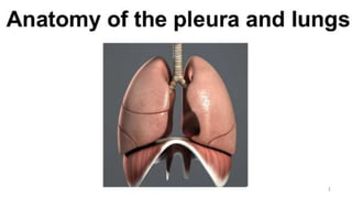
- 11. Lungs • The lungs are the essential organs of respiration. • They are situated on either side of the heart and other mediastinal contents • the lungs are soft, spongy and very elastic. • Each lung is conical in shape, covered with visceral pleura, being attached to the mediastinum only by its root • In the child, they are pink, but with age, they become dark and mottled 11
- 12. 12
- 13. Anatomical features of the lungs • Each lung has an apex, base, three borders and two surfaces • Apex each lung has a blunt apex, which projects upward into the neck for about 1 in. (2.5 cm) above the clavicle; • Base is concave, and rests upon the upper surface of the diaphragm ; • The costal surface is smooth and convex which corresponds to the concave chest wall; • The medial surface has a posterior vertebral part and anterior mediastinal part. The vertebral part lies in contact with the sides of the thoracic vertebrae and intervertebral discs The mediastinal part is deeply concave, and related to the mediastinal content which causes impressions on this surface. The hilum, where various structures enter or leave the lung lies on this surface • The anterior border is thin and overlaps the heart;it is here on the left lung that the cardiac notch is found. • The posterior border is thick and lies beside the vertebral column. • Inferior border 13
- 14. 14
- 15. 15
- 16. The mediastinal part of the medial surface • The mediastinal part is deeply concave, and related to the mediastinal content which causes impressions on this surface. • Also it contains the hilum, where various structures enter or leave the lung lies on this surface 16
- 18. Bronchopulmonary Segments The bronchopulmonary segments are the anatomic, functional, and surgical units of the lungs. Each lobar (secondary) bronchus, which passes to a lobe of the lung, gives off branches called segmental (tertiary) bronchi. Each segmental bronchus passes to a structurally and functionally independent unit of a lung lobe called a Broncho pulmonary segment, which is surrounded by connective tissue. 18
- 19. 19
- 20. 20
- 21. Alveoli of lung • They are tiny air containing sacs and the functional unit of lung,where gaseous exchange occurs between the blood and atmospheric air. • Number:Adult lung contains about 150 million alveoli in each lung. • Lining epithelium:Simple squamous epithelium. So,basically lung is an air containing organ 21
- 22. Blood Supply of the Lungs • The bronchi, the connective tissue of the lung, and the visceral pleura receive their blood supply from the bronchial arteries, which are branches of the descending aorta. • The bronchial veins drain into the azygos and hemiazygos veins. • The alveoli receive deoxygenated blood from the terminal branches of the pulmonary arteries. The oxygenated blood leaving the alveolar capillaries drains into the tributaries of the pulmonary veins, to empty into the left atrium of the heart. 22
- 23. Nerve Supply of the Lungs At the root of each lung is a pulmonary plexus composed of efferent and afferent autonomic nerve fibers. The plexus is formed from branches of the sympathetic trunk and receives parasympathetic fibers from the vagus nerve. The sympathetic efferent fibers produce bronchodilatation and vasoconstriction. The parasympathetic efferent fibers produce bronchoconstriction, vasodilatation, and increased glandular secretion. Afferent impulses derived from the bronchial mucous membrane and from stretch receptors in the alveolar walls pass to the central nervous system in both sympathetic and parasympathetic nerves. 23
- 25. Seminar on Ultrasonogram of Lung Presented by- Dr. Tania Kabir Resident,Phase A,R-12 Department of Radiology & Imaging BSMMU 25
- 26. Ultrasonogram of lungs • Lung US has dramatically increased in popularity over the last decade and is routinely performed at the patient’s bedside, specially in the emergency department and the intensive care unit. As this modality becomes an imaging staple, the diagnostic radiologist should be fluent in lung US performance and interpretation to maintain relevance and assist the ordering clinician. This seminar will provide an overview of lung US to equip the knowledge needed to interpret this modality 26
- 27. Ultrasonogram of lungs:continue… • Meta-analysis suggest that lung US has higher sensitivity and specificity compared with chest radiography for evaluation of some pulmonary conditions, including pneumonia, pleural effusion, pneumothorax and pulmonary edema. 27
- 28. Lung US vs Chest Radiography 28 Ref:Lung Ultrasound: The Essentials Thomas J. Marini, MD • Deborah J. Rubens, MD • Yu T. Zhao, BA • Justin Weis, MD • Timothy P. O’Connor, MD • William H. Novak, MD • Katherine A. Kaproth-Joslin, MD, PhD
- 29. Advantages of lung US • It is radiation free • Low cost • Simple procedure • Portable • Allow real time examination of pulmonary structures • Can be done on severely ill or ICU patients • Can guide interventional procedures, including thoracocentesis and biopsy with less complication 29
- 30. Limitations • It is an operator dependent process, so its quality varies by practitioner • Another limitation is the time needed to perform the examination, complete lung US can take 20 minutes • Technically challenging: Due to rib shadow, built of the patient etc. • Can only detect pathology that reaches the lung periphery 30
- 31. Indications of lung US • Pleural pathology • Pericardial pathology • Shortness of breath • Cyanosis • Cough • Shock • Basically as a part of a physical exam in symptomatic patient 31
- 33. This discussion will review…. • Control and functions of the US machine • Techniques for acquiring images • Interpretation of key lung US findings 33
- 34. Before each scan… 1. Choose your probe 2. Select your exam pre-set 3. Adjust your gain 4. Set your depth 34
- 35. Before each scan….. 1.Choose your probe 35
- 36. 36
- 37. Before each scan… 1. Choose your probe 2. Select your exam pre-set 37
- 38. Exam Pre-set 38
- 39. Before each scan… 1. Choose your probe 2. Select your exam pre-set 3. Adjust your gain 39
- 40. 40
- 41. 41
- 42. Before each scan… 1. Choose your probe 2. Select your exam pre-set 3. Adjust your gain 4. Set your depth 42
- 43. 43
- 44. 44
- 45. Image acquisition • The supine position is perfect for scanning the anterior chest,whereas the lateral chest may be examined in the semi-supine position.The ideal position for scanning the posterior chest is with the patient sitting on the bed,his/her back turned to the operator. • Complete lung US involves examining each hemithorax in the anterior,lateral and posterior lung zones.These three lung zones again can be divided into 06 zones numerically.They are zone 1,2,3,4,5 and 6 • All lung fields should also be examined in transverse and longitudinal orientation 45
- 46. Image acquisition:continue • The probe must be held perpendicular to the skin to ensure perpendicular orientation to the pleura. 46
- 48. Lung zones 48
- 49. Longitudinal vs Transverse approach 49
- 50. 50
- 51. Physics of lung US • Lung US is predominantly artifact based,in contrast to other US examinations in which anatomy is directly visualized.Most US waves are reflected at the pleura in an air filled lung owing to the acoustic impedance mismatch at the air and soft tissue interface that results in a hyperechoic pleural line.Thus,the air filled lung parenchyma cannot be directly visualized at US. • When the air content decreases and lung density increases due to the presence of exudate,transudate,collagen or blood in the lung,the acoustic mismatch between the lung and the surrounding tissues is lowered,the US beam can be partly reflected at deeper zones and creates some artifacts through which we can interpret the clinical condition. 51
- 52. Physics of lung US:continue… • When the air content further decreases,such as in lung consolidations,the acoustic window on the lungs becomes completely open,and the lung may be directly visualized as a solid parenchyma,like the liver or spleen 52
- 53. Some important lung findings….. 53
- 54. Pleural line • A pleural line is a hyperechoic reflection formed by the difference in acoustic impedance between the pleura-lung surface interface.It represents the parietal pleura • It appears as a smooth,regular and relatively straight hyperechoic line.Blurring,irregularities,interruption of continuity or absence of pleural line indicates abnormalities. 54
- 55. Longitudinal lung view: Pleural line 55
- 56. The Bat sign • The Bat sign refers to the characteristic appearance of the pleural line along with the adjacent ribs.The ribs resemble the wings of the bat,while the pleural line which lies about half a centimeter below the ribs mimics the body of the bat 56
- 57. Longitudinal lung view: Pleural line 57
- 58. Longitudinal lung view: Bat sign 58
- 60. A-lines • An A-line is a type of reverberation artifact caused by multiple reflections of the pleura when the probe is perpendicular to the ribs for scanning. • A-lines are situated below the pleural line and present as a series of smooth,clear,regular and equidistant hyperechoic parallel lines. • The echoes of the A-lines gradually diminish as they move deeper into the lung field where they ultimately disappear 60
- 61. A-lines:continue • The A-lines predominates in normal air filled lungs.Thus,A-lines means air lines • Pathologic conditions with air filled lungs,such as asthma,COPD,mild viral illness and pulmonary embolism also have A-line artifact 61
- 64. 64
- 65. 65
- 66. B-lines • A single B-line is a type of linear hyperechoic reflection of an artifact caused by an US wave encountering the alveolar gas-liquid interface • B-lines arise from and are vertical to the pleural line • They are well defined,laserlike,spread downwards to the edge of the screen without fading and move in syncronusly with the lung sliding.So,they are also called lung rockets 66
- 67. B-lines:continue • B-line artifacts replace normal A-lines. • < 3 B-lines per intercostal space is normal whereas >3 B-lines per intercostal space is pathologic • B-lines are diffusely present in pulmonary edema,pulmonary fibrosis and pnemonitis 67
- 68. 68
- 69. B lines 69
- 70. Z -Line Z-lines (reverberation artifacts)could be randomly found in any part of the lungs during lung US exam and are likely to be caused by short-paths reverberations between the parietal pleural and the endothoracic fascia. Because of the extra-pulmonary location, they are often seen as static vertical artifacts which do not move with lung sliding. Z-lines do not have any clinical significance except that they could easily be misinterpreted as B-lines 70
- 71. Lung sliding • In a real time US,the pleural line moves in a to- and fro- pattern,synchronized with respiratory movement.This kind of movement is called lung sliding 71
- 72. Lung Sliding 72
- 73. Sandy beach/Sea shore sign • Now if we put a longitudinal scan of sliding lung under M-mode US,as the tissues superficial to the pleural line remains stationary,they show smooth horizontal lines. • Deep to the pleura,the lung motion interrupts the lines,creating a finely interrupted granular or “sandy” pattern.This normal pattern is called the “sea shore sign” as it depicts the boundary between the stationary chest wall (ocean) and moving lung (sand) 73
- 74. Lung sliding:M mode Sea shore sign 74
- 76. Bar code sign • If lung sliding is absent as in pneumothorax,and we examined at M-mode US,here the structures above and below the pleural line are both stationary,so they shows smooth,horizontal,uninterrupted lines.This appearance is called ‘Bar code’ sign. • The key to identify this sign is that everything above the pleural line and below the pleural line are identical 76
- 77. 77
- 78. Absent lung sliding:M mode Bar code sign 78
- 79. Lung point • Lung point is a transition between the pneumothorax or air in the pleural space and normal lung.The appearance of an alternate area where lung sliding is present and then absent is called a lung point. • It is a pathognomonic sign of pneumothorax and can accurately locate the position of the gas boundary when a mild-moderate pneumothorax is present 79
- 81. Consolidation • In consolidation,during lung US,lung fields may have a tissue like density.A completely consolidated lung mimics the solid appearance of the liver;this is known as ‘Hepatization’. • Lung consolidation may be accompanied by air bronchogram which manifested as hyperechoic foci within consolidated lung 81
- 82. 82
- 84. Pleural effusion • US directly images pleural fluid.Simple effusions commonly present as anechoic fluid in the posterior dependent part of lung • However it should be reminded that loculated pleural effusion will be present in the non dependent part of the lung • Complex pleural fluid collections,including chronic effusions,malignant effusions,haemothorax and empyema are heterogenous in appearance on US depending on the extent of debris,septations and pleural thickening 84
- 85. 85
- 86. COVID-19 infection • Viral infections,including COVID-19 can also be assessed and monitored using lung US.Lung US can diagnose,stratify risk and monitor COVID-19 infection,although findings can lack specificity. • B-line artifact of varying severity,consolidation and pleural irregularities have all been visualized in COVID-19 infection • In areas of focal ground glass opacity,diffuse confluent B- lines are present with loss of A-lines. 86
- 87. COVID-19 infection:continue • During the recovery phase,the B-lines decrease and the A-lines typically return. • It is important to note that,lung US performed on patients with COVID-19 presents a risk to the operator.This risk can be minimized with proper use of protective equipment 87
- 88. COVID-19 infection on lung US. A, Anteroposterior chest radiograph, B, axial chest CT image, and C, lung US image from the same 74-year-old man, who tested positive for COVID-19 5 days prior to imaging. The chest radiograph shows bilateral peripheral opacity, which presents with a ground-glass appearance on the chest CT image. Lung US imaging in this patient demonstrated numerous B-lines throughout the parenchyma 88
- 89. Summary • For years,chest radiography and chest CT have been the staples of regular thoracic diagnostic imaging.As a versatile and highly accurate imaging modality,lung US has the potential to substantially alter the thoracic diagnostic imaging milieu for the better 89
- 90. Summary: continue • But as the use of lung US increases,diagnostic radiologists stand at a crossroads.Should we leave lung US to clinicians?Or,might we stake our own claim to the modality?We feel that there is a role for both clinicians and radiologists in performing and interpreting lung US. • Routine use as a point-of-care screening tool in the emergency department may be performed by the clinician.In more complicating cases,however a radiologist may offer expertise that is best suited to a formal diagnostic examination 90
- 91. Take home message… • Lung US is predominantly artifact based as opposed to most other US examination • The A-line artifact is seen in air filled lung • B-line artifact is seen in conditions like pulmonary edema and or fibrosis • Pleural effusions and consolidation are directly visualized • For a complete examination of the deeper thoracic structures,chest radiography and/or CT imaging are preferred. 91
- 92. References • Jacobsen N, Pietersen PI, Nolsoe C, Konge L, Graumann O, Laursen CB. Clinical Applications of Contrast-Enhanced Thoracic Ultrasound (CETUS) Compared to Standard Reference Tests: A Systematic Review. Ultraschall Med 2020. 10.1055/a-1143- 3141. Published online April 7, 2020. • Rudan I, Tomaskovic L, Boschi-Pinto C, Campbell H; WHO Child Health Epidemiology Reference Group. Global estimate of the incidence of clinical pneumonia among children under five years of age. Bull World Health Organ 2004;82(12):895–903. • Walker CLF, Rudan I, Liu L, et al. Global burden of childhood pneumonia and diarrhoea. Lancet 2013;381(9875):1405–1416. • Wardlaw T, Salama P, Johansson EW, Mason E. Pneumonia: the leading killer of children. Lancet 2006;368(9541):1048–1050 • Volpicelli G, Lamorte A, Villén T. What’s new in lung ultrasound during the COVID-19 pandemic. Intensive Care Med 2020;46(7):1445–1448. • Sorensen B, Hunskaar S. Point-of-care ultrasound in primary care: a systematic review of generalist performed point-of-care ultrasound in unselected populations. Ultrasound J 2019;11(1):31. • Blaivas M, Pawl R. Analysis of lawsuits filed against emergency physicians for point-of-care emergency ultrasound examination performance and interpretation over a 20-year period. Am J Emerg Med 2012;30(2):338–341. • Stolz L, O’Brien KM, Miller ML, Winters-Brown ND, Blaivas M, Adhikari S. A review of lawsuits related to point-of-care emergency ultrasound applications. West J Emerg Med 2015;16(1):1–4. • American College of Radiology. Practice Parameters for Documentation and Reporting. https://www.acr.org/Clinical- Resources/Practice-Parametersand-Technical-Standards/Practice-Parameters-for-Documentation-andReporting. Published 2020. Accessed June 2020. • European Society of Radiology (ESR). Good practice for radiological reporting. Guidelines from the European Society of Radiology (ESR). Insights Imaging 2011;2(2):93–96 92
- 93. References • Lung ultrasound: contusions, interstitial syndrome, A, B, C, E, I and Z-lines explored https://www.vetpracticesupport.com/lung-ultrasound- contusions-interstitial-syndrome-a-b-c-e-i-and-z-lines-explored • J Ultrasound Med. 2009 Feb;28(2):163-74.Sonographic interstitial syndrome: the sound of lung water. Soldati G1, Copetti R, Sher https://onlinelibrary.wiley.com/doi/full/10.7863/jum.2009.28.2.163?sid=nlm%3Apubmed • J Intensive Care. 2016; 4(1): 57.Lung ultrasound—a primary survey of the acutely dyspneic patient Francis Chun, Yue Lee /https://www.ncbi.nlm.nih.gov/pmc/articles/PMC5007698/ • Chest. 2012 May;141(5):1177-1183. doi: 10.1378/chest.11-0208. Epub 2011 Oct 20.Diagnostic accuracy of ultrasonography in the acute assessment of common thoracic lesions after trauma.Hyacinthe AC1, Broux C1, Francony G1, Genty C2, Bouzat P1, Jacquot C1, Albaladejo P1, Ferretti GR3, Bosson JL4, Payen JF1. • Emergency (Tehran). 2018; 6(1): e55Screening performance of Ultrasonographic B-lines in Detection of Lung Contusion following Blunt Trauma; a Diagnostic Accuracy Study Saeed Abbasi,1 Hossein Shaker,1 Fariba Zareiee,1 Davood Farsi,1 Peyman Hafezimoghadam,1 Mahdi Rezai,1 Babak Mahshidfar,1 and Mani Mofidi1,*https://www.ncbi.nlm.nih.gov/pmc/articles/PMC6289153/ • Egyptian Journal of Chest Diseases and Tuberculosis Volume 64, Issue 2, April 2015, Pages 469-475 Role of chest ultrasonography in the diagnosis of lung contusion SadiaHelmya,Bassem,Beshayb,MohamedAbdel, HadybAbdel,menamMansour https://www.sciencedirect.com/science/article/pii/S0422763814200719#b0025 • Lung Ultrasound: The Essentials,Thomas J. Marini , Deborah J. Rubens, Yu T. Zhao, Justin Weis, Timothy P. O’Connor, William H. Novak, Katherine A. Kaproth-Joslin • Lung Ultrasound: The EssentialsThomas J. Marini, MD • Deborah J. Rubens, MD • Yu T. Zhao, BA • Justin Weis, MD • Timothy P. O’Connor, MD • William H. Novak, MD • Katherine A. Kaproth-Joslin, MD, PhD 93
- 94. 94 • Thanks for Your Kind Attention