Download to read offline
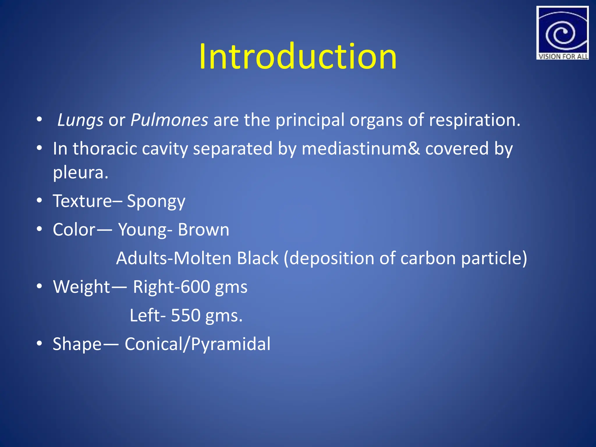

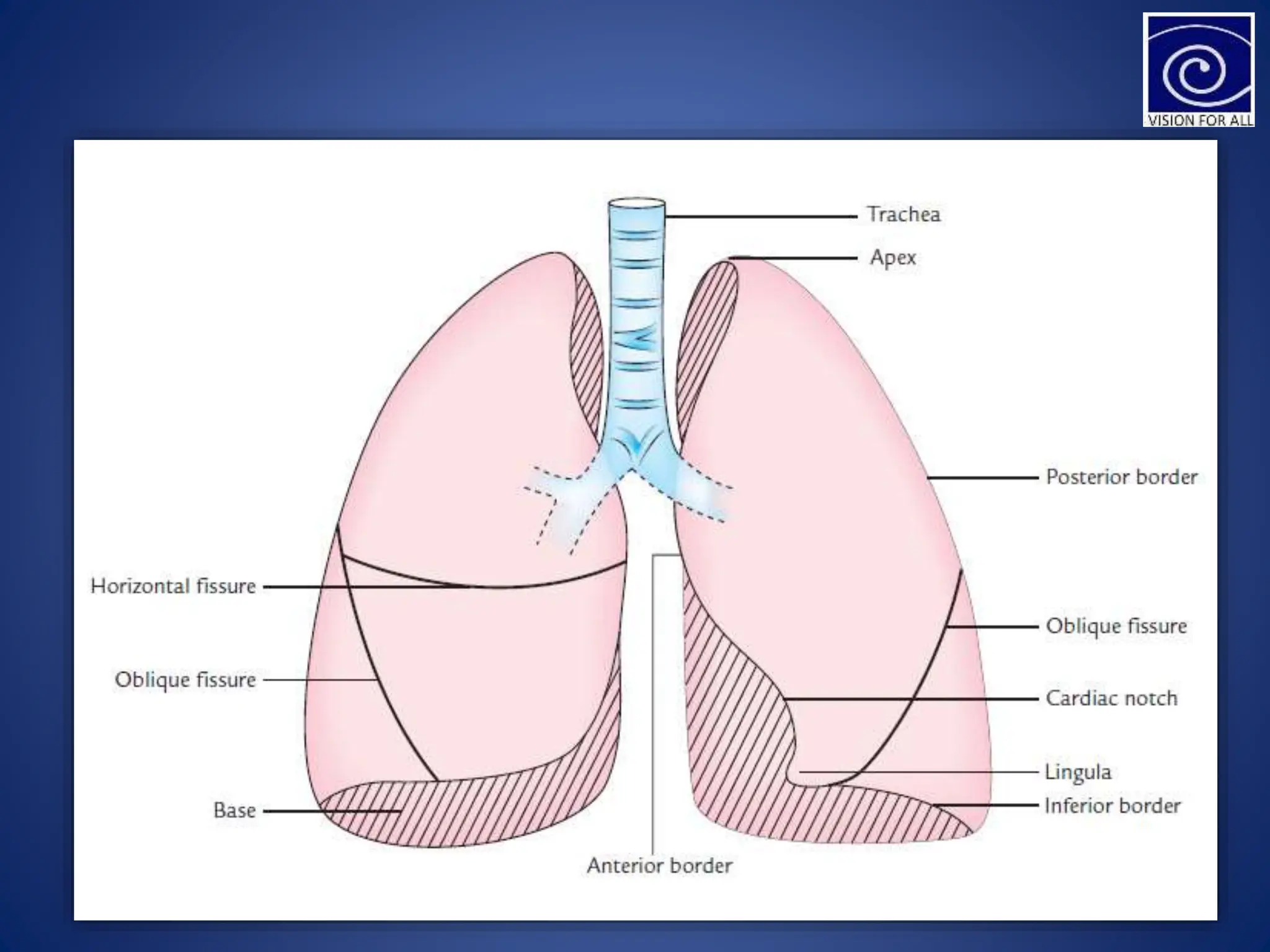


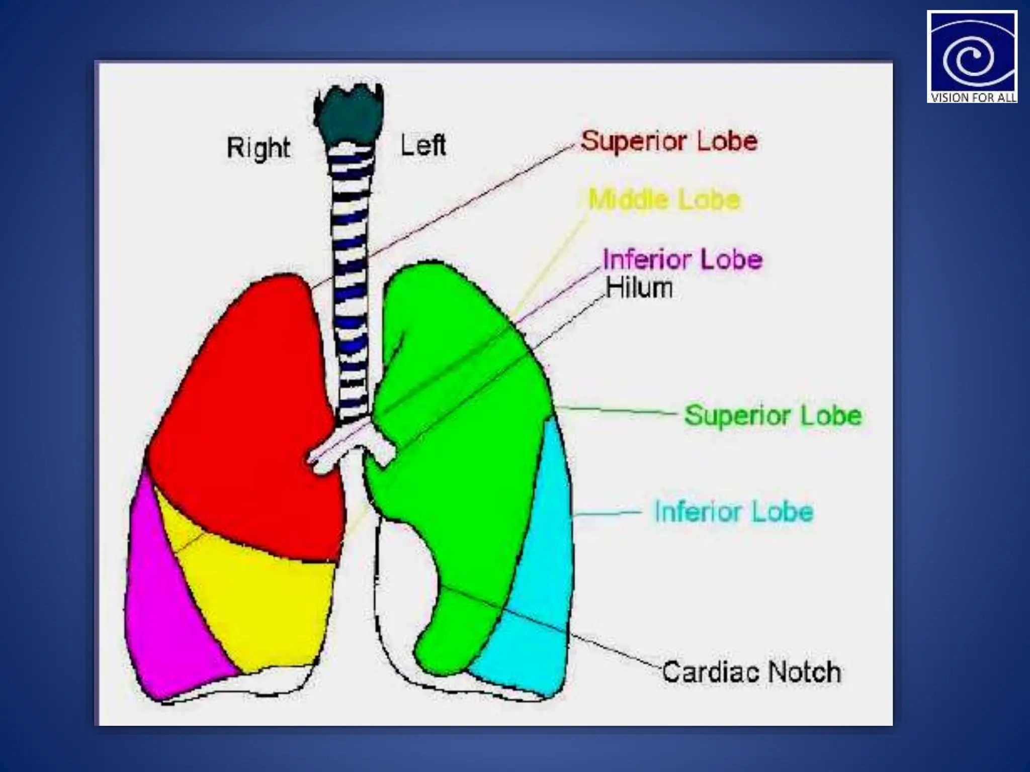


The lungs are the principal organs of respiration located in the thoracic cavity. Each lung is cone-shaped with an apex, base, and borders. The right lung has three lobes separated by two fissures, while the left lung has two lobes separated by one fissure. The root of each lung contains the bronchus, pulmonary vessels and nerves and connects to the mediastinum. The lungs receive arterial blood supply from the pulmonary and bronchial arteries and venous drainage occurs through the pulmonary and bronchial veins. Lymphatic drainage is through vessels in the lungs that drain to tracheobronchial and bronchomediastinal lymph nodes. Nerve supply is from the vagus nerve and thoracic spinal nerves.


