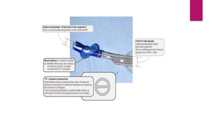
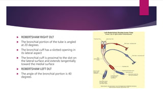
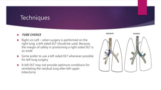
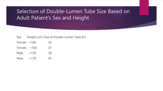
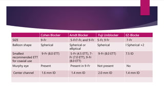
LUNG ISOLATION
TECHNIQUES
MODERATOR- DR PREETHI
PRESENTER- DR JAWABULLA
 Lung-isolation techniques are primarily designed to facilitate OLV in patients undergoing
cardiac, thoracic, mediastinal, vascular, esophageal, or orthopedic procedures involving
the chest cavity.
 Lung isolation is also used to protect the lung from soiling by the contralateral lung in
such cases as bronchopleural fistula, pulmonary hemorrhage, and whole-lung lavage.
Indication for lung isolation
 Thoraxic procedures
Deflated lung provides better operating conditions and reduced trauma during thoraxic procedures
 Control of contamination or hemorrhage
A lung isolation device can prevent infected material from one lung contaminating the other lung
 Unilateral pathology
A bronchopleural fistula/bronchocutaneous fistula may have such a low resistance to gas flow that most of
the tidal volume passes through it making it impossible to adequately ventilate the other lung
An other indication for lung isolation is when lung have markedly different compliance or airway resistance
such as that which occurs following single lung transplantation or unilateral injury
Anatomical consideration
 The right mainstem bronchus is shorter, straighter, have a larger diameter than the
left
 It takes off at an angle of 25 degrees in adult
 The left main stem bronchus diverges from median plane at a 45 degree angles
 The right lobe upper bronchus takeoff is very close to the origin of the right
mainstem bronchus
 These anatomical features – it is usually easier to intubate the right mainstem
bronchus than left but difficult to place a tube in the right main stem bronchus
without obstructing the upper lobe orifice
Lung isolation can be achieved by three different methods:
 DLTs- The most common technique
 Bronchial blockers
 Single-lumen endobronchial tubes (SLTs)
Doble lumen tubes
 Most commonly used to provide lung isolation
 A DLT is essentially two single lumen tubes
bonded together and designated either as right or
left sided, depending on which mainstem
bronchus is designed to fit
 The tracheal lumen is designed to terminate above
the carina
 The distal portion of lumen is angled to fit into the
appropriate mainstem bronchus
 The internal lumen of each tube is a D shaped with
straight side of the D in the middle of the tube
 The bronchial cuff for right sided tube varies in shape . Some tubes the cuff has a
slot to allow ventilation of the right upper lobe
 Most manufacturers color the bronchial cuff blue. They also use blue markings on
the pilot balloon
 Few DLTs have a carinal hook to aid in proper placement and minimize tube
movements after placement- potential problems- difficulty during intubation,
trauma to airway, malposition of the tube because of the hook, interference with
bronchial closure during pneumonectomy, hook can break off and become lost in
the bronchial tree
Right sided tubes
A right sided DLT is acceptably
positioned if the upper lobe
ventilation opening or the space
between the two cuffs is aligned
with the right upper lobe orifice
Left sided tubes
The outermost acceptable position
for a left sided DLT is when the
bronchial cuff is just below the
carina
If the tube were more proximal ,
the bronchial cuff could obstruct
the trachea or the contralateral
mainstem bronchus
The most acceptable distal portion
is when the bronchial segment is at
the proximal edge of the upper
lobe bronchial orifice
More distal insertion would result in obstruction of the upper
lobe bronchus
Sizing
 Adult DLTs comes in size 35,37,39,41 Fr (the French scale is the external diameter
times three)
 For younger patients 26,28,32 Fr tubes
SPECIFIC TUBES
 CARLENS DOUBLE LUMEN TUBE
 Is intended to be inserted into the left mainstem
bronchus
 It has a carinal hook
 Useful with massive hemoptysis when verification of
tube placement is especially difficult
 WHITE DOUBLE LUMEN TUBE
 Designed to fit the right mainstem bronchus
 It has a carinal hook
 The cuff for the right mainstem bronchus is
circumferential superior to the upper lobe bronchus
and continues distally behind the opening
 ROBERTSHAW RIGHT DLT
 The bronchial portion of the tube is angled
at 20 degrees
 The bronchial cuff has a slotted opening in
its lateral aspect
 The bronchial cuff is proximal to the slot on
the lateral surface and extends tangentially
toward the medial surface
 ROBERTSHAW LEFT DLT
 The angle of the bronchial portion is 40
degrees
 BRONCHO- CATH RIGHT SIDED TUBE
 Has a bronchial cuff that is roughly the shape of an
S or slanted doughnut
 The cuff edges nearest upper lobe bronchus is
close to the trachea than part of the cuff touching
the medial bronchial wall
 The end of the bronchial segment has no bevel
 BRONCHO-CATH LEFT SIDED TUBE
 Bronchial portion of the tube is at an angle of
approx-.45 degress
 The bronchial portion has a curved tip
 SHER-I-BRONCH RIGHT SIDED DLT
 SHER-I-BRONCH LEFT SIDED DLT
 SILBRONCHO DLT
Techniques
 TUBE CHOICE
 Right v/s Left – when surgery is performed on the
right lung, a left sided DLT should be used. Because
the margin of safety in positioning a right sided DLT is
so small
 Some prefer to use a left sided DLT whenever possible
for left lung surgery
 A left DLT may not provide optimum conditions for
ventilating the residual lung after left upper
lobectomy
Right sided dlt
 Although a left-sided DLT is used more commonly for most elective thoracic
procedures, there are specific clinical situations in which the use of a right-sided DLT is
indicated
 Because the right mainstem bronchus is shorter than the left bronchus, and because
the right upper lobe bronchus originates at a distance of 1.5 to 2 cm from the carina,
techniques using right endobronchial intubation must take into account the location
and potential for obstruction of the orifice of the right upper lobe bronchus.
 The right-sided DLT incorporates a modified cuff and slot on the endobronchial lumen
that allows ventilation for the right upper lobe
 Before placing a right DLT, the patients chest Xray or CT scan can be closely examined
to identify a right upper lobe bronchus take off, which would make it difficult use a
right DLT
KARTAGENERS – WHICH INCLUDES COMPLETE SITUS
INVERSUS AND A LONGER THAN NORMAL RIGHT
MAINSTEM BRONCHUS
Indications
 □ Distorted anatomy of the entrance of left mainstem bronchus
 □ External or intraluminal tumor compression
 □ Descending thoracic aortic aneurysm
 □ Site of surgery involving the left mainstem bronchus
 □ Left lung transplantation
 □ Left-sided tracheobronchial disruption
 □ Left-sided pneumonectomy*
□ Left-sided sleeve resection
 SIZE- appropriate sized DLT is critical to minimize the frequency of complications
 DLT that is too small – fail to provide lung isolation or may require bronchial
volumes and pressures that could produce mucosal ischemia
 It can result in the tube advancing too far into the bronchus, a higher level of auto
PEEP or barotrauma
 Undersized tube may be more likely to be displaced
 Ventilation and suctioning are more difficult with a small tube
 Large DLT – result in less resistance to gas flow , facilitate suctioning and passage
of a fiberscope and reduce the risk of advancing DLT too far into bronchus but
may result in trauma
 A tube is oversized if the bronchial lumen will not fit into the bronchus or there is
no air leak with the bronchial cuff deflated
 Not more than 3cc of air in the bronchial cuff should be required to create a seal
 When there is a high risk that fluids will seep past the bronchial cuff , a smaller
DLT should be used
 In children, age not weight is a predictor of bronchial diameter, and the right main
bronchial diameter is significantly larger than the left
 The optimal size of DLT is easily selected if the diameters of the patients main
bronchus and the DLT tip are known
 Diameter of mainstem bronchus – difficult to determine – considerable variations
in mainstem bronchial diameters.Age, sex, gender, weight and height – weak
predictive value when selecting proper DLT
SIZE OF DLT FOR CHILDREN
AGE
(YEARS)
DLT
(FRENCH)
8 TO 10 26
10 TO 12 26-28
12 TO 14 32
14 TO 16 35
Selection of Double-Lumen Tube Size Based on
Adult Patient’s Sex and Height
Sex Height (cm) Size of Double-Lumen Tube (Fr)
Female <160 35
Female >160 37
Male <170 39
Male >170 41
PREPARING THE DLT
 The tracheal and bronchial cuff should be inflated and checked for leaks and
symmetrical cuff inflation- each inflation tube is associated with proper cuff
 The cuff and stylet should be lubricated and stylet placed inside the bronchial
lumen, making certain it does not extend beyond the tip
 Connector should be assembled to the tube
Insertion
 The DLT is advanced through the larynx with the angled tip directed anteriorly
 After the bronchial cuff has passed the cords, the tube is turned 90 degrees so
that the bronchial portion point towards the appropriate bronchus
 Leaving the stylet in place for the entire intubation procedure rather than
removing it once the bronchial cuff has passed the vocal cords may result in more
accurate placement
 A DLT is most accurately placed by inserting a fiberscope into the bronchial lumen
and directing it into the appropriate bronchus under vision
 Concurrent videolaryngoscopy maybe required to elevate supraglottic tissues to
facilitate passing the DLT through the glottic opening after the fiberscope is in the
trachea
 A DLT with a carinal hook should be inserted with the bronchial segment
anteriorly until the bronchial cuff passes the cords. It should then be rotated 180
degrees so that the hook is anterior and advanced until the hook passes the vocal
cords
 The tube is then advanced until the hook engages in carina
 Inserting Bronchial portion into the bronchus can be performed blindly after
insertion through the vocal cords- blind method- correct depth of insertion may
be difficult to determine
 In adults- there is correlation between the ideal depth of insertion and patients
height
 Can be estimated from chest Xray
 Advancing the tube with bronchial cuff partially inflated until an increase in
resistance is felt may prevent inserting the tube too deeply
Cuff inflation
 Once the tip is thought to be in a mainstem bronchus , the tracheal cuff should be
inflated like that of a tracheal tube
 The bronchial cuff should be inflated with small incremental volumes until an
airtight seal is just attained
 The total volume should be less than 3ml
CONFIRMING POSITION
 Its essential because the tube may not perform properly if incorrectly positioned
 Position should be checked after insertion , after repositioning the patient, before
beginning one lung ventilation.
 Most frequent DLT movement is during lateral decubitus positioning .Movement is
usually outward or distal migration
 AUSCULTATORY TECHNIQUES-
 Auscultation detects DLT malposition only part of the time because breath sounds
can be transmitted from one region of lungs
 A DLT may function satisfactorily although not in an ideal position
CLAMPING METHODS
 LEFT SIDED TUBES- with only the tracheal cuff inflated and the tracheal lumen
connected to the breathing system, both lungs should be auscultated in axillary
regions and upper lung fields to detect differences
 The bronchial cuff should then be inflated and both lumens connected to the
breathing system- Auscultation should then be repeated
 Next attachment between the breathing system and the tracheal lumen should be
occluded and the tracheal lumen opened to atmosphere
 Breath sounds should be heard only over the left lung. If breath sounds are heard
bilaterally, the tube is too high in trachea
 If breath sounds are heard only over the right lung , the bronchial lumen is on the
right side- bronchial cuff should be deflated, tube withdrawn until distal end is
above the carina, rotated, reinserted
 The attachment breathing system and the bronchial lumen should then be
clamped and ventilated through the tracheal lumen
 Breath sounds should be heard only over the right lung
 If there is marked resistance to ventilation, tube is either far into left bronchus or
not deep enough
 If tube is not deep enough in the bronchus, breath sounds will be present
bilaterally
Positioning Of DLT
 Auscultation alone is unreliable for
confirmation of proper DLT placement.
 Auscultation and bronchoscopy should both
be used each time a DLT is placed and again
when the patient is repositioned.
 Fiberoptic bronchoscopy is performed first
through the tracheal lumen to ensure that
the endobronchial portion of the DLT is in
the left bronchus and that there is no
bronchial cuff herniation over the carina
after inflation.
 Through the tracheal view, the blue endobronchial cuff ideally should be seen
approximately 5 to 10 mm below the tracheal carina in the left bronchus.
 It is crucial to identify the take-off of the right upper lobe bronchus through the
tracheal view.
 Going inside this right upper lobe with the bronchoscope should reveal three
orifices (apical, anterior, and posterior). This is the only structure in the
tracheobronchial tree that has three orifices.
 In the supine patient, the take-off of the right upper lobe is normally on the lateral
wall of the right mainstem bronchus at the 3- to 4-o’clock position in relation to
the main carina.
 Broncho-Cath tubes from Mallinckrodt have a radiopaque line encircling the tube.
This line is proximal to the bronchial cuff and can be useful while positioning a
left-sided DLT.
 The radiopaque marker is 4 cm from the distal tip of the endobronchial lumen.
 This marker reflects white during fiberoptic visualization and, when positioned
slightly above the tracheal carina, should provide the necessary margin of safety
for positioning into the left mainstem bronchus.
 The next observation with the fiberoptic bronchoscope is made through the
endobronchial lumen to check for patency of the tube and determination of
margin of safety.
 The orifices of both the left upper and lower lobes must be identified to avoid
distal impaction in the left lower lobe and occlusion of the left upper lobe
Problems related to DLT
 The most common problems and complications associated with the use of a DLT
are malpositioning and airway trauma .
1.A malpositioned DLT will fail to allow collapse of the lung causing gas trapping
during positive pressure ventilation or it may partially collapse the ventilated or
dependent lung causing hypoxemia
-A common cause of malpositioning - dislodgement of the endobronchial cuff
because of the overinflation , surgical manipulation of the bronchus or extension of
the head and neck during or after patient positioning
2. Airway trauma and rupture of the membranous part of the trachea or the bronchus
 Airway trauma can occur from an oversized DLT or when an undersized DLT
migrates distally into the lobar bronchus and the main body of the DLT comes into
the bronchus, producing laceration or rupture of the airway
 Airway damage during the use of DLTs can present an unexpected air leak,
subcutaneous emphysema, massive airway bleeding into the lumen of the DLT
3. Another potential problem – development of tension pneumothorax in the
dependent, ventilated lung during OLV
BRONCHIAL BLOCKERS
 Alternative method to achieve lung separation involves mainstem bronchus to
allow lung collapse distal to the occlusion
 Bronchial blockers can also be used selectively achieve lobar collapse
 Currently there are several blockers available for lung isolation
These devices are either
within a modified SLT as an enclosed bronchial
blocker Torque Control Blocker Univent
-are used independently with a conventional
SLT, such as the Arndt wire-guided
endobronchial blocker
- the Cohen tip deflecting endobronchial
blocker
- the Fuji Uniblocker
-the EZ-Blocker
 There are specific conditions in which a bronchial blocker may be preferred to a
DLT, such as patients with previous oral or neck surgery who present with a
challenging airway and require lung separation for intrathoracic surgery.
 In these cases, the use of an SLT during an awake nasotracheal or orotracheal
intubation or via tracheostomy secures the airway, and thereafter an independent
bronchial blocker can be placed to achieve lung separation.
 Another group of patients who may benefit from the use of bronchial blockers are
those cancer patients who have undergone a previous contralateral pulmonary
resection.
 In such cases, selective lobar blockade with a bronchial blocker in the ipsilateral
side improves oxygenation and facilitates surgical exposure.
 Blockers can be advanced over a guidewire placed with a fiberoptic bronchoscope
into the required lobar bronchus.
 Bronchial blockers are most commonly used intraluminally (coaxially) with the SLT.
 Another advantage of the bronchial blockers is when postoperative mechanical
ventilation is being considered after prolonged thoracic or esophageal surgery.
 In many instances, these patients have an edematous upper airway at the end of the
procedure.
 If a bronchial blocker is used, there is no need to change the SLT and there is no
compromise of the airway if mechanical ventilation is needed in the postoperative
period.
 For standard adult 9-Fr blockers, an ETT greater than or equal to 7.0 mm ID can be
used with a bronchoscope less than 4.0 mm in diameter. Larger bronchoscopes will
require an ETT greater than 7.5 mm ID. All blockers need to be well lubricated before
placement
Wire-Guided Endobronchial Blocker (Arndt
Blocker)
The Arndt blocker has a retractable loop that is placed over the fiberoptic
bronchoscope, which is then used to guide the blocker into place.
The Arndt blockers usually advance easily into the right mainstem bronchus without
the loop.
Cohen Endobronchial Blocker
 The Cohen blocker uses a wheel located in the most proximal part of the unit that
deflects the tip of the distal part of the blocker into the desired bronchus.
 This blocker has been preangled at the distal tip to facilitate insertion into a target
bronchus
 On the distal shaft above the balloon, there is an arrow that, when seen with the
fiberoptic bronchoscope, indicates in which direction the tip deflects.
 To position the Cohen blocker, the arrow is aligned with the bronchus to be
intubated, the proximal wheel is turned to deflect the tip toward the desired side,
and then the blocker is advanced with fiberoptic guidance.
Fuji Uniblocker
 The Fuji Uniblocker is an independent blocker that is made of silicone material
and has a simple, fixed distal hockey-stick angulation to facilitate insertion.
 The blocker is simply rotated to the left or right as needed under fiberoptic
bronchoscope guidance for placement in the required bronchus.
EZ-Blocker
 The EZ-blocker is a recently introduced 7-Fr, 4-lumen catheter with a Y-shaped
bifurcation.
 Each distal end has a balloon that can be guided into the right and left main
bronchus.
 This device comes with its own multiport adaptor and is used through a 7.5 SLT.
 The end of the Y sits on the tracheal carina. Each distal end is positioned into the
right and left bronchus, and the bronchial balloon is inflated in the operative side
for lung isolation.
Cohen Blocker Arndt Blocker Fuji Uniblocker EZ-Blocke
SIZE 9-Fr 5-Fr7-Fr, and 9-Fr 5-Fr, 9 Fr 7-Fr
Balloon shape Spherical Spherical or
elliptical
Spherical l Spherical ×2
Smallest
recommended ETT
for coaxial use
9-Fr (8.0 ETT) 5-Fr (4.5 ETT), 7-
Fr (7.0 ETT), 9-Fr
(8.0 ETT)
9-Fr (8.0 ETT) 7.5 ID
Murphy eye Present Present in 9-Fr Not present No
Center channel 1.6 mm ID 1.4 mm ID 2.0 mm ID 1.4 mm ID
Complications Related to the Bronchial Blockers
 Failure to achieve lung separation because of abnormal anatomy or lack of a seal
within the bronchus has been reported.
 Inclusion of the bronchial blocker or the distal wire loop of an Arndt blocker into
the stapling line has been reported during a lobectomy - required surgical
reexploration after unsuccessful removal of the bronchial blocker after extubation.
 Another potentially dangerous complication with all bronchial blockers is that the
inflated balloon may move and lodge above the carina or be accidentally inflated
in the trachea-inability to ventilate, hypoxia, and potentially cardiorespiratory
arrest unless quickly recognized and the blocker deflated.
Single lumen bronchial tubes
 Another option for achieving lung separation is to use a single lumen tube to
intubate a mainstem bronchus
 SLT s are sometimes used in pediatric patient whose airways are too small for DLTs
or in whom a bronchial blocker cannot be used
 In the patient with massive hemoptysis, bronchial intubation with a single lumen
tube is often easiest method
 Special tubes with a single lumen, an angulated distal tip/ bronchial and cuffs at
both the tracheal and bronchial positions- longer than the conventional SLT
Difficult Airways and One-Lung Ventilation
 A number of patients requiring OLV are identified during preoperative evaluation
to have a potentially difficult airway. Others present with unexpected difficulty to
intubate after induction of anesthesia
 A patient who requires OLV might have distorted upper airway anatomy or
distorted anatomy at or beyond the tracheal carina, such as descending thoracic
aortic aneurysm compressing the entrance of the left mainstem bronchus or an
intraluminal or extraluminal tumor near the tracheobronchial bifurcation that
makes the insertion of a left-sided DLT relatively difficult or impossible.
 This can be detected by reviewing the chest radiographs and CT scans of the
chest.
 A flexible fiberoptic bronchoscopic examination is necessary to assess a distorted area
of the airway before selection of a specific tube or blocker to achieve OLV.
 In patients who require OLV and present with a difficult airway, the primary goal is to
establish an airway with an SLT placed orally with the aid of a flexible fiberoptic
bronchoscope, after appropriate airway anesthesia is achieved.
 Once the SLT is in place, an independent bronchial blocker can be passed.
 If the patient requires OLV and cannot be intubated orally, an awake nasotracheal
intubation can be performed with an SLT and, once the airway is established, then a
bronchial blocker can be used
 An alternative to achieve OLV in a patient with a difficult airway is to intubate the
patient’s trachea with an SLT; then a DLT-SLT tube-exchange catheter can be used
to replace the existing SLT with a DLT after general anesthesia is induced.
 For a DLT the exchange catheter should be at least 83 cm.
 A 14-Fr exchange catheter can be used for 41-Fr and 39-Fr DLTs and for 37-Fr /
35-Fr DLTs an 11-Fr exchange catheter is used.
 Specially designed exchange catheters for DLTs are available with a softer distal
tip to decrease the risk of distal airway trauma (e.g., Cook Exchange Catheter).
Lung-Isolation Techniques in Patients With a
Tracheostomy in Place
 Placement of a DLT through a tracheostomy stoma may be prone to malposition
because the upper airway has been shortened and the conventional DLT may be
too long.
 Before placing any lung isolation devices through a tracheostomy stoma it is
important to consider whether it is a fresh stoma (i.e., a few days old, when the
airway can be lost immediately on decannulation) versus a chronic tracheostomy.
 The alternatives to achieve OLV in a tracheostomized patient include
 (1) insertion of an SLT followed by an independent bronchial blocker passed coaxially
or externally to the SLT
 (2) the use of a disposable cuffed tracheostomy cannula with an independent
bronchial blocker
 (3) replacement of the tracheostomy cannula with a specially designed short DLT- the
Naruke DLT,
 (4) placement of a small DLT through the tracheostomy stoma
 (5) if possible, oral access to the airway for standard placement of a DLT or blocker
(this is occasionally an option in patients on prolonged mechanical ventilation for
respiratory failure or postoperative complications
HYPOXEMIA
 A major concern that influences anesthetic management for thoracic surgery is
the occurrence of hypoxemia during OLV.
 There is no universally acceptable figure for the safest lower limit of oxygen
saturation during OLV. A saturation greater than or equal to 90% (PaO2 > 60 mm
Hg) is commonly accepted, and for brief periods a saturation in the high 80%s
may be acceptable in patients without significant comorbidity.
 However, the lowest acceptable saturation will be higher in patients with organs at
risk of hypoxia because of limited regional blood flow (e.g., coronary or
cerebrovascular disease) and in patients with limited oxygen transport (e.g.,
anemia or decreased cardiopulmonary reserve).
 Previously, hypoxemia occurred frequently during OLV- period between 1950 and
1980 describe an incidence of hypoxemia (arterial saturation < 90%) of 20% to
25%, now incidence of less than 5% is reported.
 This improvement is most likely a result of several factors:
 1improved lung isolation techniques such as routine fiberoscopy to prevent lobar
obstruction from DLTs
 2 improved anesthetic drugs that cause less inhibition of HPV
 3 better understanding of the pathophysiology of OLV.
THERAPIES FOR DESATURATION DURING OLV
Severe or precipitous desaturation: Resume two-lung ventilation (if possible)
Gradual desaturation:
Ensure that delivered FiO2 is 1.0
Check position of double-lumen tube or blocker with fiberoptic bronchoscopy
Ensure that cardiac output is optimal, decrease volatile anesthetics to <1MAC
Apply a recruitment maneuver to the ventilated lung (this will transiently make the
hypoxemia worse)
Increase PEEP to the ventilated lung (except in patients with emphysematous pathology)
 Apneic oxygen insufflation of the nonventilated lung
 Apply CPAP 1–2 cm H2O to the nonventilated lung (apply a recruitment maneuver
to this lung immediately before CPAP)
 Partial ventilation techniques of the nonventilated lung
□ Intermittent positive pressure ventilation
□ Fiberoptic lobar insufflation
□ Selective lobar collapse (using a bronchial blocker)
□ Small tidal volume ventilation
 Pharmacologic manipulations - Eliminating known potent vasodilators such as
nitroglycerin, halothane, and large doses of other volatile anesthetics will improve
oxygenation during OLV
 Mechanical restriction of the blood flow to the nonventilated lung- the surgeon
can directly compress or clamp the blood flow to the nonventilated lung. This can
be done temporarily in emergency desaturation situations or definitively in cases
of pneumonectomy or lung transplantation. Another technique of mechanical
limitation of blood flow to the nonventilated lung is the inflation of a pulmonary
artery catheter balloon
 Venovenous ECMO
Conclusion
 In summary, the optimal method of lung isolation will depend on a number of
factors, including the patient’s airway anatomy, the indication for lung isolation,
the available equipment, and the training of the anesthesiologist. Whatever
method of lung isolation is used, the “ABCs” of lung isolation are
 Anatomy.
 Bronchoscopy
 Chest imaging

LUNG ISOLATION TECHNIQUES and differential lung ventilation .pptx

  • 1.
    LUNG ISOLATION TECHNIQUES MODERATOR- DRPREETHI PRESENTER- DR JAWABULLA
  • 2.
     Lung-isolation techniquesare primarily designed to facilitate OLV in patients undergoing cardiac, thoracic, mediastinal, vascular, esophageal, or orthopedic procedures involving the chest cavity.  Lung isolation is also used to protect the lung from soiling by the contralateral lung in such cases as bronchopleural fistula, pulmonary hemorrhage, and whole-lung lavage.
  • 3.
    Indication for lungisolation  Thoraxic procedures Deflated lung provides better operating conditions and reduced trauma during thoraxic procedures  Control of contamination or hemorrhage A lung isolation device can prevent infected material from one lung contaminating the other lung  Unilateral pathology A bronchopleural fistula/bronchocutaneous fistula may have such a low resistance to gas flow that most of the tidal volume passes through it making it impossible to adequately ventilate the other lung An other indication for lung isolation is when lung have markedly different compliance or airway resistance such as that which occurs following single lung transplantation or unilateral injury
  • 5.
    Anatomical consideration  Theright mainstem bronchus is shorter, straighter, have a larger diameter than the left  It takes off at an angle of 25 degrees in adult  The left main stem bronchus diverges from median plane at a 45 degree angles  The right lobe upper bronchus takeoff is very close to the origin of the right mainstem bronchus  These anatomical features – it is usually easier to intubate the right mainstem bronchus than left but difficult to place a tube in the right main stem bronchus without obstructing the upper lobe orifice
  • 7.
    Lung isolation canbe achieved by three different methods:  DLTs- The most common technique  Bronchial blockers  Single-lumen endobronchial tubes (SLTs)
  • 8.
    Doble lumen tubes Most commonly used to provide lung isolation  A DLT is essentially two single lumen tubes bonded together and designated either as right or left sided, depending on which mainstem bronchus is designed to fit  The tracheal lumen is designed to terminate above the carina  The distal portion of lumen is angled to fit into the appropriate mainstem bronchus  The internal lumen of each tube is a D shaped with straight side of the D in the middle of the tube
  • 10.
     The bronchialcuff for right sided tube varies in shape . Some tubes the cuff has a slot to allow ventilation of the right upper lobe  Most manufacturers color the bronchial cuff blue. They also use blue markings on the pilot balloon  Few DLTs have a carinal hook to aid in proper placement and minimize tube movements after placement- potential problems- difficulty during intubation, trauma to airway, malposition of the tube because of the hook, interference with bronchial closure during pneumonectomy, hook can break off and become lost in the bronchial tree
  • 11.
    Right sided tubes Aright sided DLT is acceptably positioned if the upper lobe ventilation opening or the space between the two cuffs is aligned with the right upper lobe orifice Left sided tubes The outermost acceptable position for a left sided DLT is when the bronchial cuff is just below the carina If the tube were more proximal , the bronchial cuff could obstruct the trachea or the contralateral mainstem bronchus The most acceptable distal portion is when the bronchial segment is at the proximal edge of the upper lobe bronchial orifice More distal insertion would result in obstruction of the upper lobe bronchus
  • 12.
    Sizing  Adult DLTscomes in size 35,37,39,41 Fr (the French scale is the external diameter times three)  For younger patients 26,28,32 Fr tubes
  • 13.
    SPECIFIC TUBES  CARLENSDOUBLE LUMEN TUBE  Is intended to be inserted into the left mainstem bronchus  It has a carinal hook  Useful with massive hemoptysis when verification of tube placement is especially difficult  WHITE DOUBLE LUMEN TUBE  Designed to fit the right mainstem bronchus  It has a carinal hook  The cuff for the right mainstem bronchus is circumferential superior to the upper lobe bronchus and continues distally behind the opening
  • 14.
     ROBERTSHAW RIGHTDLT  The bronchial portion of the tube is angled at 20 degrees  The bronchial cuff has a slotted opening in its lateral aspect  The bronchial cuff is proximal to the slot on the lateral surface and extends tangentially toward the medial surface  ROBERTSHAW LEFT DLT  The angle of the bronchial portion is 40 degrees
  • 15.
     BRONCHO- CATHRIGHT SIDED TUBE  Has a bronchial cuff that is roughly the shape of an S or slanted doughnut  The cuff edges nearest upper lobe bronchus is close to the trachea than part of the cuff touching the medial bronchial wall  The end of the bronchial segment has no bevel  BRONCHO-CATH LEFT SIDED TUBE  Bronchial portion of the tube is at an angle of approx-.45 degress  The bronchial portion has a curved tip
  • 16.
     SHER-I-BRONCH RIGHTSIDED DLT  SHER-I-BRONCH LEFT SIDED DLT  SILBRONCHO DLT
  • 17.
    Techniques  TUBE CHOICE Right v/s Left – when surgery is performed on the right lung, a left sided DLT should be used. Because the margin of safety in positioning a right sided DLT is so small  Some prefer to use a left sided DLT whenever possible for left lung surgery  A left DLT may not provide optimum conditions for ventilating the residual lung after left upper lobectomy
  • 18.
    Right sided dlt Although a left-sided DLT is used more commonly for most elective thoracic procedures, there are specific clinical situations in which the use of a right-sided DLT is indicated  Because the right mainstem bronchus is shorter than the left bronchus, and because the right upper lobe bronchus originates at a distance of 1.5 to 2 cm from the carina, techniques using right endobronchial intubation must take into account the location and potential for obstruction of the orifice of the right upper lobe bronchus.  The right-sided DLT incorporates a modified cuff and slot on the endobronchial lumen that allows ventilation for the right upper lobe  Before placing a right DLT, the patients chest Xray or CT scan can be closely examined to identify a right upper lobe bronchus take off, which would make it difficult use a right DLT KARTAGENERS – WHICH INCLUDES COMPLETE SITUS INVERSUS AND A LONGER THAN NORMAL RIGHT MAINSTEM BRONCHUS
  • 19.
    Indications  □ Distortedanatomy of the entrance of left mainstem bronchus  □ External or intraluminal tumor compression  □ Descending thoracic aortic aneurysm  □ Site of surgery involving the left mainstem bronchus  □ Left lung transplantation  □ Left-sided tracheobronchial disruption  □ Left-sided pneumonectomy* □ Left-sided sleeve resection
  • 20.
     SIZE- appropriatesized DLT is critical to minimize the frequency of complications  DLT that is too small – fail to provide lung isolation or may require bronchial volumes and pressures that could produce mucosal ischemia  It can result in the tube advancing too far into the bronchus, a higher level of auto PEEP or barotrauma  Undersized tube may be more likely to be displaced  Ventilation and suctioning are more difficult with a small tube
  • 21.
     Large DLT– result in less resistance to gas flow , facilitate suctioning and passage of a fiberscope and reduce the risk of advancing DLT too far into bronchus but may result in trauma  A tube is oversized if the bronchial lumen will not fit into the bronchus or there is no air leak with the bronchial cuff deflated  Not more than 3cc of air in the bronchial cuff should be required to create a seal  When there is a high risk that fluids will seep past the bronchial cuff , a smaller DLT should be used
  • 22.
     In children,age not weight is a predictor of bronchial diameter, and the right main bronchial diameter is significantly larger than the left  The optimal size of DLT is easily selected if the diameters of the patients main bronchus and the DLT tip are known  Diameter of mainstem bronchus – difficult to determine – considerable variations in mainstem bronchial diameters.Age, sex, gender, weight and height – weak predictive value when selecting proper DLT
  • 23.
    SIZE OF DLTFOR CHILDREN AGE (YEARS) DLT (FRENCH) 8 TO 10 26 10 TO 12 26-28 12 TO 14 32 14 TO 16 35
  • 24.
    Selection of Double-LumenTube Size Based on Adult Patient’s Sex and Height Sex Height (cm) Size of Double-Lumen Tube (Fr) Female <160 35 Female >160 37 Male <170 39 Male >170 41
  • 25.
    PREPARING THE DLT The tracheal and bronchial cuff should be inflated and checked for leaks and symmetrical cuff inflation- each inflation tube is associated with proper cuff  The cuff and stylet should be lubricated and stylet placed inside the bronchial lumen, making certain it does not extend beyond the tip  Connector should be assembled to the tube
  • 26.
    Insertion  The DLTis advanced through the larynx with the angled tip directed anteriorly  After the bronchial cuff has passed the cords, the tube is turned 90 degrees so that the bronchial portion point towards the appropriate bronchus  Leaving the stylet in place for the entire intubation procedure rather than removing it once the bronchial cuff has passed the vocal cords may result in more accurate placement  A DLT is most accurately placed by inserting a fiberscope into the bronchial lumen and directing it into the appropriate bronchus under vision
  • 27.
     Concurrent videolaryngoscopymaybe required to elevate supraglottic tissues to facilitate passing the DLT through the glottic opening after the fiberscope is in the trachea  A DLT with a carinal hook should be inserted with the bronchial segment anteriorly until the bronchial cuff passes the cords. It should then be rotated 180 degrees so that the hook is anterior and advanced until the hook passes the vocal cords  The tube is then advanced until the hook engages in carina
  • 28.
     Inserting Bronchialportion into the bronchus can be performed blindly after insertion through the vocal cords- blind method- correct depth of insertion may be difficult to determine  In adults- there is correlation between the ideal depth of insertion and patients height  Can be estimated from chest Xray  Advancing the tube with bronchial cuff partially inflated until an increase in resistance is felt may prevent inserting the tube too deeply
  • 29.
    Cuff inflation  Oncethe tip is thought to be in a mainstem bronchus , the tracheal cuff should be inflated like that of a tracheal tube  The bronchial cuff should be inflated with small incremental volumes until an airtight seal is just attained  The total volume should be less than 3ml
  • 30.
    CONFIRMING POSITION  Itsessential because the tube may not perform properly if incorrectly positioned  Position should be checked after insertion , after repositioning the patient, before beginning one lung ventilation.  Most frequent DLT movement is during lateral decubitus positioning .Movement is usually outward or distal migration  AUSCULTATORY TECHNIQUES-  Auscultation detects DLT malposition only part of the time because breath sounds can be transmitted from one region of lungs  A DLT may function satisfactorily although not in an ideal position
  • 31.
    CLAMPING METHODS  LEFTSIDED TUBES- with only the tracheal cuff inflated and the tracheal lumen connected to the breathing system, both lungs should be auscultated in axillary regions and upper lung fields to detect differences  The bronchial cuff should then be inflated and both lumens connected to the breathing system- Auscultation should then be repeated  Next attachment between the breathing system and the tracheal lumen should be occluded and the tracheal lumen opened to atmosphere  Breath sounds should be heard only over the left lung. If breath sounds are heard bilaterally, the tube is too high in trachea
  • 32.
     If breathsounds are heard only over the right lung , the bronchial lumen is on the right side- bronchial cuff should be deflated, tube withdrawn until distal end is above the carina, rotated, reinserted  The attachment breathing system and the bronchial lumen should then be clamped and ventilated through the tracheal lumen  Breath sounds should be heard only over the right lung  If there is marked resistance to ventilation, tube is either far into left bronchus or not deep enough  If tube is not deep enough in the bronchus, breath sounds will be present bilaterally
  • 34.
    Positioning Of DLT Auscultation alone is unreliable for confirmation of proper DLT placement.  Auscultation and bronchoscopy should both be used each time a DLT is placed and again when the patient is repositioned.  Fiberoptic bronchoscopy is performed first through the tracheal lumen to ensure that the endobronchial portion of the DLT is in the left bronchus and that there is no bronchial cuff herniation over the carina after inflation.
  • 35.
     Through thetracheal view, the blue endobronchial cuff ideally should be seen approximately 5 to 10 mm below the tracheal carina in the left bronchus.  It is crucial to identify the take-off of the right upper lobe bronchus through the tracheal view.  Going inside this right upper lobe with the bronchoscope should reveal three orifices (apical, anterior, and posterior). This is the only structure in the tracheobronchial tree that has three orifices.  In the supine patient, the take-off of the right upper lobe is normally on the lateral wall of the right mainstem bronchus at the 3- to 4-o’clock position in relation to the main carina.
  • 36.
     Broncho-Cath tubesfrom Mallinckrodt have a radiopaque line encircling the tube. This line is proximal to the bronchial cuff and can be useful while positioning a left-sided DLT.  The radiopaque marker is 4 cm from the distal tip of the endobronchial lumen.  This marker reflects white during fiberoptic visualization and, when positioned slightly above the tracheal carina, should provide the necessary margin of safety for positioning into the left mainstem bronchus.
  • 37.
     The nextobservation with the fiberoptic bronchoscope is made through the endobronchial lumen to check for patency of the tube and determination of margin of safety.  The orifices of both the left upper and lower lobes must be identified to avoid distal impaction in the left lower lobe and occlusion of the left upper lobe
  • 38.
    Problems related toDLT  The most common problems and complications associated with the use of a DLT are malpositioning and airway trauma . 1.A malpositioned DLT will fail to allow collapse of the lung causing gas trapping during positive pressure ventilation or it may partially collapse the ventilated or dependent lung causing hypoxemia -A common cause of malpositioning - dislodgement of the endobronchial cuff because of the overinflation , surgical manipulation of the bronchus or extension of the head and neck during or after patient positioning
  • 39.
    2. Airway traumaand rupture of the membranous part of the trachea or the bronchus  Airway trauma can occur from an oversized DLT or when an undersized DLT migrates distally into the lobar bronchus and the main body of the DLT comes into the bronchus, producing laceration or rupture of the airway  Airway damage during the use of DLTs can present an unexpected air leak, subcutaneous emphysema, massive airway bleeding into the lumen of the DLT 3. Another potential problem – development of tension pneumothorax in the dependent, ventilated lung during OLV
  • 40.
    BRONCHIAL BLOCKERS  Alternativemethod to achieve lung separation involves mainstem bronchus to allow lung collapse distal to the occlusion  Bronchial blockers can also be used selectively achieve lobar collapse  Currently there are several blockers available for lung isolation
  • 41.
    These devices areeither within a modified SLT as an enclosed bronchial blocker Torque Control Blocker Univent -are used independently with a conventional SLT, such as the Arndt wire-guided endobronchial blocker - the Cohen tip deflecting endobronchial blocker - the Fuji Uniblocker -the EZ-Blocker
  • 42.
     There arespecific conditions in which a bronchial blocker may be preferred to a DLT, such as patients with previous oral or neck surgery who present with a challenging airway and require lung separation for intrathoracic surgery.  In these cases, the use of an SLT during an awake nasotracheal or orotracheal intubation or via tracheostomy secures the airway, and thereafter an independent bronchial blocker can be placed to achieve lung separation.
  • 43.
     Another groupof patients who may benefit from the use of bronchial blockers are those cancer patients who have undergone a previous contralateral pulmonary resection.  In such cases, selective lobar blockade with a bronchial blocker in the ipsilateral side improves oxygenation and facilitates surgical exposure.  Blockers can be advanced over a guidewire placed with a fiberoptic bronchoscope into the required lobar bronchus.  Bronchial blockers are most commonly used intraluminally (coaxially) with the SLT.
  • 44.
     Another advantageof the bronchial blockers is when postoperative mechanical ventilation is being considered after prolonged thoracic or esophageal surgery.  In many instances, these patients have an edematous upper airway at the end of the procedure.  If a bronchial blocker is used, there is no need to change the SLT and there is no compromise of the airway if mechanical ventilation is needed in the postoperative period.  For standard adult 9-Fr blockers, an ETT greater than or equal to 7.0 mm ID can be used with a bronchoscope less than 4.0 mm in diameter. Larger bronchoscopes will require an ETT greater than 7.5 mm ID. All blockers need to be well lubricated before placement
  • 45.
    Wire-Guided Endobronchial Blocker(Arndt Blocker) The Arndt blocker has a retractable loop that is placed over the fiberoptic bronchoscope, which is then used to guide the blocker into place. The Arndt blockers usually advance easily into the right mainstem bronchus without the loop.
  • 46.
    Cohen Endobronchial Blocker The Cohen blocker uses a wheel located in the most proximal part of the unit that deflects the tip of the distal part of the blocker into the desired bronchus.  This blocker has been preangled at the distal tip to facilitate insertion into a target bronchus  On the distal shaft above the balloon, there is an arrow that, when seen with the fiberoptic bronchoscope, indicates in which direction the tip deflects.  To position the Cohen blocker, the arrow is aligned with the bronchus to be intubated, the proximal wheel is turned to deflect the tip toward the desired side, and then the blocker is advanced with fiberoptic guidance.
  • 47.
    Fuji Uniblocker  TheFuji Uniblocker is an independent blocker that is made of silicone material and has a simple, fixed distal hockey-stick angulation to facilitate insertion.  The blocker is simply rotated to the left or right as needed under fiberoptic bronchoscope guidance for placement in the required bronchus.
  • 48.
    EZ-Blocker  The EZ-blockeris a recently introduced 7-Fr, 4-lumen catheter with a Y-shaped bifurcation.  Each distal end has a balloon that can be guided into the right and left main bronchus.  This device comes with its own multiport adaptor and is used through a 7.5 SLT.  The end of the Y sits on the tracheal carina. Each distal end is positioned into the right and left bronchus, and the bronchial balloon is inflated in the operative side for lung isolation.
  • 49.
    Cohen Blocker ArndtBlocker Fuji Uniblocker EZ-Blocke SIZE 9-Fr 5-Fr7-Fr, and 9-Fr 5-Fr, 9 Fr 7-Fr Balloon shape Spherical Spherical or elliptical Spherical l Spherical ×2 Smallest recommended ETT for coaxial use 9-Fr (8.0 ETT) 5-Fr (4.5 ETT), 7- Fr (7.0 ETT), 9-Fr (8.0 ETT) 9-Fr (8.0 ETT) 7.5 ID Murphy eye Present Present in 9-Fr Not present No Center channel 1.6 mm ID 1.4 mm ID 2.0 mm ID 1.4 mm ID
  • 50.
    Complications Related tothe Bronchial Blockers  Failure to achieve lung separation because of abnormal anatomy or lack of a seal within the bronchus has been reported.  Inclusion of the bronchial blocker or the distal wire loop of an Arndt blocker into the stapling line has been reported during a lobectomy - required surgical reexploration after unsuccessful removal of the bronchial blocker after extubation.  Another potentially dangerous complication with all bronchial blockers is that the inflated balloon may move and lodge above the carina or be accidentally inflated in the trachea-inability to ventilate, hypoxia, and potentially cardiorespiratory arrest unless quickly recognized and the blocker deflated.
  • 51.
    Single lumen bronchialtubes  Another option for achieving lung separation is to use a single lumen tube to intubate a mainstem bronchus  SLT s are sometimes used in pediatric patient whose airways are too small for DLTs or in whom a bronchial blocker cannot be used  In the patient with massive hemoptysis, bronchial intubation with a single lumen tube is often easiest method  Special tubes with a single lumen, an angulated distal tip/ bronchial and cuffs at both the tracheal and bronchial positions- longer than the conventional SLT
  • 52.
    Difficult Airways andOne-Lung Ventilation  A number of patients requiring OLV are identified during preoperative evaluation to have a potentially difficult airway. Others present with unexpected difficulty to intubate after induction of anesthesia  A patient who requires OLV might have distorted upper airway anatomy or distorted anatomy at or beyond the tracheal carina, such as descending thoracic aortic aneurysm compressing the entrance of the left mainstem bronchus or an intraluminal or extraluminal tumor near the tracheobronchial bifurcation that makes the insertion of a left-sided DLT relatively difficult or impossible.  This can be detected by reviewing the chest radiographs and CT scans of the chest.
  • 53.
     A flexiblefiberoptic bronchoscopic examination is necessary to assess a distorted area of the airway before selection of a specific tube or blocker to achieve OLV.  In patients who require OLV and present with a difficult airway, the primary goal is to establish an airway with an SLT placed orally with the aid of a flexible fiberoptic bronchoscope, after appropriate airway anesthesia is achieved.  Once the SLT is in place, an independent bronchial blocker can be passed.  If the patient requires OLV and cannot be intubated orally, an awake nasotracheal intubation can be performed with an SLT and, once the airway is established, then a bronchial blocker can be used
  • 54.
     An alternativeto achieve OLV in a patient with a difficult airway is to intubate the patient’s trachea with an SLT; then a DLT-SLT tube-exchange catheter can be used to replace the existing SLT with a DLT after general anesthesia is induced.  For a DLT the exchange catheter should be at least 83 cm.  A 14-Fr exchange catheter can be used for 41-Fr and 39-Fr DLTs and for 37-Fr / 35-Fr DLTs an 11-Fr exchange catheter is used.  Specially designed exchange catheters for DLTs are available with a softer distal tip to decrease the risk of distal airway trauma (e.g., Cook Exchange Catheter).
  • 55.
    Lung-Isolation Techniques inPatients With a Tracheostomy in Place  Placement of a DLT through a tracheostomy stoma may be prone to malposition because the upper airway has been shortened and the conventional DLT may be too long.  Before placing any lung isolation devices through a tracheostomy stoma it is important to consider whether it is a fresh stoma (i.e., a few days old, when the airway can be lost immediately on decannulation) versus a chronic tracheostomy.
  • 56.
     The alternativesto achieve OLV in a tracheostomized patient include  (1) insertion of an SLT followed by an independent bronchial blocker passed coaxially or externally to the SLT  (2) the use of a disposable cuffed tracheostomy cannula with an independent bronchial blocker  (3) replacement of the tracheostomy cannula with a specially designed short DLT- the Naruke DLT,  (4) placement of a small DLT through the tracheostomy stoma  (5) if possible, oral access to the airway for standard placement of a DLT or blocker (this is occasionally an option in patients on prolonged mechanical ventilation for respiratory failure or postoperative complications
  • 57.
    HYPOXEMIA  A majorconcern that influences anesthetic management for thoracic surgery is the occurrence of hypoxemia during OLV.  There is no universally acceptable figure for the safest lower limit of oxygen saturation during OLV. A saturation greater than or equal to 90% (PaO2 > 60 mm Hg) is commonly accepted, and for brief periods a saturation in the high 80%s may be acceptable in patients without significant comorbidity.  However, the lowest acceptable saturation will be higher in patients with organs at risk of hypoxia because of limited regional blood flow (e.g., coronary or cerebrovascular disease) and in patients with limited oxygen transport (e.g., anemia or decreased cardiopulmonary reserve).
  • 58.
     Previously, hypoxemiaoccurred frequently during OLV- period between 1950 and 1980 describe an incidence of hypoxemia (arterial saturation < 90%) of 20% to 25%, now incidence of less than 5% is reported.  This improvement is most likely a result of several factors:  1improved lung isolation techniques such as routine fiberoscopy to prevent lobar obstruction from DLTs  2 improved anesthetic drugs that cause less inhibition of HPV  3 better understanding of the pathophysiology of OLV.
  • 59.
    THERAPIES FOR DESATURATIONDURING OLV Severe or precipitous desaturation: Resume two-lung ventilation (if possible) Gradual desaturation: Ensure that delivered FiO2 is 1.0 Check position of double-lumen tube or blocker with fiberoptic bronchoscopy Ensure that cardiac output is optimal, decrease volatile anesthetics to <1MAC Apply a recruitment maneuver to the ventilated lung (this will transiently make the hypoxemia worse) Increase PEEP to the ventilated lung (except in patients with emphysematous pathology)
  • 60.
     Apneic oxygeninsufflation of the nonventilated lung  Apply CPAP 1–2 cm H2O to the nonventilated lung (apply a recruitment maneuver to this lung immediately before CPAP)  Partial ventilation techniques of the nonventilated lung □ Intermittent positive pressure ventilation □ Fiberoptic lobar insufflation □ Selective lobar collapse (using a bronchial blocker) □ Small tidal volume ventilation
  • 61.
     Pharmacologic manipulations- Eliminating known potent vasodilators such as nitroglycerin, halothane, and large doses of other volatile anesthetics will improve oxygenation during OLV  Mechanical restriction of the blood flow to the nonventilated lung- the surgeon can directly compress or clamp the blood flow to the nonventilated lung. This can be done temporarily in emergency desaturation situations or definitively in cases of pneumonectomy or lung transplantation. Another technique of mechanical limitation of blood flow to the nonventilated lung is the inflation of a pulmonary artery catheter balloon  Venovenous ECMO
  • 62.
    Conclusion  In summary,the optimal method of lung isolation will depend on a number of factors, including the patient’s airway anatomy, the indication for lung isolation, the available equipment, and the training of the anesthesiologist. Whatever method of lung isolation is used, the “ABCs” of lung isolation are  Anatomy.  Bronchoscopy  Chest imaging