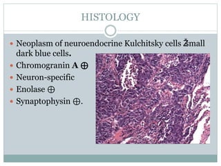
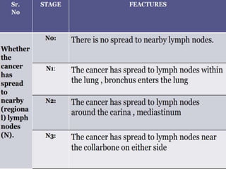
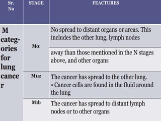
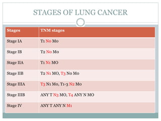
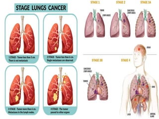
LUNG CANCER
Presented by:
Siddhartha
Masetti Mahesh
OBJECTIVES
 Lung Cancer-How common is it?
 Epidemiology
 Risk Factors
 Signs and Symptoms
 pathophysiology
 Types
 Complications
 Screening
 Diagnosis
 Treatment
 Conclusion
LUNG CANCER: HOW COMMON IS IT?
 Lung cancer is the second most common cancer in
both men and women.
 It accounts for an estimated 25% of all cancer
diagnoses.
 Lung cancer mainly occurs in older people.
 For smokers the risk is much higher.
LUNG CANCER: HOW COMMON IS IT?
 Lung cancer is the leading cause of cancer death
among both men and women in the U.S.
 Lung cancer accounts for about 1 in 4 cancer deaths
each year.
EPIDEMIOLOGY
 Lung cancer is the most commonly diagnosed cancer
worldwide, and its incidence continues to grow.
 In 2018, an estimated 2.1 million new cases of lung
cancer were diagnosed globally, accounting for
approximately 11.6% of the global cancer burden.
 An estimated 1.76 million lung cancer deaths occurred in
2018.
 According to the WHO data published in 2017 Lung
Cancers Deaths in Jamaica reached 430 or 2.51% of total
deaths. The age adjusted Death Rate is 14.26 per
100,000 of population ranks Jamaica #82 in the world
RISK FACTORS
 Smoking is the leading
risk factor for lung
cancer (78% in men, 90%
in women).
 Radon(Radioactive gas
released from breakdown
of uranium)- Second
leading Cause.
 Asbestos.
 Halogen ether exposure
 Inorganic arsenic
exposure
 Chronic interstitial
pneumonitis
 Radioisotope exposure,
ionizing radiation
 Atmospheric pollution
 Chromium, nickel
exposure
 Vinyl chloride exposure
SIGNS AND SYMPTOMS
The most common signs and symptoms of lung cancer include the
following:
 Cough
 Chest pain
 Shortness of breath
 Coughing up blood(Hemoptysis)
 Wheezing
 Hoarseness
 coin”lesion on CXR or noncalcified nodule on CT.
 Recurring infections such as bronchitis and pneumonia
 Weight loss and loss of appetite
 Fatigue
Metastatic signs and symptoms may include the
following:
 Bone pain
 Spinal cord impingement
 Neurologic problems such as headache, weakness
or numbness of limbs, dizziness, and seizures
PATHOPHYSIOLOGY
2 MAJOR TYPES OF LUNG CANCER
There are 2 major types of lung cancer:
 Small Cell Lung Cancer (SCLC)
 Non-Small Cell Lung Cancer (NSCLC)
 80 -85% of all lung cancers are NSCLC.
 If a lung cancer has characteristics of both types it is
called a mixed small cell/large cell cancer --this is not
common.
 In the lung, metastases (usually multiple lesions) are
more common than 1°neoplasms. Most often from
breast, colon,prostate, and bladder cancer.
 Sites of metastases from lung cancer:adrenals, brain,
bone (pathologic fracture),liver (jaundice,
hepatomegaly).
SMALL CELL OR OAT CELL CARCINOMA
 Location-Central.
 Undifferentiated Ž& very aggressive.
 Amplification of myc oncogenes common.
HISTOLOGY
 Neoplasm of neuroendocrine Kulchitsky cells Žsmall
dark blue cells.
 Chromogranin A ⊕
 Neuron-specific
 Enolase ⊕
 Synaptophysin ⊕.
Cont’d
 May produce ACTH (Cushing syndrome),
SIADH, or antibodies against presynaptic
Ca2+ channels (Lambert Eaton myasthenic
syndrome) or neurons (paraneoplastic myelitis,
encephalitis, subacute cerebellar degeneration).
 Managed with chemotherapy +/– radiation.
NON–SMALL CELL CARCINOMA
1. Adenocarcinoma.
2. Squamous Cell Carcinoma
3. Large cell Carcinoma
4. Bronchial carcinoid
tumor
ADENOCARCINOMA
 Location- Peripheral.
 35-40% of lung cancer .
 Most common 1° lung cancer. More common in women
than men, most likely to arise in nonsmokers.
 Activating mutations include KRAS, EGFR, and ALK.
Associated with hypertrophic osteoarthropathy
(clubbing).
 Bronchioloalveolar subtype (adenocarcinoma in
situ):CXR often shows hazy infiltrates similar to
pneumonia;
 Better prognosis.
 Bronchial carcinoid and bronchioloalveolar cell
carcinoma have lesser association with smoking
HISTOLOGY
 Glandular pattern on histology, often stains mucin
⊕ .
 Bronchioloalveolar subtype: grows along alveolar
septa apparent “thickening”of alveolar walls. Tall,
columnar cells containing mucus.
SQUAMOUS CELL CARCINOMA
 Central .
 25-30% of lung cancer .
 Arise from bronchial epithelium. Hilar mass
arising from bronchus.
 Cavitation may also occur.
 Slow growth, metastasis not common.
 Can cause Hypercalcemia (produces PTHrP).
 Histology: Keratin pearls D and intercellular
bridges
LARGE CELL CANCER
 Peripheral
 10-15% of lung cancer
 Highly anaplastic undifferentiated tumor; poor prognosis.
 Less responsive to chemotherapy; removed surgically.
 Strong association with smoking.
 Histologically, this type has sheets of
highly atypical cells(Pleomorphic giant
cells) with focal necrosis, with no
evidence of
keratinization or gland formation.
BRONCHIAL CARCINOID TUMOR
 Central or Peripheral.
 Excellent prognosis; metastasis rare.
 Symptoms due to mass effect or carcinoid
syndrome(flushing, diarrhea, wheezing).
 Nests of neuroendocrine cells;
chromogranin A ⊕.
COMPLICATIONS
 Superior vena cava/thoracic outlet syndromes.
 Pancoast tumor
 Horner syndrome
 Endocrine (paraneoplastic)
 Recurrent laryngeal nerve compression (hoarseness)
 Effusions (pleural or pericardial)
PARANEOPLASTIC SYNDROME
 Paraneoplastic syndromes occur in 10-20% of patients.
 Hypercalcemia due to squamous cell carcinoma due
PTHrP
 Cushing syndrome and Hyponatremia(SIADH) due to
small cell lung cancer.
 Paraneoplastic encephalomyelitis is due to antibodies
against Hu antigens in neurons in Small cell lung cancer
 Lambert-Eaton myasthenic syndrome is due to
Antibodies against presynaptic (P/Q-type) Ca2+
channels at NMJ in Small cell lung cancer.
 Hypertrohic osteoarthropathy
 abnormal proliferation of skin and bone at distal extremities cause
clubbing, athralgia, joint effusion, periostosis of tubular bones
 most comonly due to ADENOCARCINOMA OF LUNG.
STAGES OF CANCER
 American Joint Committee on Cancer (AJCC) TNM
system, which is based on :
STAGES OF LUNG CANCER
Stages TNM stages
Stage IA T1 N0 M0
Stage IB T2 N0 M0
Stage IIA T1 N1 MO
Stage IIB T2 N1 MO, T3 N0 M0
Stage IIIA T3 N1 M0, T1-3 N2 M0
Stage IIIB ANY T N3 MO, T4 ANY N MO
Stage IV ANY T ANY N M1
SCREENING
 The ACS identifies people with the following criteria
as suitable for lung cancer screenings:
 being 55–74 years of age
 currently smoking or having quit during the last 15 years
 have a smoking history of at least 30 pack years
 People who choose lung cancer screening need
access to a high quality screening and treatment
center.
 Doctors recommend a low dose CT scan for lung
cancer screening.
DIAGNOSTIC EVALUATION
Medical history and physical exam:-
 Blood tests:-
 A complete blood count (CBC) looks at whether patient blood
has normal numbers of different types of blood cells.
 Blood chemistry tests can help spot abnormalities in some of
patient organs, such as the liver or kidneys. For example, e.g.
high level of lactate dehydrogenase (LDH).
IMAGING TEST
 Chest x-ray:
 This is often the first test will do to look for any abnormal areas
in the lungs.
 Computed tomography (CT) scan:
 A CT scan uses to make detailed cross-sectional images of patient
body.
 can show the size, shape, and position of any lung tumors and can
help find enlarged lymph nodes.
 CT-guided needle biopsy:
 If a suspected area of cancer is deep within patient body, a CT
scan can be used to guide a biopsy needle into the suspected area.
 Positron emission
tomography (PET)
scan:-
 For this test, a form of
radioactive sugar (known
as FDG) is injected into
the blood. ▫ This
radioactivity can be seen
with a special camera.
PET/CT scan.
 Needle biopsy:-
 can often use a hollow needle to get a small sample from a
suspicious area (mass).
1. Fine needle aspiration (FNA) biopsy
2. Core biopsy.
 Bronchoscopy:-
 Bronchoscopy can help the find some tumors or blockages in
the lungs.
 Thoracoscopy:-
 spread to the spaces between the lungs and the chest wall, or to
the linings
Treatment
SURGICAL MANAGEMENT
 Lobectomy:
 In this surgery, the entire lobe
containing the tumor is
removed.
 Segmentectomy or wedge
resection:
 In these surgeries, only part of
a lobe is removed. This
approach might be used, for
example, if a person doesn’t
have enough lung function to
withstand removing the whole
lobe.
 Pneumonectomy:
 This surgery removes an entire
lung. This might be needed if
the tumor is close to the center
of the chest.
RADIATION THERAPY
 Treatment of stage I and stage II NSCLC, radiation therapy
alone is considered when surgical resection is not possible.
 Role of radiation therapy as surgical adjuvant therapy after
resection of the primary tumor is controversial.
 Radiation therapy reduces local failures in completely
resected (stages II and IIIA) NSCLC but has not been shown
to improve overall survival rates.
 Radiation therapy alone as local therapy, in patients who are
not surgical candidates, has been associated with 5-year
cancer specific survival rates of 13-39% in early-stage NSCLC
(ie, T1 and T2 disease)
 No randomized trials have directly compared radiation
therapy alone with surgery in the management of early- stage
NSCLC
CHEMOTHERAPY
 Only 30% of patients with NSCLC become eligible for
surgical resection.
 50% of patients who undergo resection experience either
a local or systemic relapse of cancer.
 80% of patients with NSCLC end up taking some sort of
chemotherapy.
 Combination chemotherapy has better survival rates
than single agent chemotherapy, which has potentially
no role in curative therapy of NSCLC.
 Adjuvant chemotherapy (after surgery) has failed to elicit
any benefits, however neoadjuvant chemotherapy (given
prior to surgery) has improved survival in patients with
Stage IIIa disease.
MOLECULAR TARGETED THERAPY
 All patients with NSCLC should have their tumor
tissue tested for mutations, such as in the genes that
code for endothelial growth factor receptor (EGFR),
KRAS, anaplastic lymphoma kinase (ALK), ROS1,
and programmed death ligand–1 (PDL-1).
 The results will help determine the patient's
eligibility for treatment with specific molecular-
targeted agents.
 EGFR mutation with Tyrosine kinase inhibitor
sensitivity : Osimertinib, Erlotinib, Afatinib,
Gefitinib, Dacomitinib
 NSCLC with BRAF mutation is treated with
dabrafenib in combination with trametinib.
 For patients with VEGF mutations, the following
agents are used in combination with chemotherapy:
Bevacizumab ,Ramucirumab.
PALLIATIVE CARE
 Palliative, or supportive care, is aimed at relieving
symptoms and improving a person’s quality of life.
 ISSUES ARE ADDRESSED IN PALLIATIVE CARE:-
 Physical.
 Emotional and coping.
 Spiritual.
 For patients with stage IV lung cancer and/or a high
symptom burden, palliative care combined with standard
oncology care should be introduced early in the
treatment course.
PREVENTION
 Prevention is the more effective modality for
decreasing the prevalence of NSCLC.
 The best way for most people to reduce their risk of
lung cancer is to not smoke and also avoid breathing
in other people's smoke.
 Avoid exposure to known cancer-causing chemicals.
 Workers exposed to asbestos or radioactive materials
should always wear required safety equipment.
 Follow a healthy diet.
CONCLUSION
 While lung cancer remains a very challenging cancer
to treat, new treatments that capitalize on advances
in our understanding of cancer. It is likely that a
more personalized approach to treatment using
biological markers and combinations of therapies
will provide better results in the future.
REFERENCES
 Joyce M Black Jane Hokanson Hawks “ Medical surgical Nursing ” 7th
edition volume no 7 Elsevier publications page number :1814-1828
 Kushi LH, Doyle C, McCullough M, et al. American Cancer Society
Guidelines on nutrition and physical activity for cancer prevention:
Reducing the risk of cancer with healthy food choices and physical activity.
CA Cancer J Clin. 2012;62:30-67
 Non-Small Cell Lung Cancer (NSCLC). (2019, November 11). Retrieved
January 22, 2020, from https://emedicine.medscape.com/article/279960-
overview#a6
 Wexler, A. (2019, September 6). Stages of lung cancer: Stages, symptoms,
and diagnosis. Retrieved January 22, 2020, from
https://www.medicalnewstoday.com/articles/316198.php#screening
Lung cancer
Lung cancer
Lung cancer

Lung cancer

  • 1.
  • 2.
    OBJECTIVES  Lung Cancer-Howcommon is it?  Epidemiology  Risk Factors  Signs and Symptoms  pathophysiology  Types  Complications  Screening  Diagnosis  Treatment  Conclusion
  • 3.
    LUNG CANCER: HOWCOMMON IS IT?  Lung cancer is the second most common cancer in both men and women.  It accounts for an estimated 25% of all cancer diagnoses.  Lung cancer mainly occurs in older people.  For smokers the risk is much higher.
  • 4.
    LUNG CANCER: HOWCOMMON IS IT?  Lung cancer is the leading cause of cancer death among both men and women in the U.S.  Lung cancer accounts for about 1 in 4 cancer deaths each year.
  • 5.
    EPIDEMIOLOGY  Lung canceris the most commonly diagnosed cancer worldwide, and its incidence continues to grow.  In 2018, an estimated 2.1 million new cases of lung cancer were diagnosed globally, accounting for approximately 11.6% of the global cancer burden.  An estimated 1.76 million lung cancer deaths occurred in 2018.  According to the WHO data published in 2017 Lung Cancers Deaths in Jamaica reached 430 or 2.51% of total deaths. The age adjusted Death Rate is 14.26 per 100,000 of population ranks Jamaica #82 in the world
  • 6.
    RISK FACTORS  Smokingis the leading risk factor for lung cancer (78% in men, 90% in women).  Radon(Radioactive gas released from breakdown of uranium)- Second leading Cause.  Asbestos.  Halogen ether exposure  Inorganic arsenic exposure  Chronic interstitial pneumonitis  Radioisotope exposure, ionizing radiation  Atmospheric pollution  Chromium, nickel exposure  Vinyl chloride exposure
  • 7.
    SIGNS AND SYMPTOMS Themost common signs and symptoms of lung cancer include the following:  Cough  Chest pain  Shortness of breath  Coughing up blood(Hemoptysis)  Wheezing  Hoarseness  coin”lesion on CXR or noncalcified nodule on CT.  Recurring infections such as bronchitis and pneumonia  Weight loss and loss of appetite  Fatigue
  • 8.
    Metastatic signs andsymptoms may include the following:  Bone pain  Spinal cord impingement  Neurologic problems such as headache, weakness or numbness of limbs, dizziness, and seizures
  • 9.
  • 10.
    2 MAJOR TYPESOF LUNG CANCER There are 2 major types of lung cancer:  Small Cell Lung Cancer (SCLC)  Non-Small Cell Lung Cancer (NSCLC)  80 -85% of all lung cancers are NSCLC.  If a lung cancer has characteristics of both types it is called a mixed small cell/large cell cancer --this is not common.  In the lung, metastases (usually multiple lesions) are more common than 1°neoplasms. Most often from breast, colon,prostate, and bladder cancer.  Sites of metastases from lung cancer:adrenals, brain, bone (pathologic fracture),liver (jaundice, hepatomegaly).
  • 12.
    SMALL CELL OROAT CELL CARCINOMA  Location-Central.  Undifferentiated Ž& very aggressive.  Amplification of myc oncogenes common.
  • 13.
    HISTOLOGY  Neoplasm ofneuroendocrine Kulchitsky cells Žsmall dark blue cells.  Chromogranin A ⊕  Neuron-specific  Enolase ⊕  Synaptophysin ⊕.
  • 14.
    Cont’d  May produceACTH (Cushing syndrome), SIADH, or antibodies against presynaptic Ca2+ channels (Lambert Eaton myasthenic syndrome) or neurons (paraneoplastic myelitis, encephalitis, subacute cerebellar degeneration).  Managed with chemotherapy +/– radiation.
  • 15.
    NON–SMALL CELL CARCINOMA 1.Adenocarcinoma. 2. Squamous Cell Carcinoma 3. Large cell Carcinoma 4. Bronchial carcinoid tumor
  • 16.
    ADENOCARCINOMA  Location- Peripheral. 35-40% of lung cancer .  Most common 1° lung cancer. More common in women than men, most likely to arise in nonsmokers.  Activating mutations include KRAS, EGFR, and ALK. Associated with hypertrophic osteoarthropathy (clubbing).  Bronchioloalveolar subtype (adenocarcinoma in situ):CXR often shows hazy infiltrates similar to pneumonia;  Better prognosis.  Bronchial carcinoid and bronchioloalveolar cell carcinoma have lesser association with smoking
  • 17.
    HISTOLOGY  Glandular patternon histology, often stains mucin ⊕ .  Bronchioloalveolar subtype: grows along alveolar septa apparent “thickening”of alveolar walls. Tall, columnar cells containing mucus.
  • 18.
    SQUAMOUS CELL CARCINOMA Central .  25-30% of lung cancer .  Arise from bronchial epithelium. Hilar mass arising from bronchus.  Cavitation may also occur.  Slow growth, metastasis not common.  Can cause Hypercalcemia (produces PTHrP).  Histology: Keratin pearls D and intercellular bridges
  • 20.
    LARGE CELL CANCER Peripheral  10-15% of lung cancer  Highly anaplastic undifferentiated tumor; poor prognosis.  Less responsive to chemotherapy; removed surgically.  Strong association with smoking.  Histologically, this type has sheets of highly atypical cells(Pleomorphic giant cells) with focal necrosis, with no evidence of keratinization or gland formation.
  • 21.
    BRONCHIAL CARCINOID TUMOR Central or Peripheral.  Excellent prognosis; metastasis rare.  Symptoms due to mass effect or carcinoid syndrome(flushing, diarrhea, wheezing).  Nests of neuroendocrine cells; chromogranin A ⊕.
  • 22.
    COMPLICATIONS  Superior venacava/thoracic outlet syndromes.  Pancoast tumor  Horner syndrome  Endocrine (paraneoplastic)  Recurrent laryngeal nerve compression (hoarseness)  Effusions (pleural or pericardial)
  • 23.
    PARANEOPLASTIC SYNDROME  Paraneoplasticsyndromes occur in 10-20% of patients.  Hypercalcemia due to squamous cell carcinoma due PTHrP  Cushing syndrome and Hyponatremia(SIADH) due to small cell lung cancer.  Paraneoplastic encephalomyelitis is due to antibodies against Hu antigens in neurons in Small cell lung cancer  Lambert-Eaton myasthenic syndrome is due to Antibodies against presynaptic (P/Q-type) Ca2+ channels at NMJ in Small cell lung cancer.  Hypertrohic osteoarthropathy  abnormal proliferation of skin and bone at distal extremities cause clubbing, athralgia, joint effusion, periostosis of tubular bones  most comonly due to ADENOCARCINOMA OF LUNG.
  • 24.
    STAGES OF CANCER American Joint Committee on Cancer (AJCC) TNM system, which is based on :
  • 27.
    STAGES OF LUNGCANCER Stages TNM stages Stage IA T1 N0 M0 Stage IB T2 N0 M0 Stage IIA T1 N1 MO Stage IIB T2 N1 MO, T3 N0 M0 Stage IIIA T3 N1 M0, T1-3 N2 M0 Stage IIIB ANY T N3 MO, T4 ANY N MO Stage IV ANY T ANY N M1
  • 29.
    SCREENING  The ACSidentifies people with the following criteria as suitable for lung cancer screenings:  being 55–74 years of age  currently smoking or having quit during the last 15 years  have a smoking history of at least 30 pack years  People who choose lung cancer screening need access to a high quality screening and treatment center.  Doctors recommend a low dose CT scan for lung cancer screening.
  • 30.
    DIAGNOSTIC EVALUATION Medical historyand physical exam:-  Blood tests:-  A complete blood count (CBC) looks at whether patient blood has normal numbers of different types of blood cells.  Blood chemistry tests can help spot abnormalities in some of patient organs, such as the liver or kidneys. For example, e.g. high level of lactate dehydrogenase (LDH).
  • 32.
    IMAGING TEST  Chestx-ray:  This is often the first test will do to look for any abnormal areas in the lungs.  Computed tomography (CT) scan:  A CT scan uses to make detailed cross-sectional images of patient body.  can show the size, shape, and position of any lung tumors and can help find enlarged lymph nodes.  CT-guided needle biopsy:  If a suspected area of cancer is deep within patient body, a CT scan can be used to guide a biopsy needle into the suspected area.
  • 34.
     Positron emission tomography(PET) scan:-  For this test, a form of radioactive sugar (known as FDG) is injected into the blood. ▫ This radioactivity can be seen with a special camera. PET/CT scan.
  • 35.
     Needle biopsy:- can often use a hollow needle to get a small sample from a suspicious area (mass). 1. Fine needle aspiration (FNA) biopsy 2. Core biopsy.  Bronchoscopy:-  Bronchoscopy can help the find some tumors or blockages in the lungs.  Thoracoscopy:-  spread to the spaces between the lungs and the chest wall, or to the linings
  • 37.
  • 39.
    SURGICAL MANAGEMENT  Lobectomy: In this surgery, the entire lobe containing the tumor is removed.  Segmentectomy or wedge resection:  In these surgeries, only part of a lobe is removed. This approach might be used, for example, if a person doesn’t have enough lung function to withstand removing the whole lobe.  Pneumonectomy:  This surgery removes an entire lung. This might be needed if the tumor is close to the center of the chest.
  • 40.
    RADIATION THERAPY  Treatmentof stage I and stage II NSCLC, radiation therapy alone is considered when surgical resection is not possible.  Role of radiation therapy as surgical adjuvant therapy after resection of the primary tumor is controversial.  Radiation therapy reduces local failures in completely resected (stages II and IIIA) NSCLC but has not been shown to improve overall survival rates.  Radiation therapy alone as local therapy, in patients who are not surgical candidates, has been associated with 5-year cancer specific survival rates of 13-39% in early-stage NSCLC (ie, T1 and T2 disease)  No randomized trials have directly compared radiation therapy alone with surgery in the management of early- stage NSCLC
  • 41.
    CHEMOTHERAPY  Only 30%of patients with NSCLC become eligible for surgical resection.  50% of patients who undergo resection experience either a local or systemic relapse of cancer.  80% of patients with NSCLC end up taking some sort of chemotherapy.  Combination chemotherapy has better survival rates than single agent chemotherapy, which has potentially no role in curative therapy of NSCLC.  Adjuvant chemotherapy (after surgery) has failed to elicit any benefits, however neoadjuvant chemotherapy (given prior to surgery) has improved survival in patients with Stage IIIa disease.
  • 43.
    MOLECULAR TARGETED THERAPY All patients with NSCLC should have their tumor tissue tested for mutations, such as in the genes that code for endothelial growth factor receptor (EGFR), KRAS, anaplastic lymphoma kinase (ALK), ROS1, and programmed death ligand–1 (PDL-1).  The results will help determine the patient's eligibility for treatment with specific molecular- targeted agents.
  • 44.
     EGFR mutationwith Tyrosine kinase inhibitor sensitivity : Osimertinib, Erlotinib, Afatinib, Gefitinib, Dacomitinib  NSCLC with BRAF mutation is treated with dabrafenib in combination with trametinib.  For patients with VEGF mutations, the following agents are used in combination with chemotherapy: Bevacizumab ,Ramucirumab.
  • 45.
    PALLIATIVE CARE  Palliative,or supportive care, is aimed at relieving symptoms and improving a person’s quality of life.  ISSUES ARE ADDRESSED IN PALLIATIVE CARE:-  Physical.  Emotional and coping.  Spiritual.  For patients with stage IV lung cancer and/or a high symptom burden, palliative care combined with standard oncology care should be introduced early in the treatment course.
  • 46.
    PREVENTION  Prevention isthe more effective modality for decreasing the prevalence of NSCLC.  The best way for most people to reduce their risk of lung cancer is to not smoke and also avoid breathing in other people's smoke.  Avoid exposure to known cancer-causing chemicals.  Workers exposed to asbestos or radioactive materials should always wear required safety equipment.  Follow a healthy diet.
  • 47.
    CONCLUSION  While lungcancer remains a very challenging cancer to treat, new treatments that capitalize on advances in our understanding of cancer. It is likely that a more personalized approach to treatment using biological markers and combinations of therapies will provide better results in the future.
  • 48.
    REFERENCES  Joyce MBlack Jane Hokanson Hawks “ Medical surgical Nursing ” 7th edition volume no 7 Elsevier publications page number :1814-1828  Kushi LH, Doyle C, McCullough M, et al. American Cancer Society Guidelines on nutrition and physical activity for cancer prevention: Reducing the risk of cancer with healthy food choices and physical activity. CA Cancer J Clin. 2012;62:30-67  Non-Small Cell Lung Cancer (NSCLC). (2019, November 11). Retrieved January 22, 2020, from https://emedicine.medscape.com/article/279960- overview#a6  Wexler, A. (2019, September 6). Stages of lung cancer: Stages, symptoms, and diagnosis. Retrieved January 22, 2020, from https://www.medicalnewstoday.com/articles/316198.php#screening