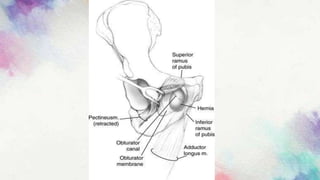
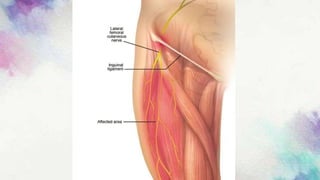
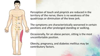
LUMBOSACRAL PLEXUS -
LESIONS AND DISEASE
-Hitesh Rohit(3rd year BPT)
What’s inside?
• Diabetic neuropathy
• Lumbosacral plexus syndrome
• Mononeuropathies of lower limb
INTRODUCTION:-
• Radiculopathy:-Radiculopathy describes a range of symptoms
produced by the pinching of a nerve root in the spinal column.
• Neuropathy:- Disease or dysfunction of one or more peripheral
nerves, typically causing numbness or weakness.
• Mononeuropathy:-Damage to a single nerve.
• Polyneuropathy:-Damage to many nerves.
• Plexus syndrome:-Damage to the whole plexus due to injuries,
tumors or autoimmune reactions leads to plexus syndrome.
DIABETIC NEUROPATHY:-
• This condition is uncommon in childhood and increases with age.
• Peripheral nerve damage is related to poor control of diabetes. This is more
common in insulin-dependent patients.
• Damage results from either metabolic disturbance with sorbitol and
fructose accumulation in axons and Schwann cells or an occlusion of the
nutrient vessels supplying nerves (vasa vasorum).
• The frequent occurrence of neuropathy with other vascular
complications – retinopathy and nephropathy – suggests that
the latter is the more usual mechanism.
• Neurological complications correlate with levels of glycosylated
haemoglobin A1C, an indicator of the long-term control of
hyperglycaemia.
• Classification:-
• 1)Polyneuropathy
• 2)Autonomic neuropathy
• 3)Cranial nerve palsy
• 4)Asymmetrical neuropathy(Diabetic amyotrophy)
• 1) Polyneuropathy:-
• Present in 30% of all diabetics, but only 10% are symptomatic.
• Distal weakness and sensory loss is usual.
• Two forms of sensory neuropathy occur – large fibre, causing ataxia and
small fibre causing a painful anaesthesia.
• 2) Autonomic neuropathy:-
• In most patients with peripheral neuropathy, some degree of
autonomic disturbance is present.
• Occasionally this predominates:
• pupil abnormalities
• loss of sweating
• orthostatic hypotension
• resting tachycardia
• gastroparesis and diarrhoea
• hypotonic dilated bladder
• impotence.
• 3)Cranial nerve palsy:-
• An oculomotor palsy, usually without pain, may occur with pupillary
sparing, which helps to differentiate from an aneurysmal cause.
• The 6th and 7th cranial nerves may also be involved in diabetes.
• 4) Asymmetrical neuropathy:-
• Much less common than polyneuropathy.
• Pain and weakness rapidly develop.
• The anterior thigh is preferentially affected with wasting of the
quadriceps, loss of the knee jerk and minimal sensory loss.
• The condition is due to anterior spinal root or plexus disease.
• Imaging the lumbar roots and plexus excludes other causes.
• Functional recovery is good.
• Treatment:-
• Improved control of diabetes is essential.
• Carbamazepine, gabapentin, pregabalin, tricyclic antidepressants or α-
adrenergic blockers, e.g. phenoxybenzene, help control pain.
• Drugs which reduce aldose reductase and halt accumulation of sorbitol
and fructose in nerves are being evaluated.
• Management of autonomic neuropathy – Improve diabetic control and
treat symptoms e.g. fludrocortisone for BP control.
• Asymmetrical neuropathies usually spontaneously recover, whereas
prognosis for symmetric neuropathies is less certain.
LUMBOSACRAL PLEXUS SYNDROME:-
The proximity of the plexus to important abdominal and
pelvic structures renders it liable to damage from
disease of these structures.
Trauma following surgery, e.g. hysterectomy, lumbar
sympathectomy or during labour. Compression from an
abdominal mass, e.g. aortic aneurysm. Infiltration from
pelvic tumour,Radiotherapy.
• Symptoms:-
• It may be unilateral or bilateral, depending upon causation.
• Weakness, sensory loss and reflex changes are dictated by the location
and extent of plexus damage.
• Pain of a severe burning quality may be present; it may be worsened by
coughing, sneezing, etc.
• In general:
• Lower plexus lesions produce:Weakness of posterior thigh (hamstring) and
foot muscles with posterior leg sensory loss.
• Upper plexus lesions produce:Weakness of hip flexion and adduction with
anterior leg sensory loss.
Lumbosacral neuritis:-
• Inflammation of the lumbosacral spinal nerves is called lumbosacral
neuritis.
• Causes of Lumbosacral neuritis:-
• It can occur due to a variety of different causes.
• Essentially, there is irritation of the nerve fibers that lie within the spinal
canal.
• This irritation can occur from infection, inflammation, compression by a
small bone spur, compression from a small spinal tumor and even
endocrine causes such as diabetes.
• A herniated lumbar disc is also a well recognised cause.
• Symptoms:-
• low back pain and shooting pains down the leg.
• some alteration in the sensation of the skin on the leg
• weakness of the muscles
• On examination, patients may find that certain movements
exacerbate their pain.
• Diagnosis:-
• Many a times, a clear-cut diagnosis can be made from history and
clinical examination alone.
• However, in some cases this may not be sufficient and patients may
require additional investigations of the spine.
• A simple x-ray of the spine will demonstrate any irregularities within the
alignment of the spinal column.
• In addition, any narrowing of the disc spaces, bone spur formation,
osteoporosis, fractures and tumors may become visible through an x-ray
alone.
• In some cases there is a requirement of CT scan and MRI or nerve
conduction study to determine the involvement of specific nerve fibers.
• Treatment:-
• The choice of treatment depends upon the cause of Lumbosacral
Neuritis.
• For example patients with diabetic neuropathy may benefit from
simple physical therapy along with multivitamin supplementation and
good diabetic control.
• Patients who have a tumor may benefit from some form of surgery
along with radiotherapy.
• Patients who have a protruded lumbar intervertebral disc may find
relief through physical therapy along with conservative treatment
options and a possible surgical correction of the defect.
LOWER LIMB MONONEUROPATHIES:-
• 1) Femoral nerve(L2,L3,L4):-
• Damaged by:
• Fractures of the upper femur
• Congenital dislocation of the hip, hip surgery
• Neoplastic infiltration
• Psoas muscle abscess
• Haematoma into iliopsoas muscle(haemophilia, anticoagulants)
• Systemic causes of mononeuropathy, e.g. diabetes
• Results in:
• Weakness of hip flexion
• Weakness of knee extension with wasting of thigh muscles
• Sensory loss over the anterior and medial aspects of the thigh
• The knee jerk is lost
• 2) Obturator nerve (L2,L3,L4):
• Damaged by:-
• Fractures of the upper femur
• Congenital dislocation of the hip, hip surgery
• Neoplastic infiltration
• Psoas muscle abscess
• Haematoma into iliopsoas muscle(haemophilia, anticoagulants)
• Systemic causes of mononeuropathy, e.g. diabetes
• During labour and occasionally as a consequence of compression by
hernia in the obturator canal.
• Results in: –
• Weakness of hip external rotation and adduction.
• The patient may complain of inability to cross the affected leg on the
other.
• Sensory loss is confined to the innermost aspect of the thigh.
• The adductor reflex is absent (adductor response to striking the
medial epicondyle).
• 3) Lateral cutaneous nerve of thigh(L2,L3):-
• Compression (entrapment) may occur at the point where it passes
between the two prongs of attachment of the inguinal ligament.
• Compression of the nerve results in uncomfortable paresthesias and
sensory impairment in its cutaneous distribution, a common
condition known as meralgia paresthetica (meros, “thigh”).
• Usually numbness and mild sensitivity of the skin are the only
symptoms, but occasionally there is a persistent distressing burning
pain.
Perception of touch and pinprick are reduced in the
territory of the nerve; there is no weakness of the
quadriceps or diminution of the knee jerk.
The symptoms are characteristically worsened in certain
positions and after prolonged standing or walking.
Occasionally, for an obese person, sitting is the most
uncomfortable position.
Obesity, pregnancy, and diabetes mellitus may be
contributory factors.
• Most of the patients with meralgia paresthetica request no treatment
once they learn of its benign character.
• Weight loss and adjustment of restrictive clothing or correction of
habitual postures that might compress the nerve are sometimes helpful.
• A few with the most painful symptoms have demanded a neurectomy or
section of the nerve, but it is always wise to perform a lidocaine block
first, so that the patient can decide whether the persistent numbness is
preferable.
• Hydrocortisone injections at the point of entrapment may have helped in
a few cases.
• 4) Sciatic nerve(L4,L5,S1,S2,S3):-
• The nerve descends between the ischial tuberosity and the greater
trochanter of the femur. In the thigh it innervates the hamstring
muscles (semitendinosus, semimembranosus and biceps).
• Damaged by:-
• Congenital or traumatic hip dislocation.
• Penetrating injuries.
• Accidental damage from ‘misplaced’ intramuscular injection.
• Entrapment at sciatic notch.
• Systemic causes of mononeuropathy
• Total hip arthroplasty
• Tumors of the pelvis (sarcomas, lipomas)
Sitting for a long period with legs
flexed and abducted (lotus
position) under the influence of
narcotics or barbiturates or lying
flat on a hard surface in a
sustained stupor may severely
injure one or both sciatic nerves or
branches thereof.
• Results in:-
• Weakness of hamstring muscles with loss of knee flexion.
• Distal foot and leg muscles are also affected.
• Sensory loss involves the outer aspect of the leg.
• The ankle reflex is absent.
Sciatica:-
• Sciatica refers to pain that radiates along the path of the sciatic nerve,
which branches from your lower back through your hips and buttocks
and down each leg.
• Typically, sciatica affects only one side of your body.
• Sciatica most commonly occurs when a herniated disk, bone spur on
the spine or narrowing of the spine (spinal stenosis) compresses part
of the nerve.
• Symptoms:-
• Radiating pain down to leg
• Numbness
• Tingling sensation
• Weakness of muscle
• Treatment:-
• Anti-inflammatory drugs
• Narcotics
• Muscle relaxants
• Steroid injections
• 5)Common peroneal nerve(L4,L5,S1,S2):-
• The nerve arises from the division of the sciatic nerve in the popliteal
fossa. It bears a close relationship with the head of the fibula as it
winds anteriorly.
• Damaged by:-
• Trauma to the head of the fibula; pressure here from kneeling,
crossing legs.
• Systemic causes of mononeuropathy, e.g. diabetes
• Results in:-
• Weakness of dorsiflexion and eversion of the foot. The patient walks
with a ‘foot drop’.
• Sensory loss involves the dorsum and outer aspect of the foot.
• Partial common peroneal nerve palsies are common with very
selective muscle weakness.
• 6) Posterior tibial nerve (L4,L5,S1,S2,S3):-
• This nerve also arises from the division of the sciatic nerve in the
popliteal fossa and descends behind the tibia, terminating in the
medial and lateral plantar nerves which innervate the small muscles
of the foot.
• The sensory branch contributes to the sural nerve.
• Damaged by:-
• Trauma in the popliteal fossa.
• Fracture of the tibia.
• Systemic causes of mononeuropathy.
• Results in:-
• Weakness of plantar flexion and inversion of the foot.
• The patient cannot stand on toes.
• Sensory loss involves the sole of the foot.
• The ankle reflex is lost.
• Tarsal tunnel syndrome:-
• The posterior tibial nerve may be entrapped below the medial
malleolus due to thickening of the tendon sheaths or the adjacent
connective tissues or by osteoarthritic changes.
• This produces a burning pain in the sole of the foot. Weakness of toe
flexion and atrophy of small muscles of the foot occur in advanced
cases.
• A prolonged sensory conduction velocity confirms the diagnosis.
• Surgical decompression is often required.
• Plantar and small digital nerves:-
• Compression of these nerves at the sole of the foot produces
localised burning pain.
• Involvement of interdigital nerves produces pain and analgesia
in adjacent halves of neighbouring toes.
Lumbosacral lesions and disease.HR

Lumbosacral lesions and disease.HR

  • 1.
    LUMBOSACRAL PLEXUS - LESIONSAND DISEASE -Hitesh Rohit(3rd year BPT)
  • 2.
    What’s inside? • Diabeticneuropathy • Lumbosacral plexus syndrome • Mononeuropathies of lower limb
  • 3.
    INTRODUCTION:- • Radiculopathy:-Radiculopathy describesa range of symptoms produced by the pinching of a nerve root in the spinal column. • Neuropathy:- Disease or dysfunction of one or more peripheral nerves, typically causing numbness or weakness. • Mononeuropathy:-Damage to a single nerve. • Polyneuropathy:-Damage to many nerves. • Plexus syndrome:-Damage to the whole plexus due to injuries, tumors or autoimmune reactions leads to plexus syndrome.
  • 4.
    DIABETIC NEUROPATHY:- • Thiscondition is uncommon in childhood and increases with age. • Peripheral nerve damage is related to poor control of diabetes. This is more common in insulin-dependent patients. • Damage results from either metabolic disturbance with sorbitol and fructose accumulation in axons and Schwann cells or an occlusion of the nutrient vessels supplying nerves (vasa vasorum).
  • 5.
    • The frequentoccurrence of neuropathy with other vascular complications – retinopathy and nephropathy – suggests that the latter is the more usual mechanism. • Neurological complications correlate with levels of glycosylated haemoglobin A1C, an indicator of the long-term control of hyperglycaemia.
  • 6.
    • Classification:- • 1)Polyneuropathy •2)Autonomic neuropathy • 3)Cranial nerve palsy • 4)Asymmetrical neuropathy(Diabetic amyotrophy)
  • 7.
    • 1) Polyneuropathy:- •Present in 30% of all diabetics, but only 10% are symptomatic. • Distal weakness and sensory loss is usual. • Two forms of sensory neuropathy occur – large fibre, causing ataxia and small fibre causing a painful anaesthesia. • 2) Autonomic neuropathy:- • In most patients with peripheral neuropathy, some degree of autonomic disturbance is present. • Occasionally this predominates:
  • 8.
    • pupil abnormalities •loss of sweating • orthostatic hypotension • resting tachycardia • gastroparesis and diarrhoea • hypotonic dilated bladder • impotence.
  • 9.
    • 3)Cranial nervepalsy:- • An oculomotor palsy, usually without pain, may occur with pupillary sparing, which helps to differentiate from an aneurysmal cause. • The 6th and 7th cranial nerves may also be involved in diabetes.
  • 10.
    • 4) Asymmetricalneuropathy:- • Much less common than polyneuropathy. • Pain and weakness rapidly develop. • The anterior thigh is preferentially affected with wasting of the quadriceps, loss of the knee jerk and minimal sensory loss. • The condition is due to anterior spinal root or plexus disease. • Imaging the lumbar roots and plexus excludes other causes. • Functional recovery is good.
  • 11.
    • Treatment:- • Improvedcontrol of diabetes is essential. • Carbamazepine, gabapentin, pregabalin, tricyclic antidepressants or α- adrenergic blockers, e.g. phenoxybenzene, help control pain. • Drugs which reduce aldose reductase and halt accumulation of sorbitol and fructose in nerves are being evaluated. • Management of autonomic neuropathy – Improve diabetic control and treat symptoms e.g. fludrocortisone for BP control. • Asymmetrical neuropathies usually spontaneously recover, whereas prognosis for symmetric neuropathies is less certain.
  • 12.
    LUMBOSACRAL PLEXUS SYNDROME:- Theproximity of the plexus to important abdominal and pelvic structures renders it liable to damage from disease of these structures. Trauma following surgery, e.g. hysterectomy, lumbar sympathectomy or during labour. Compression from an abdominal mass, e.g. aortic aneurysm. Infiltration from pelvic tumour,Radiotherapy.
  • 13.
    • Symptoms:- • Itmay be unilateral or bilateral, depending upon causation. • Weakness, sensory loss and reflex changes are dictated by the location and extent of plexus damage. • Pain of a severe burning quality may be present; it may be worsened by coughing, sneezing, etc. • In general: • Lower plexus lesions produce:Weakness of posterior thigh (hamstring) and foot muscles with posterior leg sensory loss. • Upper plexus lesions produce:Weakness of hip flexion and adduction with anterior leg sensory loss.
  • 14.
    Lumbosacral neuritis:- • Inflammationof the lumbosacral spinal nerves is called lumbosacral neuritis. • Causes of Lumbosacral neuritis:- • It can occur due to a variety of different causes. • Essentially, there is irritation of the nerve fibers that lie within the spinal canal. • This irritation can occur from infection, inflammation, compression by a small bone spur, compression from a small spinal tumor and even endocrine causes such as diabetes. • A herniated lumbar disc is also a well recognised cause.
  • 15.
    • Symptoms:- • lowback pain and shooting pains down the leg. • some alteration in the sensation of the skin on the leg • weakness of the muscles • On examination, patients may find that certain movements exacerbate their pain. • Diagnosis:- • Many a times, a clear-cut diagnosis can be made from history and clinical examination alone. • However, in some cases this may not be sufficient and patients may require additional investigations of the spine.
  • 16.
    • A simplex-ray of the spine will demonstrate any irregularities within the alignment of the spinal column. • In addition, any narrowing of the disc spaces, bone spur formation, osteoporosis, fractures and tumors may become visible through an x-ray alone. • In some cases there is a requirement of CT scan and MRI or nerve conduction study to determine the involvement of specific nerve fibers.
  • 17.
    • Treatment:- • Thechoice of treatment depends upon the cause of Lumbosacral Neuritis. • For example patients with diabetic neuropathy may benefit from simple physical therapy along with multivitamin supplementation and good diabetic control. • Patients who have a tumor may benefit from some form of surgery along with radiotherapy. • Patients who have a protruded lumbar intervertebral disc may find relief through physical therapy along with conservative treatment options and a possible surgical correction of the defect.
  • 18.
    LOWER LIMB MONONEUROPATHIES:- •1) Femoral nerve(L2,L3,L4):- • Damaged by: • Fractures of the upper femur • Congenital dislocation of the hip, hip surgery • Neoplastic infiltration • Psoas muscle abscess • Haematoma into iliopsoas muscle(haemophilia, anticoagulants) • Systemic causes of mononeuropathy, e.g. diabetes
  • 19.
    • Results in: •Weakness of hip flexion • Weakness of knee extension with wasting of thigh muscles • Sensory loss over the anterior and medial aspects of the thigh • The knee jerk is lost
  • 20.
    • 2) Obturatornerve (L2,L3,L4): • Damaged by:- • Fractures of the upper femur • Congenital dislocation of the hip, hip surgery • Neoplastic infiltration • Psoas muscle abscess • Haematoma into iliopsoas muscle(haemophilia, anticoagulants) • Systemic causes of mononeuropathy, e.g. diabetes • During labour and occasionally as a consequence of compression by hernia in the obturator canal.
  • 22.
    • Results in:– • Weakness of hip external rotation and adduction. • The patient may complain of inability to cross the affected leg on the other. • Sensory loss is confined to the innermost aspect of the thigh. • The adductor reflex is absent (adductor response to striking the medial epicondyle).
  • 23.
    • 3) Lateralcutaneous nerve of thigh(L2,L3):- • Compression (entrapment) may occur at the point where it passes between the two prongs of attachment of the inguinal ligament. • Compression of the nerve results in uncomfortable paresthesias and sensory impairment in its cutaneous distribution, a common condition known as meralgia paresthetica (meros, “thigh”). • Usually numbness and mild sensitivity of the skin are the only symptoms, but occasionally there is a persistent distressing burning pain.
  • 25.
    Perception of touchand pinprick are reduced in the territory of the nerve; there is no weakness of the quadriceps or diminution of the knee jerk. The symptoms are characteristically worsened in certain positions and after prolonged standing or walking. Occasionally, for an obese person, sitting is the most uncomfortable position. Obesity, pregnancy, and diabetes mellitus may be contributory factors.
  • 26.
    • Most ofthe patients with meralgia paresthetica request no treatment once they learn of its benign character. • Weight loss and adjustment of restrictive clothing or correction of habitual postures that might compress the nerve are sometimes helpful. • A few with the most painful symptoms have demanded a neurectomy or section of the nerve, but it is always wise to perform a lidocaine block first, so that the patient can decide whether the persistent numbness is preferable. • Hydrocortisone injections at the point of entrapment may have helped in a few cases.
  • 27.
    • 4) Sciaticnerve(L4,L5,S1,S2,S3):- • The nerve descends between the ischial tuberosity and the greater trochanter of the femur. In the thigh it innervates the hamstring muscles (semitendinosus, semimembranosus and biceps). • Damaged by:- • Congenital or traumatic hip dislocation. • Penetrating injuries. • Accidental damage from ‘misplaced’ intramuscular injection. • Entrapment at sciatic notch. • Systemic causes of mononeuropathy • Total hip arthroplasty • Tumors of the pelvis (sarcomas, lipomas)
  • 29.
    Sitting for along period with legs flexed and abducted (lotus position) under the influence of narcotics or barbiturates or lying flat on a hard surface in a sustained stupor may severely injure one or both sciatic nerves or branches thereof.
  • 30.
    • Results in:- •Weakness of hamstring muscles with loss of knee flexion. • Distal foot and leg muscles are also affected. • Sensory loss involves the outer aspect of the leg. • The ankle reflex is absent.
  • 31.
    Sciatica:- • Sciatica refersto pain that radiates along the path of the sciatic nerve, which branches from your lower back through your hips and buttocks and down each leg. • Typically, sciatica affects only one side of your body. • Sciatica most commonly occurs when a herniated disk, bone spur on the spine or narrowing of the spine (spinal stenosis) compresses part of the nerve.
  • 33.
    • Symptoms:- • Radiatingpain down to leg • Numbness • Tingling sensation • Weakness of muscle • Treatment:- • Anti-inflammatory drugs • Narcotics • Muscle relaxants • Steroid injections
  • 34.
    • 5)Common peronealnerve(L4,L5,S1,S2):- • The nerve arises from the division of the sciatic nerve in the popliteal fossa. It bears a close relationship with the head of the fibula as it winds anteriorly. • Damaged by:- • Trauma to the head of the fibula; pressure here from kneeling, crossing legs. • Systemic causes of mononeuropathy, e.g. diabetes
  • 35.
    • Results in:- •Weakness of dorsiflexion and eversion of the foot. The patient walks with a ‘foot drop’. • Sensory loss involves the dorsum and outer aspect of the foot. • Partial common peroneal nerve palsies are common with very selective muscle weakness.
  • 37.
    • 6) Posteriortibial nerve (L4,L5,S1,S2,S3):- • This nerve also arises from the division of the sciatic nerve in the popliteal fossa and descends behind the tibia, terminating in the medial and lateral plantar nerves which innervate the small muscles of the foot. • The sensory branch contributes to the sural nerve.
  • 38.
    • Damaged by:- •Trauma in the popliteal fossa. • Fracture of the tibia. • Systemic causes of mononeuropathy. • Results in:- • Weakness of plantar flexion and inversion of the foot. • The patient cannot stand on toes. • Sensory loss involves the sole of the foot. • The ankle reflex is lost.
  • 39.
    • Tarsal tunnelsyndrome:- • The posterior tibial nerve may be entrapped below the medial malleolus due to thickening of the tendon sheaths or the adjacent connective tissues or by osteoarthritic changes. • This produces a burning pain in the sole of the foot. Weakness of toe flexion and atrophy of small muscles of the foot occur in advanced cases. • A prolonged sensory conduction velocity confirms the diagnosis. • Surgical decompression is often required.
  • 41.
    • Plantar andsmall digital nerves:- • Compression of these nerves at the sole of the foot produces localised burning pain. • Involvement of interdigital nerves produces pain and analgesia in adjacent halves of neighbouring toes.