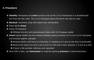
The document describes the procedure for a lumbar puncture (LP). It begins by defining an LP as the insertion of a needle into the lumbar spine to withdraw cerebrospinal fluid or inject substances for diagnostic or therapeutic purposes. It then lists common indications for an LP such as diagnosing meningitis or measuring cerebrospinal fluid pressure. The document provides details on equipment, the procedure steps, and post-care instructions for an LP. It concludes by noting potential complications.
A lumbar puncture (spinal tap) is performed in your lower back, in the lumbar region. During a lumbar puncture, a needle is inserted between two lumbar bones (vertebrae) to remove a sample of cerebro-spinal fluid. This is the fluid that surrounds your brain and spinal cord to protect them from injury.
As part of my course, I have prepared power point presentation on lumbar puncture. Books which i have referred are Sister Nancy and Poter and Perry Fundamental of nursing. I hope this ppt will be be some help to the prospect Nursing learners.
A lumbar puncture, or spinal tap, is performed to obtain cerebrospinal fluid for diagnostic purposes. It carries risks such as post-lumbar puncture headache, infection, and nerve injury. The procedure involves positioning the patient, prepping and draping the skin, administering local anesthesia, and inserting a spinal needle between lumbar vertebrae to collect CSF samples for analysis. Complications are usually minor but can include headache, back pain, bleeding, or neurological issues and may require treatment.
The document discusses central venous pressure (CVP) monitoring. It aims to explain what CVP is, the purposes and indications for monitoring it, the equipment needed, nursing roles and responsibilities, potential complications, and how to interpret CVP readings. Specifically, CVP refers to the blood pressure in the right atrium and is monitored using a catheter placed in the jugular or subclavian vein. CVP provides information about a patient's fluid balance, circulating blood volume, and right heart function. Nurses must understand how to set up monitoring equipment properly and know that abnormal CVP readings should be considered in the full clinical context of the patient.
Defines Lumbar Puncture, Discusses the Indications of Lumbar Puncture, Contraindication, and complications of Lumbar Puncture, Equipment used and preparation required for the procedure, positioning the infant and assessing the landmarks for the procedure and the procedure of Lumbar Puncture. Interpretation of CSF, and the Nursing Care Post Procedure and the important key points to remember.
The document discusses the goals, positioning, procedures, and risks of proper patient positioning using lithotomy. The goals are to maintain airway, circulation, prevent nerve damage, provide exposure, and comfort. Lithotomy positioning involves supine position with legs flexed at hips and knees and supported. It is used for perineal, vaginal, urological, rectal and gynecological procedures. Potential risks include nerve injuries and complications must be prevented through safe practices like proper stirrup placement and handling.
A brincoscopy is the direct inspection and observation of the larynx, trachea, and bronchi through flexible or rigid bronchoscope.
Flexible fiber-optic bronchoscope allows for more patient comfort and better visualization of smaller airways and the fiberoptic scope is used more frequently in current practice.
Rigid bronchoscopy is preferred for small children and endobronchial tumour resection.
The purpose of bronchoscopy has diagnostic and therapeutic uses in pulmonary conditions. Diagnostic uses include
This document provides information on central venous catheterization, including indications, contraindications, complications, techniques, and tips. It discusses the Seldinger technique for placement and locations for catheter insertion, including the internal jugular, subclavian, and femoral veins. Precautions are outlined for each approach. Ultrasound guidance is becoming standard to visualize the vein and compress it during insertion.
A lumbar puncture, or spinal tap, is a procedure where cerebrospinal fluid is collected from the lower back for diagnostic purposes. It is indicated for conditions like meningitis, subarachnoid hemorrhage, and certain CNS diseases. Contraindications include infected skin at the needle site, brain abnormalities, or increased intracranial pressure. The procedure involves locating the interspace between vertebrae, inserting a needle at a slight upward angle, and checking for fluid return. Potential complications are local pain, infection, bleeding, spinal fluid leak, or spinal headache.
This document discusses lumbar puncture, also known as a spinal tap. It begins by listing the objectives of understanding the indications and contraindications for lumbar puncture, the technique for insertion, and potential complications. The document then provides details on the indications for lumbar puncture such as diagnosing central nervous system infections or disorders. Contraindications like increased intracranial pressure or skin infections are also outlined. The procedure, necessary equipment, pre- and post-procedure care, and potential complications like headache or bleeding are then described.
Thoracentesis (thor-a-sen-tee-sis) is a procedure that is done to remove a sample of fluid from around the lung.
The lung is covered with a tissue called the pleura. The inside of the chest is also lined with pleura.
The space between these two areas is called the pleural space.
This space normally contains just a thin layer of fluid, however, some conditions such as pneumonia, some types of cancer, or congestive heart failure may cause excessive fluid to develop (pleural effusion).
Thoracentesis, also known as pleural fluid analysis, is a procedure in which a needle is inserted through the back of the chest wall into the pleural space (a space that exists between the two lungs and the anterior chest wall) to remove fluid or air.
Pleural fluid analysis is the microscopic and chemical lab analysis of the fluid obtained during thoracentesis.
IndDiagnostic: determination of pleural effusion etiology (e.g. transudative versus exudative) usually requires the removal of 50 to 100mL of pleural fluid for laboratory studies. Most new effusions require diagnostic thoracentesis, an exception being a new effusion with a clear clinical diagnosis (e.g. CHF) with no evidence for superimposed pleural space infection
Therapeutic: reduce dyspnea and respiratory compromise in patients with large pleural effusions. This is typically achieved by removing a much larger volume of fluid compared to the diagnostic thoracentesis
ications
Endotracheal intubation involves placing a flexible plastic tube into the trachea to maintain an open airway or administer drugs. It is used to administer oxygen, remove secretions, ventilate the lungs, and treat respiratory failure. Indications include CNS depression, neuromuscular disease, chest injuries, airway obstruction, and aspiration risk. The procedure requires a laryngoscope, ET tube, suction equipment, and securing the tube once placed to ventilate the lungs. Complications can include injury and intubation in the wrong airway.
This document defines and discusses arterial blood gas analysis. It is a blood test taken from an artery that measures oxygen and carbon dioxide levels in the blood. It is used to evaluate respiratory status, metabolic status, and acid-base balance. Common sites for extraction are the radial, brachial, and femoral arteries. The procedure involves performing Allen's test before extraction to ensure adequate blood flow after puncture. Components measured include pH, oxygen saturation, bicarbonate, and base excess. Normal values for several components are provided. Sources of error in results can include air bubbles in the syringe or an inadvertent venous sample.
A myelogram is an imaging test that uses fluoroscopy and contrast dye to evaluate the spinal cord and nerve roots. It involves injecting contrast dye into the spinal canal and tilting the patient to track the flow of dye. Myelograms are usually performed to investigate spinal stenosis or masses, and can help guide surgery or radiation planning. Newer non-ionic contrast dyes are used instead of older oil-based dyes due to reduced risk of side effects like arachnoiditis. A myelogram is followed by CT imaging to provide additional diagnostic information.
Thoracentesis is a procedure to drain fluid from the pleural space around the lungs. It involves inserting a large bore needle through the chest wall under local anesthesia. The fluid is drained to diagnose the cause of excess fluid or provide relief from symptoms. Nurses prepare equipment, position the patient, monitor them during the procedure, and assess for complications like pneumothorax afterwards. Proper documentation and care of any insertion site is also important. Thoracentesis can help determine the cause of pleural effusions and relieve symptoms like shortness of breath.
There are numerous types of brain surgery. The type used is based on the area of the brain and the condition being treated.
Brain surgery is a critical and complicated process. The type of brain surgery done depends highly on the condition being treated.
Intracranial surgery refers to various medical procedures that involve repairing structural problems in the brain.
Craniotomy
A craniotomy involves making an incision in the scalp and creating a hole known as a bone flap in the skull. The hole and incision are made near the area of the brain being treated.
During open brain surgery, it is done to remove tumors, clip off an aneurysm, drain blood or fluid from an infection & remove abnormal brain tissue
Decompressive craniectomy
It is a neurosurgical procedure in which part of the skull is removed to allow a swelling brain room to expand without being squeezed. It is performed on victims of traumatic brain injury, stroke and other conditions associated with raised intracranial pressure.
This document provides information about arterial blood gas (ABG) analysis, including the nurse's role in ABG testing. It discusses the importance of ABG interpretation for critically ill patients. The document outlines the six-step process for ABG interpretation and lists common acid-base disorders. It also defines an ABG test, describes normal ABG values, indications for testing, sample collection points and techniques, equipment used, and potential complications of ABG sampling. The overall goal is to help nurses understand ABGs and properly perform arterial punctures to obtain blood samples.
Nurses role in arterial puncture and abg analysisStephy Stanly
The document discusses arterial blood gas analysis and interpretation. It provides information on equipment and procedures for arterial blood sampling and analysis. Normal ranges are provided for pH, PCO2, PO2, oxygen saturation, and HCO3. Types of acid-base imbalances are explained including compensated and uncompensated respiratory and metabolic acidosis and alkalosis. Examples of arterial blood gas results are given and their acid-base interpretations.
The CVP catheter is an important tool used to assess right ventricular function and systemic fluid status. Normal CVP is 2-6 mm Hg. CVP is elevated by : overhydration which increases venous return.
This document discusses central venous pressure (CVP), including indications for CVP monitoring, measurement, waveform interpretation, and techniques for central venous cannulation. It notes that CVP can be used to assess intravascular volume status, right ventricular function, and is indicated for major procedures involving fluid shifts. The internal jugular vein and subclavian vein are common access sites, and ultrasound guidance can help with cannulation. Potential complications include arterial puncture, pneumothorax, and infection.
Craniotomy
A craniotomy involves making an incision in the scalp and creating a hole known as a bone flap in the skull. The hole and incision are made near the area of the brain being treated.
During open brain surgery, it is done to remove tumors, clip off an aneurysm, drain blood or fluid from an infection & remove abnormal brain tissue
Decompressive craniectomy
It is a neurosurgical procedure in which part of the skull is removed to allow a swelling brain room to expand without being squeezed. It is performed on victims of traumatic brain injury, stroke and other conditions associated with raised intracranial pressure.
Increased intracranial pressure is defined as cerebrospinal fluid pressure greater than 15 mm Hg.
Infections
Tumors
Stroke
Aneurysm
Epilepsy
Seizures
Hydrocephalus
Hypertensive brain injury
Hypoxemia
Meningitis
Due to etiological factors
Components of ICP is disturbed- brain tissue, CSF, blood volume
An increase in the volume of ANY ONE component must be accompanied by a reciprocal decrease in one of the other components.
When this volume-pressure relationship becomes unbalanced, ICP increases.
The document describes the components and uses of endotracheal tubes, which are curved plastic or latex tubes used for intubation to provide an airway for mechanical ventilation or respiratory support. It details the parts of the ET tube including the proximal end, central portion with markers, and distal end, as well as types, sizing, complications, and uses. The document also provides information on ambu bags, including their parts and mechanism for providing intermittent positive pressure ventilation.
This document provides information on intercostal tube insertion and the nursing responsibilities associated with chest drainage systems. It defines an intercostal tube as a drainage tube inserted into the pleural cavity to remove air, blood, or fluid. Tubes can range from 6 to 40 French in size. Chest tubes are used to drain the pleural space after procedures like pneumothorax, hemothorax, thoracotomy, or chest trauma. The document outlines the principles of chest drainage systems, types of systems, the insertion procedure, post-care for the patient and equipment, and the nurse's ongoing responsibilities in monitoring the system.
1. Tracheostomy is a surgical opening in the front of the trachea below the larynx that creates an airway. It is performed to maintain a clear airway, remove secretions, and enable ventilation.
2. Tracheostomy tubes can be plastic, metal, cuffed, or fenestrated. Speaking valves allow vocalization without finger occlusion using a silicone diaphragm that opens for inhalation and closes for exhalation.
3. Complications include bleeding, infection, tube obstruction, aspiration, and tracheal stenosis. Care involves cleaning the stoma and tube, suctioning secretions, providing humidification, and assessing for ineffective airway clearance or
Central venous catheterization and venous cut down techniques were presented. Central venous catheterization involves placing lines into large neck, chest, or groin veins and should only be done aseptically in operating rooms or high dependency units. It has indications for monitoring, infusing irritant drugs, pacing, dialysis, and emergencies. Sites include the subclavian, internal jugular, and femoral veins, each with advantages and disadvantages. Ultrasound guidance is becoming standard. Complications include infections, arterial puncture, and pneumothorax. Venous cut down is an open surgical technique to access veins and remains useful when other methods fail or are unavailable.
A tracheostomy is an opening in the neck into the trachea that aids breathing. Tracheostomy care includes cleaning the tracheostomy site and changing dressings and inner tubes. The nurse must follow sterile technique, assess the tracheostomy site for signs of infection, clean the inner cannula and site, replace dressings and ties securely, and document the procedure. Tracheostomy care promotes airway patency, prevents infection, and provides patient comfort.
Thoracentesis is a procedure to drain excess fluid from the pleural space between the lungs and chest wall. It involves inserting a needle through the chest wall under local anesthesia to remove fluid for analysis or to relieve symptoms like shortness of breath. Precautions are taken before and during the procedure to monitor vital signs and breathing. After the procedure, the patient is observed for complications and a chest x-ray may be taken to evaluate the drainage.
The document discusses the lumbar puncture procedure, which involves inserting a needle into the lumbar subarachnoid space to obtain cerebrospinal fluid samples. It describes the anatomy of the lumbar spine, indications for lumbar puncture including diagnostic testing for conditions like meningitis, contraindications, potential complications, the equipment needed, and steps for performing and monitoring the procedure. Normal cerebrospinal fluid components and values are also outlined.
The document provides information on performing lumbar punctures, including indications, contraindications, equipment, procedures, complications, and normal CSF findings. It describes how to position the patient laterally or sitting and insert the spinal needle between vertebrae to collect CSF samples. Potential complications discussed are headache, apnea, back pain, bleeding, infection, and brainstem herniation. Precautions and treatments for complications are also outlined.
Myelography is a radiographic examination that uses contrast medium injected into the space surrounding the spinal cord and brain to detect pathology. The contrast medium allows visualization of the spinal cord, nerve roots, and surrounding structures. Potential risks include headache, vomiting, and nerve damage from the injection. The procedure involves injecting non-ionic contrast medium into the subarachnoid space, typically at the lumbar region, and taking fluoroscopic or radiographic images of the spine. Patients are monitored afterwards for complications and instructed to rest.
This document outlines postoperative care considerations following craniotomy. It describes monitoring vital signs and neurological status, maintaining normal intracranial pressure, preventing infections and aspiration, managing pain, avoiding constipation, and providing patient and family education. Close observation is needed to monitor for complications like hemorrhage, edema, seizures, and infection.
This document provides information on nursing care for a child undergoing a lumbar puncture procedure. It defines a lumbar puncture as a procedure where a needle is inserted into the spinal canal to collect cerebrospinal fluid for diagnostic testing. The document outlines the indications, contraindications, necessary equipment, step-by-step procedure, normal test results, potential complications, and the nurse's responsibilities before, during, and after the procedure.
Lumbar puncture (other name are spinal tap;spinal puncture;thecal puncture and rachiocentesis) is a procedure that is often performed in the emergency department by inserting needle into fluid within the spinal canal to obtain information about the cerebrospinal fluid (CSF).
Although usually used for diagnostic purposes to rule out potential life-threatening conditions for example bacterial meningitis or subarachnoid hemorrhage,
it is also sometimes used for therapeutic purposes for example treatment of pseudo tumor cerebri.
This document provides information on spinal anesthesia. It begins by defining spinal anesthesia as a single injection of local anesthetic into the subarachnoid space, usually at the lumbar level of L3-L4. It then lists several advantages of spinal anesthesia including reduced costs, increased patient satisfaction, and benefits for patients with respiratory disease, diabetes, or who are elderly. Potential complications are also outlined such as bradycardia, nausea and vomiting, and hypotension. Proper patient positioning and administration technique are emphasized to perform the block safely and effectively.
A lumbar puncture, or spinal tap, is a procedure that involves inserting a needle into the lumbar subarachnoid space to withdraw cerebrospinal fluid (CSF) for diagnostic purposes. It is commonly performed to diagnose conditions like meningitis, subarachnoid hemorrhage, and malignancies. The procedure involves positioning the patient on their side, cleaning the puncture site, inserting the spinal needle between two lumbar vertebrae to access the CSF, collecting CSF samples, and monitoring the patient afterwards for complications. Analysis of the CSF samples can provide information to diagnose various neurological and infectious conditions.
This document provides information on regional anesthesia techniques. It defines regional anesthesia as applying local anesthetic around a peripheral nerve to reduce or prevent impulse transmission without central nervous system depression. It discusses the advantages of spinal anesthesia such as reduced costs, improved patient satisfaction and suitability for certain procedures and patient populations. Potential complications of spinal anesthesia like hypotension, nausea and vomiting, and high spinal blockade are also outlined, as well as treatments for issues like hypotension. Contraindications and techniques for spinal anesthesia are provided.
Lumbar puncture, also known as a spinal tap, is a medical procedure where a needle is inserted into the spinal cord in the lumbar region to collect cerebrospinal fluid (CSF) for diagnostic testing. The main purpose is to diagnose diseases of the central nervous system. It involves positioning the patient laterally, prepping and draping the skin, administering local anesthesia, inserting a spinal needle between vertebrae to collect CSF, and observing postoperative care like keeping the patient lying down. Potential complications include headache, back pain, infection, bleeding, and nerve damage.
This document provides information about myelography, a radiographic examination of the spinal cord. It involves injecting contrast medium to detect spinal cord pathology. The spinal cord extends from the brain down the back and is protected by three meningeal layers. Cerebrospinal fluid surrounds and cushions the spinal cord. A myelogram is performed by puncturing the subarachnoid space and injecting contrast medium before taking radiographic images. Risks include reaction to the contrast medium, increased intracranial pressure, or aggravating existing conditions like arachnoiditis. Patients must stop certain medications beforehand and remain on bed rest afterwards.
The document describes several medical cases and procedures. A 54-year-old patient is seen for CLL in remission. Susan Oster is admitted with septicemia, respiratory failure, and acute hepatic failure due to septicemia. An operative report describes a diagnostic thoracentesis and pleural biopsies performed on Mara Bell Lee to investigate an undiagnosed pleural effusion.
Spinal anaesthesia involves injecting local anaesthetic into the subarachnoid space to block spinal nerves. It was first introduced in the late 1800s. The spinal cord and nerves are surrounded by meninges including the dura, arachnoid and pia mater. Cerebrospinal fluid flows in the subarachnoid space. Spinal anaesthesia is performed using a small needle inserted between vertebrae to access this space and inject anaesthetic. The level and extent of nerve blockade depends on factors like drug used, dose, patient positioning and anatomy. It provides anaesthesia for surgeries below the level of injection while sparing consciousness above.
The document discusses discography, a minimally invasive diagnostic imaging test where contrast material is injected into one or more spinal discs under x-ray guidance to help determine if a specific disc is the source of back pain. It covers the normal anatomy of discs, indications for the procedure, contraindications, techniques, potential findings, and risks. Discography provides information that can help evaluate patients with chronic back pain.
Lumbar puncture and bone marrow aspirationPratik Kumar
This document provides information on lumbar puncture and bone marrow aspiration procedures. For lumbar puncture, it describes the indications such as diagnostic evaluation of infections and inflammation or tumors. It outlines the contraindications, equipment, patient positioning, needle insertion technique, and complications. For bone marrow aspiration, it similarly outlines the indications, contraindications, procedural steps, and potential complications.
Techniques of the spinal anaesthesia.pptxMinaz Patel
The document discusses the techniques of spinal anaesthesia, including anatomical considerations of the spinal column and meninges, steps in administering a spinal block such as patient positioning, choosing the local anaesthetic, and monitoring the patient. Potential complications of spinal anaesthesia are also reviewed.
Cardiac catheterization in children requires careful preparation and monitoring to ensure safety. Nutritional and fluid requirements differ for infants versus older children due to faster stomach emptying and dehydration risks in infants. Premedication, sedation, and local anesthesia are used but carry risks like hypoglycemia that require monitoring. Vascular access is typically through the femoral vein but other approaches like internal jugular may be needed. Catheters and wires of varying sizes and stiffness are used to obtain pressure measurements, samples, and images to evaluate hemodynamics and diagnose conditions. Accurate interpretation of pressure tracings is important to identify abnormalities.
These lecture slides, by Dr Sidra Arshad, offer a quick overview of the physiological basis of a normal electrocardiogram.
Learning objectives:
1. Define an electrocardiogram (ECG) and electrocardiography
2. Describe how dipoles generated by the heart produce the waveforms of the ECG
3. Describe the components of a normal electrocardiogram of a typical bipolar lead (limb II)
4. Differentiate between intervals and segments
5. Enlist some common indications for obtaining an ECG
6. Describe the flow of current around the heart during the cardiac cycle
7. Discuss the placement and polarity of the leads of electrocardiograph
8. Describe the normal electrocardiograms recorded from the limb leads and explain the physiological basis of the different records that are obtained
9. Define mean electrical vector (axis) of the heart and give the normal range
10. Define the mean QRS vector
11. Describe the axes of leads (hexagonal reference system)
12. Comprehend the vectorial analysis of the normal ECG
13. Determine the mean electrical axis of the ventricular QRS and appreciate the mean axis deviation
14. Explain the concepts of current of injury, J point, and their significance
Study Resources:
1. Chapter 11, Guyton and Hall Textbook of Medical Physiology, 14th edition
2. Chapter 9, Human Physiology - From Cells to Systems, Lauralee Sherwood, 9th edition
3. Chapter 29, Ganong’s Review of Medical Physiology, 26th edition
4. Electrocardiogram, StatPearls - https://www.ncbi.nlm.nih.gov/books/NBK549803/
5. ECG in Medical Practice by ABM Abdullah, 4th edition
6. Chapter 3, Cardiology Explained, https://www.ncbi.nlm.nih.gov/books/NBK2214/
7. ECG Basics, http://www.nataliescasebook.com/tag/e-c-g-basics
These lecture slides, by Dr Sidra Arshad, offer a simplified look into the mechanisms involved in the regulation of respiration:
Learning objectives:
1. Describe the organisation of respiratory center
2. Describe the nervous control of inspiration and respiratory rhythm
3. Describe the functions of the dorsal and respiratory groups of neurons
4. Describe the influences of the Pneumotaxic and Apneustic centers
5. Explain the role of Hering-Breur inflation reflex in regulation of inspiration
6. Explain the role of central chemoreceptors in regulation of respiration
7. Explain the role of peripheral chemoreceptors in regulation of respiration
8. Explain the regulation of respiration during exercise
9. Integrate the respiratory regulatory mechanisms
10. Describe the Cheyne-Stokes breathing
Study Resources:
1. Chapter 42, Guyton and Hall Textbook of Medical Physiology, 14th edition
2. Chapter 36, Ganong’s Review of Medical Physiology, 26th edition
3. Chapter 13, Human Physiology by Lauralee Sherwood, 9th edition
Does Over-Masturbation Contribute to Chronic Prostatitis.pptxwalterHu5
In some case, your chronic prostatitis may be related to over-masturbation. Generally, natural medicine Diuretic and Anti-inflammatory Pill can help mee get a cure.
Adhd Medication Shortage Uk - trinexpharmacy.comreignlana06
The UK is currently facing a Adhd Medication Shortage Uk, which has left many patients and their families grappling with uncertainty and frustration. ADHD, or Attention Deficit Hyperactivity Disorder, is a chronic condition that requires consistent medication to manage effectively. This shortage has highlighted the critical role these medications play in the daily lives of those affected by ADHD. Contact : +1 (747) 209 – 3649 E-mail : sales@trinexpharmacy.com
Travel vaccination in Manchester offers comprehensive immunization services for individuals planning international trips. Expert healthcare providers administer vaccines tailored to your destination, ensuring you stay protected against various diseases. Conveniently located clinics and flexible appointment options make it easy to get the necessary shots before your journey. Stay healthy and travel with confidence by getting vaccinated in Manchester. Visit us: www.nxhealthcare.co.uk
Our backs are like superheroes, holding us up and helping us move around. But sometimes, even superheroes can get hurt. That’s where slip discs come in.
2. LUMBAR PUNCTURE (LP)
Also known as spinal tap
It is a medical procedure where cerebrospinal fluid (CSF) that
surrounding the brain and spinal cord was collected.
During a lumbar puncture, a needle is carefully inserted into lower part
of the spine (lumbar area).
Sample of CFS were collected in order to determine a condition that
may affect brain, spinal cord or other part of nervous system
The samples are studied for
Colour
Blood cell counts
Protein
Glucose and other substances.
Some of the sample may be put into a container with a growth
substance. This is called a culture. If any bacteria or fungi grow in the
culture, an infection may be present.
The pressure of the CSF also is measured during the procedure.
3. A normal CSF result are as shown in table below:
4. The position of LP can be either in sitting or side lying (lateral decubitus position )
Patient such as infant and younger children is position in side lying with their
hips, knees and chin in flex position
Sitting position is an alternative position for children that are capable to remain
still and do not give resistance.
Site for puncture are located at or below L3-L4
L3-L4, L4-L5 and L5-S1 are all possible site for LP
L4-L5 is the most common site since it has largest space when flexed.
(Criner et. al. ,2010)
5. INDICATION
A lumbar puncture is done to:
1. Diagnosis of
CNS Infection e.g. meningitis, encephalitis and CNS syphilis
Cancer
Bleeding in area around the brain or spinal cord (subarachnoid hemorrhage)
2. Evaluation and diagnosis of demyelinating or inflammatory CNS processes
such as
Multiple sclerosis
Guillain-Barré syndrome.
3. Infusion of anaesthetic, chemotherapy or contrast agent into spinal canal
6. CONTRAINDICATION
1. Increase intracranial pressure
2. Patient with coagulation disorder or on anticoagulant therapy
3. Cutaneous infection at the site of procedure
4. Patient with history of back pain, lower extremity neuralgia or
sciatica
(Criner et. al. ,2010)
7. COMPLICATION
Although complication or effect of LP is not common but there is a possible risk
that may happen right after LP such as:
1. Headache due to persistent CSF leak
May resolve within 6-8 hours of bed rest, intake of fluid and analgesic
2. Localize back pain without neurological abnormalities
Can be treated with acetaminophen and applied heat
In rare cases infection or bleeding may occur
(Christopher & Henretig ,2008)
8. PREPARATION
1. Pre treatment evaluation
I. Subjectives
History of pancytopenia, anticoagulation or aspirin use, renal insufficiency,
disseminated intravascular coagulation, liver dysfunction, seizures; cerebral
bleeding, head trauma or back surgery should be elicited.
Review of systems: Headache, confusion, altered mental status, nuchal rigidity,
fever, bleeding, lower extremity or back pain, difficulty with elimination or
ambulation.
II. Patient evaluation
General appearances, vital signs, fever.
Complete a focused neurological and mental status examination. Assess for focal
neurologic findings. Evaluate for evidence of increased intracranial pressure: high
blood pressure, widening pulse pressure, papilledema, and decreased level of
consciousness. Evaluate for evidence of local infection or metabolic abnormalities.
III. Diagnostic
Review for any previous lumbar puncture (MRI, CT results, if applicable).
As indicated, current CBC with differential, PT/PTT, platelets, and/or other
chemistries as needed.
9. 2. Patient preparation
I. After providing the purpose, risk and benefit and step of the procedure,
obtain inform consent from patient or family.
II. Perform the time out and document in patient record
III. The most important step is positioning the patient. The lateral decubitus
position may be used; firm bed, head on pillow, head flexed with chin on the
chest, legs maximally flexed toward the head. Alternatively, the patient may
be sitting, flexed forward and supported by stable table or assistant
3. Basic equipment
Mask
Sterile glove
Gown
Skin prep solution
Sterile towel
Small basin
4 x 4 gauze sponge
Sponge forceps
Local anesthetic
4-5 sterile test tube with stoppers
22-25 gauge needles
Spinal needles
Manometer and
3 way stopcock
(Kupesic S. ,2017)
11. 4. Procedure
Identify interspaces and mark puncture site at the L4-L5 interspaces in a perpendicular
line from the iliac crest. The L3-L4 interspace above this level may also be used.
Recheck landmark, prep with iodine then clorhexidine
Place sterile drape
Using 1% lidocaine
Infiltrate the skin and subcutaneous tissue with 22-25 gauge needle
Insert spinal needle into the midway between spinous process of L3-L4 or L4-L5 interspace
and directed slightly cephalad
Advance slowly until there is a decrease in resistance or a pop as the dura is penetrated
Remove the stylet and wait 2 sec to look for CSF and if none, advance 1-2 mm at a time
If bone is felt partially withdraw and reposition
Once CSF is seen, use manometer to read the opening pressure in lateral decubitus.
12. Kupesic S. ,2017
When CSF flow established, rotate the needle 90 degrees counter-clock wise for patients
in the lateral decubitus position.
If the patient is in the sitting position no adjustment is needed.
Remove 1-2ml of CSF in each of the four tubes.
Replace the stylet fully into the spinal needle before withdrawing the needle (it help to
avoid aspiration of the nerve root and subarachnoid tissues on withdrawal)
Remove the needle in one motion
Keep the gauze ready in the opposite hand to apply on the puncture site for a short time
Place Band Aid at the site
5. Post-procedure
Cleanse procedure area using povidone iodine solution and place dry sterile dressing
Advise patient to lie supine for ½-1 hour and to increase oral fluids over the next 12-24
hours.
Assess patient for any adverse reactions to procedure.
14. REFERENCE
Plavsic, S. K. (2017). Urgent procedures in medical practice. New Delhi: Jaypee
Brothers Medical .
Criner, G. J., Barnette, R. E., & DAlonzo, G. E. (2010). Critical Care Study Guide
Text and Review. New York, NY: Springer New York.
K. Christopher & Henretig F. M. (2008). Textbook of Pediatric Emergency
Procedures. Lippincott Williams & Wilkins
Editor's Notes
Identify – mark landmark – prep with iodine – drape – infiltrate using lidocaine – insert needle – measure pressure CSF – establish CSF flow – remove CSF into tube – remove needle – band aid at site