
Recommended
Recommended
More Related Content
What's hot
What's hot (20)
Similar to Liver Histology Guide
Similar to Liver Histology Guide (20)
Recently uploaded
Recently uploaded (20)
Liver Histology Guide
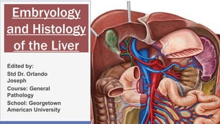
- 1. Embryology and Histology of the Liver Edited by: Std Dr. Orlando Joseph Course: General Pathology School: Georgetown American University 1
- 2. Embryology of the Liver and Biliary Apparatus 2
- 3. Overview 3
- 4. Alimentary System The liver is part of the alimentary system consisting of digestive tract(mouth to anus) associated glands and organs and endoderm. During the 3rd and 4th weeks, cephalocaudal and lateral folding of the embryo converts the trilaminar germ disc into an elongated cylinder, and the resulting endodermal gut tube consists of the cranial foregut, a midgut open to the yolk sac via the vitelline duct, and a hindgut The endoderm of the primordial gut forms most of the gut, epithelium, and glands. The primordial gut is initially closed at its cranial end by the oropharyngeal membrane and at its caudal end by the cloacal membrane The foregut is the region that will give rise to the liver and biliary apparatus 4
- 5. 5 Development of the Germ Layers
- 6. 6 A, Transverse view of a 4-week embryo showing the relationship of the primordial gut to the omphaloenteric duct.
- 7. Liver& Biliary Apparatus The liver, gallbladder, and biliary duct system arise as a ventral outgrowth, the hepatic diverticulum, from the distal part of the foregut early in the fourth week The diverticulum extends into the septum transversum, a mass of splanchnic mesoderm separating the pericardial and peritoneal cavities. The septum forms the ventral mesogastrium in this region. The hepatic diverticulum enlarges rapidly and divides into two parts as it grows between the layers of the ventral mesogastrium, or mesentery of the dilated portion of the foregut and the future stomach. The larger cranial part of the hepatic diverticulum is the primordium of the liver ; the smaller caudal part cystic diverticulum7
- 8. The proliferating endodermal cells form interlacing cords of hepatocytes and give rise to the epithelial lining of the intrahepatic part of the biliary apparatus. The hepatic cords anastomose around endothelium-lined spaces, the primordia of the hepatic sinusoids. The fibrous and hematopoietic tissue and Kupffer cells of the liver are derived from mesenchyme in the septum transversum. The liver grows rapidly from the 5th to 10th weeks and fills a large part of the upper abdominal cavity The quantity of oxygenated blood flowing from the umbilical vein into the liver determines the development and functional segmentation of the liver. Initially, the right and left lobes are approximately the same size, but the right lobe soon becomes larger. 8
- 9. Hematopoiesis (formation and development of various types of blood cells) begins in the liver during the sixth week, giving the liver a bright reddish appearance. By the ninth week, the liver accounts for approximately 10% of the total weight of the fetus. Bile formation by hepatic cells begins during the 12th week. The small caudal part of the cystic diverticulum becomes the gallbladder, and the stalk of the diverticulum forms the cystic duct Initially, the extrahepatic biliary apparatus is occluded with epithelial cells, but it is later canalized because of vacuolation resulting from degeneration of these cells.9
- 10. The stalk of the diverticulum connecting the hepatic and cystic ducts to the duodenum becomes the bile duct. The bile entering the duodenum through the bile duct after the 13th week gives the meconium (intestinal discharges of the fetus) a dark green color. 10
- 11. Biliary Development During week 7-8 the biliary development of the liver is formed from the hepatic diverticulum by the ductal plate The ductal plate is a primitive biliary epithelium which develops in mesenchyme adjacent to portal vein branches (periportal hepatoblasts). During liver development it is extensively reorganized (ductal plate remodeling) within the developing liver to form the intrahepatic bile ducts (IHBD). 11
- 12. Ventral Mesentery The ventral mesentery, a thin, double-layered membrane gives rise to: The lesser omentum, passing from the liver to the lesser curvature of the stomach (hepatogastric ligament) and from the liver to the duodenum (hepatoduodenal ligament) The falciform ligament, extending from the liver to the ventral abdominal wall The umbilical vein passes in the free border of the falciform ligament on its way from the umbilical cord to the liver. The ventral mesentery, derived from the mesogastrium, also forms the visceral peritoneum of the liver. The liver is covered by peritoneum, except for the bare area, which is in direct contact with the diaphragm 12
- 13. 13 Median section of caudal half of an embryo at the end of the fifth week, showing the liver and associated ligaments. The arrow indicates the communication of the peritoneal cavity with the extraembryonic coelom.
- 14. Formation of Alimentary system 14
- 15. Histology of the Liver 15
- 16. Parts of the Liver Basic Features Portal tract Portal triad Hepatic venule Hepatocyte Kupffer cells Hepatic cords and sinusoids Histologic examination of the Liver Hematoxylin and eosin stain Trichrome stain Reticular stain Periodic acid-Schiff Reticulin stain Prussian stain Immunohistochemistry Electron microscope 16 Classical Lobule Portal Lobule Acinus Lobule Zones of the Liver Liver Arrangements Periporal Zone(3) Intermediate Zone(2) Central Zone(1)
- 19. Adult Liver 19
- 20. Liver Basic Features 20 Hepatic Cords Space of Disse Kupffer cells Portal triad Terminal Hepatic venule Sinusoids HepatocytesPortal Tract
- 21. Liver Stroma 21
- 24. 24
- 25. 25
- 28. Portal Lobule 28
- 30. Liver Acinus 30
- 31. Zones of Liver 31 Peri-Portal Zone Intermediate Zone Central Zone Bile Drainage Blood Flow Central Vein Portal Vein Bile Duct Hepatic Artery
- 32. Bile and Blood flow of the liver 32
- 33. Liver Connective Tissue 33 Peri-Portal Zone Portal Vein Bile Duct Hepatic Artery Portal Tract
- 36. PAS Stain for Glycogen 36 Hepatocytes Sinusoids Glycogen rich
- 38. Biliary System 38 Bile ductule Canal of Herings Cholangiocytes
- 40. References Moore.L.Keith PHD.Persuad.T.V.N.PHD.Torschia.G.Mark PHD.Developing Human.10th Edition. Elsevier Inc. Chapter 11.Foregut.Dvelopmen of liver and biliary ducts,page271-218. Douglas.F.Paulsen(PHD).Histology and cell biology examination and boards review.5th edition.Mc.Graw Hill Medical.Langes.Chapter 16.Glands associated with the digestive system.The liver and bile duct.page 234-238. The University of Utah Eccles Health Sciences Library.Mercer university school of medicine. The Internet Pathology Laboratory or Medical Education.Anatomy and Histology.Normal histology.tissue gallbladder and liver.Histology of the liver and gallbladder.Retrived from. http://library.med.utah.edu/WebPath/HISTHTML/NORMAL/NORMAL.html The University of Western Australia. School of Anatomy and Human Biology.Blue histology.Notes.accessory digestive glands.histology of the liver and gallbladder.retrived from http://www.lab.anhb.uwa.edu.au/mb140/ Southern Illinois University School of Medicine.gastrointestinal system.Liver and gallbladder,bile duct,bile canniculi.retrived .Histology of the liver and biliary system and gallbladder.retrived from http://www.siumed.edu/~dking2/erg/liver.htm. Dr. med. H. Jastrow.Electron micrscpoic atlas of cells,tiisues and organs in the internet.Hepatocyte.Retrived from https://www.uni- mainz.de/FB/Medizin/Anatomie/workshop/EM/externes/Wartenberg/Leber3.jpg 40
Editor's Notes
- Initially, this duct attaches to the ventral aspect of the duodenal loop; however, as the duodenum grows and rotates, the entrance of the bile duct is carried to the dorsal aspect of the duodenum
- Ductal plate (arrows) developing around the portal vein mesenchyme in the liver of a 10-week-old embryo. There is extramedullary hematopoiesis in the sinusoids.
- Photomicrograph of fetal liver (Gp. A), showing continuation of sinusoidal capillaries (a) lined by squamous epithelium with Portal traid). islands of extramedullary hematopoiesis along with the liver cords.(precursors for RBCS).Also can be lymphocytes am Megakaryocytes
- The bulk of the liver consists of epithelial hepatocytes arranged into cords, separated by vascular sinusoids.
- The liver has a thin capsule of dense connective tissue, and a visceral (inferior) layer of peritoneal mesothelium, and is divided into left and right lobes.
- Portal areas (also called portal triads or portal canals) are located at the corners of liver lobules. Portal areas are normally surrounded by much larger areas packed with hepatic cords and sinusoids. Show blood flow and bile drainage. Each portal area contains three (hence the term portal triad) more-or-less conspicuous tubular structures all wrapped together in connective tissue. a branch of the bile duct a branch of the portal vein a branch of the hepatic artery
- A liver acinus encompasses the liver tissue that is served by a single terminal branch of the hepatic artery. These small vessels extend out from portal areas, along the boundaries between adjacent lobules. An acinus is typically diamond-shaped in cross section, with a hepatic arteriole crossing the center and with central veins at the two opposite corners. The acinus includes triangular portions of two adjacent lobules.
- The connective tissue of portal tracts consists of mainly collagen type I, which appears as thick, deep blue fibers on trichrome stain.
- A.Hepatic sinusoids lined by reticulin fibers in a normal liver (reticulin stain). B. Hepatocytes are arranged into cords, separated by vascular sinusoids lined by a fenestrated endothelium. Beneath the endothelium (i.e., between the endothelium and the hepatocytes) is a narrow "space of Disse", where thin reticular fibers (a form of collagen) provide support.
- Look at this picture, in which the network of fibres has been stained histochemically. This shows the central hepatic vein, and the supporting reticulin fibres, which are made of type III collagen.