Lipid droplets are organelles found in nearly all cell types that consist of a neutral lipid core surrounded by a phospholipid monolayer. They function in the storage, transport, and metabolism of lipids. Lipid droplets form through a budding process from the endoplasmic reticulum, facilitated by proteins embedded in the ER membrane. Perilipin proteins then coat the surface of lipid droplets, protecting the stored lipids from degradation. During periods of energy demand, phosphorylation of perilipin allows for lipase-mediated breakdown of lipids from the droplets. In addition to energy storage, lipid droplets are involved in other processes like protein storage and maturation, vitamin storage, and stress response.
This document provides an overview of high performance liquid chromatography (HPLC). It describes HPLC as a type of liquid chromatography that uses small particle sizes as the stationary phase to separate compounds dissolved in a solution. The key components of an HPLC system are a solvent delivery pump, injector, column, detectors, and data collection system. Different types of columns include normal phase, reverse phase, size exclusion, and ion exchange. Factors that affect HPLC separation include column length and temperature, flow rate, particle size, and mobile phase properties. HPLC is used for quantitative and qualitative analysis in various applications like analyzing drugs, pollutants, and food/drug products.
This document discusses solvent extraction, which is a versatile separation method used in analytical chemistry. It can be used to separate, purify, enrich, and analyze both tracer and macro amounts of metal ions. The key principles discussed include the phase rule, which describes solvent extraction as a two-phase system, and the Nernst distribution law, which defines the partition or distribution coefficient. Different types of extraction systems are classified, including chelate extraction involving complex formation, extraction by solvation, and ion-pair formation. Factors that affect metal complex stability such as ligand basicity and ring size are also outlined.
Mass spectrometry deals with the study of charged molecules and fragment ions produced from a sample exposed to ionizing conditions. It works by bombarding samples with electron beams or chemical ions, which causes the samples to form positively charged molecular or fragment ions. These ions are then separated based on their mass-to-charge ratio, producing a spectrum that can be used to determine molecular weights, identify unknown compounds, and detect impurities. Mass spectrometry is a versatile analytical technique with applications in pharmaceutical analysis, proteomics, and other areas.
Presentation about Gas Chromatography (GC) mainly used in analytical techniques with brief description of its components.
Used mainly in R&D in chemical industries.
The Power Point Presentation includes The Size Exclusion Chromatography and Its Method. These Slides may be helpful for master of science students. The Syllabus for the slides was prepared by following as KSV, Gandhinagar. Paper Code is CH-AC-302, Unit-01
Graphite Furnace Atomic Absorption Spectroscopy (GFAAS) is a type of atomic absorption spectroscopy that uses a graphite-coated furnace to vaporize samples. Small aliquots of up to 100 microliters of aqueous samples are placed into the graphite tube and heated to high temperatures to break chemical bonds and produce free ground-state atoms. The amount of light absorbed by these atoms is proportional to the concentration of the element of interest. GFAAS offers greater sensitivity and lower detection limits than flame atomic absorption spectroscopy due to the higher temperatures achieved in the graphite furnace. It has applications in analyzing low metal concentrations in water samples and quantifying elements like beryllium in blood and serum.
Solvent extraction is a separation process that uses immiscible solvents to transfer a solute from an aqueous solution to an organic solution. It relies on the differential solubility of compounds in two different immiscible solvents. Key aspects of solvent extraction include the Nernst distribution law, which states that the concentration ratio of a solute between the two solvents is constant at equilibrium. Solvent extraction methods include simple, multiple, continuous, and countercurrent extraction. It has various applications such as extracting uranium and plutonium from nuclear waste, separating metal ions, and recovering heat-sensitive compounds, antibiotics, and proteins.
Detectors are the brain of any chromatograhic system. It help us to record the chromatogram based on certain characteristics of the analyte and help us in identifying that compound both qualitatively and quantitatively.
MALDI is a soft ionization technique used in mass spectrometry to analyze large biomolecules. It works by co-crystallizing the analyte sample with a UV-absorbing matrix. A laser is used to excite the matrix, causing desorption and ionization of the analyte molecules. The ions are then analyzed by a mass spectrometer, typically a time-of-flight instrument. Careful sample preparation is important for reproducibility and performance. MALDI is widely used in pharmaceutical analysis and DNA sequencing due to its ability to characterize large organic and biomolecules.
Detectors in Gas Chromatography are devices used to detect and measure compounds eluting from the GC column. The document discusses several common detectors including:
- The Flame Ionization Detector (FID), one of the most widely used, responds to carbon-containing compounds. It is sensitive, destructive to samples, and provides a linear response.
- The Thermal Conductivity Detector (TCD) responds to differences in thermal conductivity between carrier gas and eluting compounds. It is non-destructive but has low sensitivity.
- Other detectors discussed are specific to certain functional groups like nitrogen/phosphorus (NPD), flame photometric (sulfur and phosphorus), electron capture (
1. The document describes how to use flame atomic absorption spectroscopy to determine the concentration of calcium in bottled water samples.
2. Flame atomic absorption spectroscopy works by aspirating and atomizing liquid samples using a flame, then measuring the absorption of light at characteristic wavelengths to detect specific metals.
3. The technique requires samples to be aspirated and mixed with combustible gases and ignited in a flame between 2100-2800°C to atomize the metals, which will absorb light from a hollow cathode lamp at wavelengths specific to each metal.
The document discusses hydrogen peroxide stabilization using chemical stabilizers. It aims to find a stabilizer that restricts the auto-decomposition of hydrogen peroxide without catalytic poisoning. Stabilizers are chemical compounds that delay the auto-catalytic decomposition of hydrogen peroxide, improving storage stability. Sodium stannate and 8-hydroxyquinoline are discussed as effective stabilizers that deactivate catalytic metal ions and increase the shelf life of hydrogen peroxide solutions. The decomposition rate of hydrogen peroxide is analyzed under various conditions like temperature, pH, concentration, and presence of catalytic impurities. Direct titration and gas evolution methods are also described to determine the decomposition rate.
This document provides an introduction and overview of gas liquid chromatography (GLC) and high performance liquid chromatography (HPLC). It defines chromatography as a technique that separates components of a mixture based on differences in affinity for a stationary and mobile phase. GLC uses an inert gas as the mobile phase and a liquid stationary phase, while HPLC uses high pressure to push a liquid mobile phase through a column. The document describes the basic instrumentation, principles, and applications of these techniques.
This document discusses inductively coupled plasma-optical emission spectroscopy (ICP-OES), a technique used to detect chemical elements. ICP-OES uses inductively coupled plasma to produce excited atoms and ions that emit electromagnetic radiation at wavelengths specific to each element. The plasma is generated by inductive coupling from cooled electrical coils operating at megahertz frequencies, reaching temperatures of 6000-10,000 K. Sample solutions are nebulized and injected into the argon plasma, where atoms are excited and emit light proportional to their concentration, which is measured by a spectrometer. Typical applications include environmental testing, food and drinks analysis, materials testing, and healthcare.
This document provides an overview of Raman spectroscopy. It discusses the principle behind Raman spectroscopy, which involves scattering of monochromatic light when it interacts with a sample. It describes the typical instrumentation used, including lasers as the light source and spectrometers to analyze the scattered light. The key differences between Raman and IR spectroscopy are outlined. Various types of Raman techniques and applications are also summarized, such as its use in analyzing inorganic, organic and biological samples.
This document discusses normal phase chromatography and reversed phase chromatography. Normal phase chromatography uses a polar stationary phase like silica gel and a non-polar mobile phase. The most popular packing material is silica gel, which separates samples based on interactions between polar silanol groups on the silica surface and the sample. Reversed phase chromatography became popular in the 1970s with the introduction of alkyl chains bonded to the silica surface. This reversed the elution order so polar compounds elute first. Reversed phase is now the most common technique and uses a non-polar stationary phase like C18-bonded silica and a polar mobile phase like water.
Affinity chromatography is a method used to purify biomolecules like proteins and nucleic acids based on specific interactions between the biomolecule and a ligand immobilized on a solid support. When a mixture is passed through the column, the target biomolecule will bind to the ligand while other molecules pass through. The bound biomolecule can then be separated by changing conditions like pH or introducing a competing molecule to displace it. Affinity chromatography offers highly specific purification of target molecules in a single step.
Spectral interferences in atomic absorption spectroscopy can occur due to overlapping absorption lines from different elements, molecular absorption from flame gases or sample-derived molecules, or scattering from particles. Chemical interferences involve the formation of non-volatile compounds between the analyte and interfering ions or the formation of oxides/hydroxides. Physical interferences affect the rate of sample uptake and atomization efficiency due to variations in parameters like gas flow rates, sample viscosity, solids content, or flame temperature.
Reversed phase chromatography is an adsorption technique used to separate nonpolar substances. It works by having a nonpolar stationary phase and a polar mobile phase, opposite of normal phase chromatography. Molecules like proteins, peptides, and nucleic acids can be separated using reversed phase chromatography. The separation depends on the hydrophobic binding of solutes from the mobile phase to the hydrophobic ligands attached to the stationary phase. Common stationary phases use silica beads with attached alkyl hydrocarbon chains of varying lengths. Gradient elution with mixtures of water and organic solvents like acetonitrile or methanol is typically used for separation. Reversed phase chromatography has applications in preparative purification of proteins, peptides, and other biomolecules.
This document provides an overview of atomic emission spectrophotometry (AES) and atomic absorption spectrophotometry (AAS). It discusses the principles, instrumentation, applications in pharmaceutical analysis, and examples of quantitation for each technique. AES works by exciting the atoms of an element, which then emit light at characteristic wavelengths. AAS analyzes samples by measuring the absorption of light from a lamp, with higher concentrations absorbing more light. Both techniques can be used to quantify elements in samples like infusion solutions and identify metallic impurities.
(1) Coprecipitation occurs when substances normally soluble under certain conditions are carried down by a precipitate. This can be problematic in chemical analysis if undesired impurities coprecipitate with the analyte.
(2) There are four types of coprecipitation: surface adsorption, mixed-crystal formation, occlusion, and mechanical entrapment. Surface adsorption and mixed-crystal formation are equilibrium processes, while occlusion and mechanical entrapment arise from crystal growth kinetics.
(3) Surface adsorption involves contaminants adsorbing to the surface of precipitates like coagulated colloids. Mixed-crystal formation replaces ions in a crystal lattice with contaminant ions. Occlusion traps foreign ions
The document provides an overview of mass spectrometry, including its basic principles, components, working principle, and various applications. Mass spectrometry involves ionizing chemical compounds and separating the resulting ions based on their mass-to-charge ratio, producing a mass spectrum that can be used to determine the elemental or isotopic composition of a sample. Key components include an ion source, mass analyzer, and detector. Common ionization methods are also described, such as electron impact, chemical ionization, electrospray ionization, and matrix-assisted laser desorption/ionization.
This document provides information about mass spectrometry including definitions, principles, components, and methods of ionization. It defines mass spectrometry as a technique that ionizes chemical species and sorts them by mass-to-charge ratio. The key components of a mass spectrometer are described as the inlet system, ion source, mass analyzer, and detector. Common ionization methods like MALDI and electrospray ionization are explained in terms of how they work to ionize samples for analysis.
The document provides information about mass spectrometry including:
- Mass spectrometry is a powerful analytical technique that uses instruments called mass spectrometers to identify molecules by breaking them into ionized fragments and measuring their mass-to-charge ratios.
- The basic components of a mass spectrometer are the sample inlet, ionization source, mass analyzer, and ion detector. Common ionization sources are electrospray ionization, matrix-assisted laser desorption/ionization, and electron ionization. Common mass analyzers are quadrupoles, ion traps, and time-of-flight.
- Mass spectrometry has a variety of applications and has undergone significant technological developments since its invention in the early 20th
This document provides an overview of gas chromatography. It discusses the basic components and process, including the stationary and mobile phases, columns, detectors, and applications. Specifically, it explains that gas chromatography separates components based on their partitioning between a stationary and mobile phase, which are typically a liquid and gas. It also outlines the key parts of a gas chromatography instrument, such as the column, carrier gas, sample introduction, and common detectors like the flame ionization detector. Finally, it notes some examples of how gas chromatography is used, such as in polymer analysis, reaction studies, and determining molecular properties.
This presentation gives an overview of Lipid Rafts, how it was discovered, its importance and the future research in this area,Feel free to comment and ask any questions
This document summarizes the current understanding of lipid rafts. Lipid rafts are microdomains in cell membranes enriched in cholesterol and glycosphingolipids that exist as distinct liquid-ordered regions resistant to extraction by nonionic detergents. While lipid rafts vary in size and composition, they are generally small (100-200nm) and may constitute a large fraction of the plasma membrane. Lipid rafts contain certain proteins involved in cell signaling and are thought to play a role in regulating signal transduction.
This document summarizes lipids and phospholipids. It defines lipids as biological compounds that are insoluble in water but soluble in organic solvents. It describes how phospholipids are the major components of biological membranes, consisting of a glycerol backbone, fatty acids, a phosphate group, and an alcohol group. The document also summarizes the classification of lipids into glycerophospholipids, sphingolipids, and glycolipids. It provides details on the synthesis, structure, and metabolism of phospholipids.
structure and function of the cell envelope of gram negative bacteria.Muhammad Ajmal
The document summarizes the structure and function of the cell envelope of Gram-negative bacteria. It discusses three main layers: (1) the cytoplasmic or inner membrane composed of phospholipids, proteins and carbohydrates, (2) the peptidoglycan cell wall composed of polysaccharides and cross-linked peptide chains, and (3) the outer membrane containing phospholipids, lipopolysaccharides and porin proteins. Between the inner and outer membranes is the periplasmic space containing the peptidoglycan layer and degradative enzymes. The cell envelope protects bacteria from their environment while allowing selective transport of molecules.
Proteins are made up of amino acid monomers linked together by peptide bonds. They are synthesized through transcription and translation processes involving DNA, RNA and ribosomes. Proteins have four levels of structure - primary, secondary, tertiary and quaternary - which determine their shape and function. Proteins perform many essential roles in cells and organisms such as catalysis, structure, defense and transport.
LIPID CHEMISTRY Question Bank vnd.ms-powerpoint&rendition=1-4.pptLogesh Kannan
The document contains 15 questions related to lipid chemistry. It discusses essential fatty acids like linoleic acid and alpha-linolenic acid. It also discusses arachidonic acid and how it is a precursor for prostaglandins. The document discusses products of arachidonic acid metabolism, importance of prostaglandins, iodine number, saponification number, amphipathic lipids, phospholipid components and functions, pulmonary surfactant structure and importance, and biological actions of prostaglandins.
This document discusses the structure, formation, and functions of triglycerides and phospholipids. It explains that triglycerides are formed from glycerol and three fatty acids bonded together via ester bonds. Triglycerides function to store fatty acids for energy and transport lipids through the blood. Phospholipids have a phosphate-containing polar head and two fatty acid tails, allowing them to form lipid bilayers that act as semi-permeable membranes surrounding cells and organelles.
16 October 2023 Chemistry of Lipids- Lecture 3.pptxKritikaMishra43
1. Phospholipids are important structural components of cell membranes that regulate permeability and signaling. They are required for energy generation in mitochondria.
2. Dipalmitoyl phosphatidylcholine is the major component of pulmonary surfactant and is essential for reducing surface tension in the lungs and preventing respiratory distress syndrome.
3. Phospholipases hydrolyze phospholipids to release fatty acids involved in eicosanoid synthesis and cell signaling through generation of diacylglycerol and inositol triphosphate second messengers.
Lipids play several crucial roles in biological membranes:
1. Phospholipids form lipid bilayers that provide structural integrity and compartmentalization for cells and organelles. The hydrophobic tails orient inward while hydrophilic heads face outward.
2. Lipids help create distinct compartments that allow for compartmentalization of processes and organelles, facilitating functions like ATP production.
3. Lipids regulate membrane permeability and homeostasis, and help anchor integral membrane proteins to provide a stable environment for their functions like ion transport.
4. Certain lipids contribute to processes like cell recognition and adhesion through roles such as glycolipid-mediated cell-cell recognition.
This document provides information on compound lipids, including phospholipids, glycolipids, and lipoproteins. It discusses:
1) Phospholipids are esters of fatty acids containing a phosphoric acid group and nitrogenous bases. They have an amphipathic structure with a polar head and nonpolar tails, and include glycerophospholipids and sphingophospholipids.
2) Glycolipids contain a ceramide backbone linked to simple or complex carbohydrates. Major types include cerebrosides, sulfatides, globosides, and gangliosides.
3) Lipoproteins are composed of a lipid core surrounded by a phospholipid shell and ap
Role of fat body in insect metabolism.pptKHARIKARAN
The fat body plays a major role in insect metabolism. It is a loose tissue distributed throughout the insect body that is responsible for energy storage and release. The fat body stores energy as glycogen and triglycerides and is involved in immunity, detoxification, and yolk protein production. It is composed of different cell types, including trophocytes that store and secrete organic substances, oenocytes near the cuticle, mycetocytes that contain symbiotic microorganisms, chromatocytes that accumulate fats, and urocytes that store urate granules. The fat body undergoes changes during metamorphosis and is important for overwintering insects to manage their lipid energy stores.
The structure of the cell membrane, the phospholipid layer distinguished to the break down of protein and the lipid layer. Their structural components and the molecular basis of it.
Proteins are composed of chains of amino acids linked together by peptide bonds. There are 20 common amino acids that make up proteins. The sequence of amino acids is determined by the DNA sequence. Proteins have four levels of structure: primary, secondary, tertiary, and quaternary. Proteins serve many important functions in the body such as catalysis, muscle contraction, cytoskeleton structure, transport, cell signaling, and immunity.
This presentation provides an overview of the endoplasmic reticulum (ER), including its structure, functions, and regulation. Key points include: the ER is a network of tubules and sacs that synthesizes proteins and lipids, stores calcium, and aids protein folding; its shape is maintained by membrane-shaping proteins and interactions with other organelles; and the ER adapts in response to stresses through signaling pathways like the unfolded protein response. The presentation was given to undergraduate students to provide foundational knowledge about this important intracellular organelle.
The document describes several types of organelles found within cells and their functions. It discusses the nucleus and its nuclear envelope, chromosomes, and components. It also describes mitochondria as the energy powerhouses of cells that produce ATP through oxidative phosphorylation. Other organelles discussed include the endoplasmic reticulum, vesicles, peroxisomes, ribosomes, lysosomes, the Golgi apparatus, and plastids.
Lipids are organic compounds that are insoluble in water but soluble in organic solvents. They include fats, oils, waxes, phospholipids, and steroids. Lipids serve important biological functions like energy storage, signaling, and as structural components of cell membranes. Cholesterol is an important lipid that is a precursor for steroid hormones and bile acids. It is essential for membrane structure and fluidity. Different fatty acids have various health impacts depending on whether they are saturated, monounsaturated, or polyunsaturated. Polyunsaturated fatty acids like omega-3 and omega-6 are particularly important for eicosanoid production. Phospholipids are major components of biological membranes and are involved in processes like blood
Physiological And Pathological Systems Within The...Deb Birch
Redox signalling is an important electron transfer process that regulates many physiological and pathological systems in the circulatory system. It is usually induced by reactive oxygen species and can alter cell processes. Redox signalling involves thiol-based redox couples that regulate imbalances in redox potentials and are linked to changes in redox potentials. Cysteine is an amino acid that can undergo oxidative modifications to perform different functions as part of redox signalling.
This document compares and contrasts the key characteristics of Gram-negative and Gram-positive bacteria. Gram-negative bacteria have a thin peptidoglycan layer and outer membrane containing lipopolysaccharides, making them more resistant to antibiotics. They produce endotoxins and are susceptible to physical disruption. In contrast, Gram-positive bacteria have a thick peptidoglycan layer without an outer membrane or lipopolysaccharides. They produce exotoxins and are less resistant to physical disruption and antibiotics.
Here are the key organelle markers and their associated enzymes:
1. Mitochondria - ATP synthase (located in the inner mitochondrial membrane)
2. Lysosome - Cathepsin (lysosomal storage disorders occur due to deficiencies in cathepsin enzymes)
3. Golgi body - Galactosyltransferase (involved in glycoconjugate synthesis in the Golgi)
4. Microsomes (Endoplasmic Reticulum fragments) - Glucose-6-phosphatase (involved in glucose metabolism in the smooth ER)
This presentation was provided by Steph Pollock of The American Psychological Association’s Journals Program, and Damita Snow, of The American Society of Civil Engineers (ASCE), for the initial session of NISO's 2024 Training Series "DEIA in the Scholarly Landscape." Session One: 'Setting Expectations: a DEIA Primer,' was held June 6, 2024.
The simplified electron and muon model, Oscillating Spacetime: The Foundation...RitikBhardwaj56
Discover the Simplified Electron and Muon Model: A New Wave-Based Approach to Understanding Particles delves into a groundbreaking theory that presents electrons and muons as rotating soliton waves within oscillating spacetime. Geared towards students, researchers, and science buffs, this book breaks down complex ideas into simple explanations. It covers topics such as electron waves, temporal dynamics, and the implications of this model on particle physics. With clear illustrations and easy-to-follow explanations, readers will gain a new outlook on the universe's fundamental nature.
हिंदी वर्णमाला पीपीटी, hindi alphabet PPT presentation, hindi varnamala PPT, Hindi Varnamala pdf, हिंदी स्वर, हिंदी व्यंजन, sikhiye hindi varnmala, dr. mulla adam ali, hindi language and literature, hindi alphabet with drawing, hindi alphabet pdf, hindi varnamala for childrens, hindi language, hindi varnamala practice for kids, https://www.drmullaadamali.com
Main Java[All of the Base Concepts}.docxadhitya5119
This is part 1 of my Java Learning Journey. This Contains Custom methods, classes, constructors, packages, multithreading , try- catch block, finally block and more.
How to Fix the Import Error in the Odoo 17Celine George
An import error occurs when a program fails to import a module or library, disrupting its execution. In languages like Python, this issue arises when the specified module cannot be found or accessed, hindering the program's functionality. Resolving import errors is crucial for maintaining smooth software operation and uninterrupted development processes.
This slide is special for master students (MIBS & MIFB) in UUM. Also useful for readers who are interested in the topic of contemporary Islamic banking.
Executive Directors Chat Leveraging AI for Diversity, Equity, and InclusionTechSoup
Let’s explore the intersection of technology and equity in the final session of our DEI series. Discover how AI tools, like ChatGPT, can be used to support and enhance your nonprofit's DEI initiatives. Participants will gain insights into practical AI applications and get tips for leveraging technology to advance their DEI goals.
বাংলাদেশের অর্থনৈতিক সমীক্ষা ২০২৪ [Bangladesh Economic Review 2024 Bangla.pdf] কম্পিউটার , ট্যাব ও স্মার্ট ফোন ভার্সন সহ সম্পূর্ণ বাংলা ই-বুক বা pdf বই " সুচিপত্র ...বুকমার্ক মেনু 🔖 ও হাইপার লিংক মেনু 📝👆 যুক্ত ..
আমাদের সবার জন্য খুব খুব গুরুত্বপূর্ণ একটি বই ..বিসিএস, ব্যাংক, ইউনিভার্সিটি ভর্তি ও যে কোন প্রতিযোগিতা মূলক পরীক্ষার জন্য এর খুব ইম্পরট্যান্ট একটি বিষয় ...তাছাড়া বাংলাদেশের সাম্প্রতিক যে কোন ডাটা বা তথ্য এই বইতে পাবেন ...
তাই একজন নাগরিক হিসাবে এই তথ্য গুলো আপনার জানা প্রয়োজন ...।
বিসিএস ও ব্যাংক এর লিখিত পরীক্ষা ...+এছাড়া মাধ্যমিক ও উচ্চমাধ্যমিকের স্টুডেন্টদের জন্য অনেক কাজে আসবে ...
বাংলাদেশ অর্থনৈতিক সমীক্ষা (Economic Review) ২০২৪ UJS App.pdf
lipid droplet.pptx
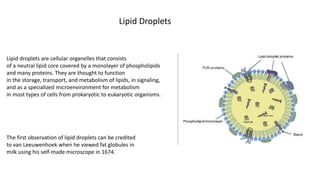
1. Lipid droplets are cellular organelles that consists
of a neutral lipid core covered by a monolayer of phospholipids
and many proteins. They are thought to function
in the storage, transport, and metabolism of lipids, in signaling,
and as a specialized microenvironment for metabolism
in most types of cells from prokaryotic to eukaryotic organisms.
Lipid Droplets
The first observation of lipid droplets can be credited
to van Leeuwenhoek when he viewed fat globules in
milk using his self-made microscope in 1674.
2.
3. In which cells and tissues, we can see them
In the last 10 years, established isolation methods and
advanced proteomic technology have allowed scientists
to study the protein composition of lipid droplets from
many cell types and tissues, such as fibroblasts , epithelia
, adipocytes , hepatocytes ,
macrophages , pancreatic cells , mammary glands
, livers , white adipose tissues , and skeletal
muscles , as well as from many other popular model
organisms, including plants , insects , yeast
green algae , bacteria
6. Exact mechanism of formation of lipid droplets is still unknown, it is proposed that they bud off the membrane of the
endoplasmic reticulum.
1. Acetyl co A is made from pyruvate dehydrogenase and mitochondria.
2. Acetyl co A converts malonyl co A by ACC.(acetyl co A carboxylase )
3. Malonyl co A converts to free fatty acids(FFA) by FAS ( fatty acid synthase)
4. Glycerol and FFA come together to the ER membrane . Inside ER lumen they conjugate together.
5. ER embedded proteins such as GPAT4 & DGAT1/2 facilitate this process.
6. Those proteins condense FFA in glycerol and make TAG(tri acyl glycerol) & DAG(di acyl glycerol).
7. ER membrane makes lenses . TAG & DAG accumulate in to the ER membrane lenses.
8. FITM2 & BSCL2 are proteins which are found in neck of ER membrane budding lenses( most initial step of lipid
droplet).
9. Those proteins facilitates the movement of TAG & DAG in to the lenses and are very important for the formation of
lipid droplet.
10. Budding ER membrane is covered by perilipin proteins( plin1/2), that stabilize membrane of lipid droplet and
protect from lipases.
The formation of droplets is not spontaneous but is facilitated by ER proteins and specific lipids. Seipin, a protein that resides at ER–
droplet junctions, functions early to stabilize the nascent structures, ensures droplets of correct morphology, prevents the
accumulation of phosphatidic acid at the ER–droplet junction, and prevents ectopic budding into the nucleus.
7. Lipid droplet degradation
• In starvation the ATP concentration of cells are low and AMP concentration is high.
• That is signaling AMPK proteins.(AMP kinase).
• When AMP binds to AMPK ,AMPK activates and it inhibit the perilipin1/2 proteins of lipid droplets.
• Perilipin 1/2 inhibit the degradation of lipid droplets.
• So after inhibiting perilipin proteins degradation of lipid droplets is started.
As well as
• In G proteins activation activate adenylyl cyclase protein.
• adenylyl cyclase turns to ATP then cyclic AMP(cAMP )
• CAMP binds PKA(protein kinase A)
• PKA deactivate perilipin 1/2 .then starts lipid droplets degradation.
And also,
• There is CMA(chaperone mediated autophagy), cytosolic proteins(HSC70) that are then targeted to
lysosomes and directly translocated across the lysosome membrane for degradation. And these poteins
inhibits perilipin1/2 of lipid droplets .
8.
9. Perilipin
(PLIN)
• Perilipin, also known as lipid droplet-associated protein, Perilipin 1, or PLIN, is a protein that, in humans, is
encoded by the PLIN gene. The perilipins are a family of proteins that associate with the surface of lipid
droplets. Phosphorylation of perilipin is essential for the mobilization of fats in adipose tissue.
• Perilipin acts as a protective coating from the body’s natural lipases, such as hormone-sensitive lipase, which
break triglycerides into glycerol and free fatty acids for use in metabolism, a process called lipolysis. In humans,
perilipin is expressed in three different isoforms, A, B, and C, and perilipin A is the most abundant protein
associated with the adipocyte lipid droplets.
• Perilipin is hyperphosphorylated by PKA following β-adrenergic receptor activation.Phosphorylated perilipin
changes conformation, exposing the stored lipids to hormone-sensitive lipase-mediated lipolysis. Although PKA
also phosphorylates hormone-sensitive lipase, which can increase its activity, the more than 50-fold increase in
fat mobilization (triggered by epinephrine) is primarily due to perilipin phosphorylation
• Perilipin is part of a gene family with five currently-known members. In vertebrates, closely related genes
include adipophilin (also known as adipose differentiation-related protein or Perilipin 2), TIP47 (Perilipin 3),
Perilipin 4 and Perilipin 5 (also called MLDP, LSDP5, or OXPAT). Insects express related proteins, LSD1 and LSD2,
in fat bodies. The yeast Saccharomyces cerevisiae expresses PLN1 (formerly PET10), that stabilizes lipid
droplets and aids in their assembly.
10. PET 10
Pet10p is a yeast lipid droplet protein of unknown function. We show that it binds specifically to and is
stabilized by droplets containing triacylglycerol (TG). Droplets isolated from cells with a PET10 deletion
strongly aggregate, appear fragile, and fuse in vivo when cells are cultured in oleic acid. Pet10p binds early to
nascent droplets, and their rate of appearance is decreased in pet10Δ. Moreover, Pet10p functionally
interacts with the endoplasmic reticulum droplet assembly factors seipin and Fit2 to maintain proper droplet
morphology. The activity of Dga1p, a diacylglycerol acyltransferase,
and TG accumulation were both 30–35% lower in the absence of Pet10p.
Pet10p contains a PAT domain,a defining property of perilipins, which was not previously known to exist in
yeast. We propose that the core functions of Pet10p and other perilipins extend beyond protection from
lipases and include the preservation of droplet integrity as well as collaboration with seipin and Fit2 in
droplet assembly and maintenance. Pet10p, an established lipid droplet protein in yeast, which binds to TG-
containing droplets where it is stabilized, is important for droplet integrity, influences protein distribution,
facilitates droplet formation, contributes to TG reservoirs, and collaborates with seipin and Fit2 proteins to
maintain normal droplet size. Pet10p and Sps4p both contain a PAT domain, and complementation of pet10Δ
with human Plin2 and Plin3 indicates that Pet10p is indeed a yeast perilipin.
11. Seipin
is a homo-oligomeric integral membrane protein in the endoplasmic reticulum (ER) that concentrates at junctions
with cytoplasmic lipid droplets (LDs). Alternatively, seipin can be referred to as Bernardinelli-Seip congenital
lipodystrophy type 2 protein (BSCL2), and it is encoded by the corresponding gene of the same name, i.e. BSCL2. At
protein level, seipin is expressed in cortical neurons in the frontal lobes, as well as motor neurons in the spinal cord.
It is highly expressed in areas like the brain, testis and adipose tissue.Seipin's function is still unclear but it has been
localized close to lipid droplets, and cells knocked out in seipin which have anomalous droplets. Hence, recent
evidence suggests that seipin plays a crucial role in lipid droplet biogenesis.
12. RESEARCH
The research described herein began with an analysis of proteins associated with droplets enriched in
one of the core lipids, TG or SE. We report that Pet10p, a known droplet surface protein with unknown
function (Athenstaedt et al., 1999), preferentially localizes to TG droplets, where it is protected from
degradation. Pet10p is required for droplet integrity; in its absence, droplet structure is less stable,
exhibiting severe aggregation in vitro. In addition, loss of the protein results in droplet fusion in vivo
when cells are cultured with oleic acid (OA). PET10-null cells accumulate less TG, which for the
acyltransferase Dga1p is accompanied by a loss of enzyme activity and movement from the ER to
droplets. Pet10p localizes to droplets early in their assembly and promotes droplet formation. We
identify a PAT domain in Pet10p and consider it a yeast perilipin, and we propose that the activities of
Pet10p reflect important functions of the perilipin classes.
13. How its work
Lipid droplets play a role in neutral lipid transport between cellular organelles.
Lipids can be synthesize in cytoplasm and transferred to the ER then stored in lipid droplets.
Play and active role in lipid synthesis.
Lipid synthetic enzymes have been found on lipid droplets.
In plant cells lipid droplets storage the energy within the organelle. that is the primary function of the plant lipid droplets.
Starvation or high energy demand activities various lipases are releasing lipids as FFA that can enter the beta oxidation cycle.
Lipid droplets are commonly associated in adipose tissues they found in nearly all the cell type.
Lipid droplets of adipose tissues are obviously responsible for the obesity.
In brain tissue LDs are formed on astrocytes in response to ROS stress.
Bona fide LD diseases (neutral lipid storage diseases ) are associated with myopathy ,autism, lost of hearing
And eyesight .
Lipid droplets are responsible for neuron degenerative diseases.
Abnormal LD dynamics can be observed in Alzheimer's disease and Huntington’s disease .
14.
15. • Lipid droplets have important functions beyond energy homeostasis.
• They store vitamins, signaling precursors, and other hydrophobic molecules.
• They mitigate some harmful effects of ER and oxidative stress. lipid handling roles of droplets include the
storage of hydrophobic vitamin and signaling precursors, and the management of endoplasmic reticulum
and oxidative stress.
• They function in protein maturation, storage, and turnover.•
• They are motile and can exist in the nucleus. Other potential roles of lipid droplets may be connected with
their intracellular motility and, in some cases, their nuclear localization.