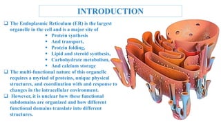
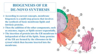
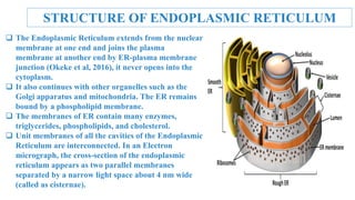
Presentation on
Endoplasmic Reticulum
Course Code: BGE-251
Course Title: Cytology & Cytogenetics
Presented by
Jannatul Ferdous Ritu (20BGE001)
Tasfiah Tasnim (20BGE006)
Fahmida Galeba (20BGE007)
Md Mostafizur Rahman (20BGE008)
Zannatul Ferdoush (20BGE009)
Presented to
Imdadul Haque Shohag
Lecturer,
Department of Biotechnology And
Genetic Engineering, BSMRSTU
CONTENTS
 Introduction
 Invention Of ER
 Biogenesis Of ER
 Structure
 ER Shaping Proteins
i. ER Tubules
ii. ER Sheets
iii. ER Microtubule Interactions
 Changes In ER Structure During Mitosis
 Changes In ER During Oocyte Maturation
And Fertilization
 Components Of Er
 Cisternae
 Vesicles
 Tubules
 Types Of Endoplasmic Reticulum
 Smooth Endoplasmic Reticulum (SER)
 Rough Endoplasmic Reticulum (RER)
 Protein Synthesis And Folding
 Lipid Biogenesis
 Calcium (Ca 2+) Metabolism
 Regulation Of ER Shape And Function
 Closing Remarks
INTRODUCTION
 The Endoplasmic Reticulum (ER) is the largest
organelle in the cell and is a major site of
 Protein synthesis
 And transport,
 Protein folding,
 Lipid and steroid synthesis,
 Carbohydrate metabolism,
 And calcium storage
 The multi-functional nature of this organelle
requires a myriad of proteins, unique physical
structures, and coordination with and response to
changes in the intracellular environment.
 However, it is unclear how these functional
subdomains are organized and how different
functional domains translate into different
structures.
DISCOVERY OF ER
The Endoplasmic Reticulum (ER) was first observed in the late 19th century by
light microscopy.
1945: Keith R. Porter, Albert Claude, and Ernest F. Fullam visualize the ER
using electron microscopy and describe it as a "lace-like reticulum."
1960s: George Palade, a Romanian-American cell biologist, contributes
significantly to understanding the ER’s role in protein synthesis and folding.
1980s: the concept of ER quality control emerges, highlighting the ER’s role in
ensuring proper protein folding and preventing misfolding.
 the 1990s: the unfolded protein response (up) is identified as a signaling
pathway that helps cells cope with ER stress.
BIOGENESIS OF ER
DE-NOVO SYNTHESIS
According to current concepts, membrane
biogenesis is a multi-step process that involves
the synthesis of basic membrane lipids and
intrinsic proteins.
Then the addition of the other components, such
as enzymes, sugars, or lipids occurs sequentially.
The insertion of protein into the ER membrane is
independent of that of the lipids. Some proteins
of the ER are formed by the ribosomes in the
cytosol which then become inserted into the
membrane.
STRUCTURE OF ENDOPLASMIC RETICULUM
 The Endoplasmic Reticulum extends from the nuclear
membrane at one end and joins the plasma
membrane at another end by ER-plasma membrane
junction (Okeke et al, 2016), it never opens into the
cytoplasm.
 It also continues with other organelles such as the
Golgi apparatus and mitochondria. The ER remains
bound by a phospholipid membrane.
 The membranes of ER contain many enzymes,
triglycerides, phospholipids, and cholesterol.
 Unit membranes of all the cavities of the Endoplasmic
Reticulum are interconnected. In an Electron
micrograph, the cross-section of the endoplasmic
reticulum appears as two parallel membranes
separated by a narrow light space about 4 nm wide
(called as cisternae).
COMPONENTS OF
ENDOPLASMIC RETICULUM
Based on morphology, endoplasmic reticulum is made up of 3 types of structures
a)Cisternae- These are long, flattened, sac-like unbranched tubules, arranged parallel forming
bundles, each tubule is 40-50 µm thick
b) Vesicles-These are usually rounded or ovoid structures, 25-500 mµ thick in diameter,
surrounded by a unit membrane, often occur isolated in the cytoplasm
c) Tubules are of diverse shape and are irregularly branched, 50-100 mµ in diameter.
These are found in cells that are active in the synthesis of glycerides. These are also found in big
pigmented epithelial cells of the retina, involved in the metabolism of Vitamin
– A (Retinol).
The membrane consists of 3 layers a plasma membrane, a middle thin and transparent layer of
phospholipids, and an outer and inner dense layer of proteins. d) Perinuclear space is the space
present between E R and Nucleus. There is a close relationship between perinuclear space,
cisternae, and cell sap
It is basically of two functionally distinct types:
1. Smooth Endoplasmic Reticulum (SER)
2. and Rough Endoplasmic Reticulum
(RER) on the absence or presence of
ribonucleoprotein granules (Ribosomes)
 Agranular or Smooth Endoplasmic Reticulum
(SER) that does not have Ribosomes attached to
its surface Granular
 or Rough ER (RER) The endoplasmic reticulum
bears ribosomes on its surface. SER and RER
change into each other as per the needs of the
cell.
TYPES OF ENDOPLASMIC
RETICULUM
MAIN FUNCTION OF
ENDOPLASMIC RETICULUM
 Synthesis and Translocation of secretory proteins across the RER
membrane.
 Integration of proteins into the plasma membrane.
 Folding and modification of proteins in the RER lumen.
 Synthesis of phospholipids and steroids on the cytosolic side of the SER
membrane.
 Storage of calcium ions in the SER lumen and their regulated release
into the cytosol.
 Carbohydrate metabolism
 Detoxification of drugs
CHANGES IN ER STRUCTURE
DURING MITOSIS
 The ER is a dynamic organelle that undergoes extensive remodeling
during mitosis to facilitate chromosome segregation and cytokinesis12.
 The mitotic ER morphology depends on the activity of cyclin-dependent
kinases (cdks), especially cyclin a, which phosphorylates and regulates
several ER-shaping proteins, such as reep3, reep4, and rtn423.
 The ER also interacts with other organelles, such as the Golgi, the
mitochondria, and the endosomes, during mitosis, and these interactions
may influence the ER structure and function CDK
CHANGES IN ER DURING OOCYTE
MATURATION AND FERTILIZATION
 ER movement: these cortical ER clusters become highly mobile during oocyte
maturation, moving around the oocyte's cytoplasm.
 This movement is thought to be important for distributing ER components and
ensuring efficient protein folding.
 Er-yolk platelet association: in some species, the ER becomes closely associated
with yolk platelets, large organelles that store proteins and lipids for the
developing embryo.
 Er Ca 2+ storage: the ER becomes a major storage site for Ca 2+ ions during
oocyte maturation.
 This fragmentation is thought to facilitate the movement of ER components
within the oocyte and to provide access to the ER for newly synthesized proteins.
ER CHANGES IN RESPONSE TO
ER STRESS
 The endoplasmic reticulum (ER) is a critical organelle responsible for
protein synthesis and folding.
 Cellular responses to ER stress are collectively known as the unfolded
protein response (UPR).
 Key changes in the ER during ER stress: UPR Activation: The three primary
UPR sensors, IRE1, ATF6, and PERK, are activated in response to ER stress.
 Apoptosis Induction: Prolonged or severe ER stress can trigger apoptosis, a
programmed cell death mechanism, to eliminate cells that are unable to
adapt to the stress.
PROTEIN SYNTHESIS AND
FOLDING OF ER
 One of the major functions of the ER is to serve as a site for protein synthesis for
secreted and integral membrane proteins, as well as a subpopulation of cytosolic
proteins.
 Translation of secretory or integral membrane proteins initiates in the cytosol,
then ribosomes containing these mRNAs are recruited to the ER membrane via a
signal sequence within the amino terminus of the nascent polypeptide that is
recognized and bound by the signal recognition particle (SRP).
 Interestingly, there are several connections to the activation of ER stress response
pathways and pathological human conditions.
 Several neurodegenerative protein misfolding diseases, such as Alzheimer's
disease, activate ER stress response pathways.
 How ER stress response pathways play a role in these pathologies is an active
area of research and various components of the stress response pathways are
being investigated as potential therapeutic targets.
LIPID BIOGENESIS OF ER
 While the ER is a major site of protein
synthesis, it is also a site of bulk membrane
lipid biogenesis, which occurs in the
endomembrane compartment that includes the
ER and Golgi apparatus.
 This region, known as the ER-Golgi
intermediate compartment (ERGIC), is rich in
tubules and vesicles.
 The trans-Golgi network has traditionally been
viewed as the main sorting station in the cell
where cytosolic cargo adaptors are recruited to
bind, indirectly or directly, and transport
proteins or lipids.
CALCIUM (Ca 2+) METABOLISM
 Finally, while the ER is a major site of synthesis and transport of a variety of biomolecules, it is also
a major store of intracellular Ca 2+
 The ER contains several calcium channels, ryanodine receptors, and inositol 1,4,5-trisphosphate
(IP3) receptors (IP3R) that are responsible for releasing Ca 2+
 If ER stores of Ca 2+ are rapidly depleted through the IP3 receptor (IP3R)-mediated release
mechanism for Ca 2+ entry into the cell is activated, known as store-operated Ca 2+
 After ER luminal Ca 2+ release-activated channels (CRAC) allow for the uptake of extracellular Ca
2+ into the ER lumen to restore Ca 2+ in the cytoplasm, but sense and respond to changes in luminal
Ca 2+
 Calcium is a widespread signaling molecule that can affect diverse processes including localization,
function, and association of proteins, either with other proteins, organelles, or nucleic acids.
 Release of Ca 2+ can result in a wave of Ca 2+ from the source of release or a spatially restricted
wave from clustered channels known as a Ca 2+
 One of the most well-studied Ca 2+ Release events occurs at fertilization following sperm entry but
also occurs during muscle contraction and secretion as well as neuronal processes including
neurotransmitter release.
FIG: CALCIUM (Ca 2+) METABOLISM
REGULATION OF ER SHAPE
AND FUNCTION
 The shape and distribution of these ER domains are regulated by a variety of
integral membrane proteins and interactions with other organelles and the
cytoskeleton.
 While it is generally known how the basic shapes of ER sheets and tubules are
determined, it is relatively unclear how changes in shape or the ratio of sheets to
tubules occur in response to specific cellular signals.
 Here, we will discuss what is known about how the structures of ER are formed,
how the dynamics of the ER are regulated, and how these dynamics change in
response to cell cycle state and cellular cues.
CLOSING REMARKS
 The ER is a complex organelle that plays a pivotal role in protein and lipid
synthesis, calcium storage, and stress response.
 Several proteins play a role in the proper formation of the different structures
of the peripheral ER including the nuclear envelope, sheets, and tubules.
 Recent work on several different human diseases has highlighted a role for
several different ER-shaping proteins in diverse diseases such as Alzheimer's
and Hereditary Spastic Paraplegia (HSP).
 The strong link of ER-shaping proteins to hereditary human diseases
highlights the need for further research into the basic biology of the ER and
how this biology changes in response to changes in the cellular environment.
BIBLIOGRAPHY
 Signal integration in the endoplasmic reticulum unfolded protein
response. David Ron, Peter Walter
 The endoplasmic reticulum by George E. Palade, M.Ed. (From the
Rockefeller institute for medical research)
 The endoplasmic reticulum: structure, function, and response to
cellular signaling Dianne s. Schwarz1,2,3 • Michael D. Blower1,2
 Cellular and molecular life sciences dianne s. Schwarz1,2,3 • michael
D. Blower1,2
endoplasmic reticulum

endoplasmic reticulum

  • 2.
    Presentation on Endoplasmic Reticulum CourseCode: BGE-251 Course Title: Cytology & Cytogenetics Presented by Jannatul Ferdous Ritu (20BGE001) Tasfiah Tasnim (20BGE006) Fahmida Galeba (20BGE007) Md Mostafizur Rahman (20BGE008) Zannatul Ferdoush (20BGE009) Presented to Imdadul Haque Shohag Lecturer, Department of Biotechnology And Genetic Engineering, BSMRSTU
  • 3.
    CONTENTS  Introduction  InventionOf ER  Biogenesis Of ER  Structure  ER Shaping Proteins i. ER Tubules ii. ER Sheets iii. ER Microtubule Interactions  Changes In ER Structure During Mitosis  Changes In ER During Oocyte Maturation And Fertilization  Components Of Er  Cisternae  Vesicles  Tubules  Types Of Endoplasmic Reticulum  Smooth Endoplasmic Reticulum (SER)  Rough Endoplasmic Reticulum (RER)  Protein Synthesis And Folding  Lipid Biogenesis  Calcium (Ca 2+) Metabolism  Regulation Of ER Shape And Function  Closing Remarks
  • 4.
    INTRODUCTION  The EndoplasmicReticulum (ER) is the largest organelle in the cell and is a major site of  Protein synthesis  And transport,  Protein folding,  Lipid and steroid synthesis,  Carbohydrate metabolism,  And calcium storage  The multi-functional nature of this organelle requires a myriad of proteins, unique physical structures, and coordination with and response to changes in the intracellular environment.  However, it is unclear how these functional subdomains are organized and how different functional domains translate into different structures.
  • 5.
    DISCOVERY OF ER TheEndoplasmic Reticulum (ER) was first observed in the late 19th century by light microscopy. 1945: Keith R. Porter, Albert Claude, and Ernest F. Fullam visualize the ER using electron microscopy and describe it as a "lace-like reticulum." 1960s: George Palade, a Romanian-American cell biologist, contributes significantly to understanding the ER’s role in protein synthesis and folding. 1980s: the concept of ER quality control emerges, highlighting the ER’s role in ensuring proper protein folding and preventing misfolding.  the 1990s: the unfolded protein response (up) is identified as a signaling pathway that helps cells cope with ER stress.
  • 6.
    BIOGENESIS OF ER DE-NOVOSYNTHESIS According to current concepts, membrane biogenesis is a multi-step process that involves the synthesis of basic membrane lipids and intrinsic proteins. Then the addition of the other components, such as enzymes, sugars, or lipids occurs sequentially. The insertion of protein into the ER membrane is independent of that of the lipids. Some proteins of the ER are formed by the ribosomes in the cytosol which then become inserted into the membrane.
  • 7.
    STRUCTURE OF ENDOPLASMICRETICULUM  The Endoplasmic Reticulum extends from the nuclear membrane at one end and joins the plasma membrane at another end by ER-plasma membrane junction (Okeke et al, 2016), it never opens into the cytoplasm.  It also continues with other organelles such as the Golgi apparatus and mitochondria. The ER remains bound by a phospholipid membrane.  The membranes of ER contain many enzymes, triglycerides, phospholipids, and cholesterol.  Unit membranes of all the cavities of the Endoplasmic Reticulum are interconnected. In an Electron micrograph, the cross-section of the endoplasmic reticulum appears as two parallel membranes separated by a narrow light space about 4 nm wide (called as cisternae).
  • 8.
    COMPONENTS OF ENDOPLASMIC RETICULUM Basedon morphology, endoplasmic reticulum is made up of 3 types of structures a)Cisternae- These are long, flattened, sac-like unbranched tubules, arranged parallel forming bundles, each tubule is 40-50 µm thick b) Vesicles-These are usually rounded or ovoid structures, 25-500 mµ thick in diameter, surrounded by a unit membrane, often occur isolated in the cytoplasm c) Tubules are of diverse shape and are irregularly branched, 50-100 mµ in diameter. These are found in cells that are active in the synthesis of glycerides. These are also found in big pigmented epithelial cells of the retina, involved in the metabolism of Vitamin – A (Retinol). The membrane consists of 3 layers a plasma membrane, a middle thin and transparent layer of phospholipids, and an outer and inner dense layer of proteins. d) Perinuclear space is the space present between E R and Nucleus. There is a close relationship between perinuclear space, cisternae, and cell sap
  • 9.
    It is basicallyof two functionally distinct types: 1. Smooth Endoplasmic Reticulum (SER) 2. and Rough Endoplasmic Reticulum (RER) on the absence or presence of ribonucleoprotein granules (Ribosomes)  Agranular or Smooth Endoplasmic Reticulum (SER) that does not have Ribosomes attached to its surface Granular  or Rough ER (RER) The endoplasmic reticulum bears ribosomes on its surface. SER and RER change into each other as per the needs of the cell. TYPES OF ENDOPLASMIC RETICULUM
  • 10.
    MAIN FUNCTION OF ENDOPLASMICRETICULUM  Synthesis and Translocation of secretory proteins across the RER membrane.  Integration of proteins into the plasma membrane.  Folding and modification of proteins in the RER lumen.  Synthesis of phospholipids and steroids on the cytosolic side of the SER membrane.  Storage of calcium ions in the SER lumen and their regulated release into the cytosol.  Carbohydrate metabolism  Detoxification of drugs
  • 11.
    CHANGES IN ERSTRUCTURE DURING MITOSIS  The ER is a dynamic organelle that undergoes extensive remodeling during mitosis to facilitate chromosome segregation and cytokinesis12.  The mitotic ER morphology depends on the activity of cyclin-dependent kinases (cdks), especially cyclin a, which phosphorylates and regulates several ER-shaping proteins, such as reep3, reep4, and rtn423.  The ER also interacts with other organelles, such as the Golgi, the mitochondria, and the endosomes, during mitosis, and these interactions may influence the ER structure and function CDK
  • 12.
    CHANGES IN ERDURING OOCYTE MATURATION AND FERTILIZATION  ER movement: these cortical ER clusters become highly mobile during oocyte maturation, moving around the oocyte's cytoplasm.  This movement is thought to be important for distributing ER components and ensuring efficient protein folding.  Er-yolk platelet association: in some species, the ER becomes closely associated with yolk platelets, large organelles that store proteins and lipids for the developing embryo.  Er Ca 2+ storage: the ER becomes a major storage site for Ca 2+ ions during oocyte maturation.  This fragmentation is thought to facilitate the movement of ER components within the oocyte and to provide access to the ER for newly synthesized proteins.
  • 13.
    ER CHANGES INRESPONSE TO ER STRESS  The endoplasmic reticulum (ER) is a critical organelle responsible for protein synthesis and folding.  Cellular responses to ER stress are collectively known as the unfolded protein response (UPR).  Key changes in the ER during ER stress: UPR Activation: The three primary UPR sensors, IRE1, ATF6, and PERK, are activated in response to ER stress.  Apoptosis Induction: Prolonged or severe ER stress can trigger apoptosis, a programmed cell death mechanism, to eliminate cells that are unable to adapt to the stress.
  • 14.
    PROTEIN SYNTHESIS AND FOLDINGOF ER  One of the major functions of the ER is to serve as a site for protein synthesis for secreted and integral membrane proteins, as well as a subpopulation of cytosolic proteins.  Translation of secretory or integral membrane proteins initiates in the cytosol, then ribosomes containing these mRNAs are recruited to the ER membrane via a signal sequence within the amino terminus of the nascent polypeptide that is recognized and bound by the signal recognition particle (SRP).  Interestingly, there are several connections to the activation of ER stress response pathways and pathological human conditions.  Several neurodegenerative protein misfolding diseases, such as Alzheimer's disease, activate ER stress response pathways.  How ER stress response pathways play a role in these pathologies is an active area of research and various components of the stress response pathways are being investigated as potential therapeutic targets.
  • 15.
    LIPID BIOGENESIS OFER  While the ER is a major site of protein synthesis, it is also a site of bulk membrane lipid biogenesis, which occurs in the endomembrane compartment that includes the ER and Golgi apparatus.  This region, known as the ER-Golgi intermediate compartment (ERGIC), is rich in tubules and vesicles.  The trans-Golgi network has traditionally been viewed as the main sorting station in the cell where cytosolic cargo adaptors are recruited to bind, indirectly or directly, and transport proteins or lipids.
  • 16.
    CALCIUM (Ca 2+)METABOLISM  Finally, while the ER is a major site of synthesis and transport of a variety of biomolecules, it is also a major store of intracellular Ca 2+  The ER contains several calcium channels, ryanodine receptors, and inositol 1,4,5-trisphosphate (IP3) receptors (IP3R) that are responsible for releasing Ca 2+  If ER stores of Ca 2+ are rapidly depleted through the IP3 receptor (IP3R)-mediated release mechanism for Ca 2+ entry into the cell is activated, known as store-operated Ca 2+  After ER luminal Ca 2+ release-activated channels (CRAC) allow for the uptake of extracellular Ca 2+ into the ER lumen to restore Ca 2+ in the cytoplasm, but sense and respond to changes in luminal Ca 2+  Calcium is a widespread signaling molecule that can affect diverse processes including localization, function, and association of proteins, either with other proteins, organelles, or nucleic acids.  Release of Ca 2+ can result in a wave of Ca 2+ from the source of release or a spatially restricted wave from clustered channels known as a Ca 2+  One of the most well-studied Ca 2+ Release events occurs at fertilization following sperm entry but also occurs during muscle contraction and secretion as well as neuronal processes including neurotransmitter release.
  • 17.
    FIG: CALCIUM (Ca2+) METABOLISM
  • 18.
    REGULATION OF ERSHAPE AND FUNCTION  The shape and distribution of these ER domains are regulated by a variety of integral membrane proteins and interactions with other organelles and the cytoskeleton.  While it is generally known how the basic shapes of ER sheets and tubules are determined, it is relatively unclear how changes in shape or the ratio of sheets to tubules occur in response to specific cellular signals.  Here, we will discuss what is known about how the structures of ER are formed, how the dynamics of the ER are regulated, and how these dynamics change in response to cell cycle state and cellular cues.
  • 19.
    CLOSING REMARKS  TheER is a complex organelle that plays a pivotal role in protein and lipid synthesis, calcium storage, and stress response.  Several proteins play a role in the proper formation of the different structures of the peripheral ER including the nuclear envelope, sheets, and tubules.  Recent work on several different human diseases has highlighted a role for several different ER-shaping proteins in diverse diseases such as Alzheimer's and Hereditary Spastic Paraplegia (HSP).  The strong link of ER-shaping proteins to hereditary human diseases highlights the need for further research into the basic biology of the ER and how this biology changes in response to changes in the cellular environment.
  • 20.
    BIBLIOGRAPHY  Signal integrationin the endoplasmic reticulum unfolded protein response. David Ron, Peter Walter  The endoplasmic reticulum by George E. Palade, M.Ed. (From the Rockefeller institute for medical research)  The endoplasmic reticulum: structure, function, and response to cellular signaling Dianne s. Schwarz1,2,3 • Michael D. Blower1,2  Cellular and molecular life sciences dianne s. Schwarz1,2,3 • michael D. Blower1,2