SlideShare a Scribd company logo
FOLLICULAR DEVELOPMENT IN THE INFANT
                  HUMAN OVARY
  SUE    LINTERN-MOORE,                HANNAH PETERS, G. P. M. MOORE
                                     and M. FABER
          The Finsen   Laboratory,   The Finsen      Institute, Copenhagen, Denmark
                            {Received    12th November      1973)
    Summary. The morphology and growth pattern of human ovarian
    follicles has been studied between birth and 9 years of age. Follicles
    have been classified according to their morphology, diameter, the
    diameter of the oocyte and the number of granulosa cells in the widest
    cross-section. Nine major classes of follicle were recognized. The smallest,
    Class B follicles, contained a non-growing oocyte and were surrounded
    by a single layer of flattened granulosa cells. The largest, Class F
    follicles, which were up to 6 mm in diameter, contained an oocyte which
    had completed growth (80 g=mm) and a large fluid-filled antrum. The
    range of follicles and the pattern of oocyte growth in relation to follicle
    growth found in the ovary was independent of age during childhood.
       Follicular growth and atresia are discussed in the light of current
    concepts of gonadal and pituitary function during infancy and child-
    hood.
                                     INTRODUCTION
The    morphological changes which occur within the human ovary during
infancy and childhood have been described in detail. Maturation of the ovary
is indicated by increases in ovarian weight and size, flattening of the surface
epithelium, development of the tunica albugínea ovarii, changes in the orienta¬
tion and quantity of connective tissue in the stroma and increased vasculariza-
tion (Stevens, 1903; Shaw, 1925; Simkins, 1932; Forbes, 1942; Delson, Lubin,
& Reynolds, 1949; Sauramo, 1954; Boyd & Hamilton, 1955; Curtis, 1962;
Merrill, 1963; Potter, 1963; Mossman, Koering & Ferry, 1964; Van Wagenen
& Simpson, 1965; Valdes-Dapena, 1967; Mossman & Duke, 1973).
   The most striking characteristic of the infant ovary is the presence of
follicles in all stages of growth and atresia. The occurrence of antral follicles
(both microscopic and macroscopic) from 7 months after conception onwards
has been reported on numerous occasions. Spivak (1934) reviewed accounts
dating from the early 18th century of 'cystic follicles' in newborn ovaries.
More recently, this subject has been dealt with by Govan & Mukherjee (1950),
Polhemus (1953), Ober & Bernstein (1955), Ahlvin & Bauer (1957), Curtis
(1962), Kraus & Neubecker (1962), Winter (1962) and Merrill (1963).
  *
    Present address: Department of   Zoology,   Australian National   University,   Canberra   ACT,
Australia.
                                                53
54                                    Sue Lintern-Moore       et   al.
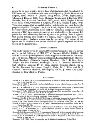
     In the present      study, the quantitative patterns of oocyte and follicular
growth     are   investigated during the period of infancy. Using this information,
a classification for the follicle types found in the infant ovary has been derived.
The classification is intended to form a basis for studies of the dynamics of
follicular growth in the human ovary.

                                MATERIALS AND METHODS
Ovaries were obtained at autopsy from infants and children ranging in age
from birth to 9 years. The specimens were fixed in Bouin's solution, embedded
in paraffin wax and sectioned at 7 µ . Sections were stained with either Harris
haematoxylin and eosin or Heidenhain's Azan.
   In order to determine the incidence and sizes of antral follicles in ovaries
at different ages, an initial survey was made of sectioned material obtained
from 400 subjects. From these, a group was selected consisting of nineteen

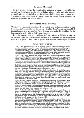
                                    EXPLANATION OF PLATE 1
       Fig. 1. Class A human oocytes (oo—9 to 25 ß diam.) in the transition stages of meiotic
       prophase. At birth, these were found either in the extreme outer cortex abutting the
       surface epithelium (s.e.) (Fig. la) or in the medullary region of the ovary (Fig. lb).
       After 2 months of age, Class A follicles were less abundant. They were mainly found in
       medullary nests, surrounded by cells which resembled those of the rete ovarii and were
       encapsulated by a complete basement membrane. Age 3 days; 910.
       Fig. 2. Class (right) and B/C (left) oocyte nuclei which have entered the resting stage of
       diplotene. Class follicles were surrounded by flattened granulosa cells with highly baso-
       philic nuclei. Class B/C follicles had both flattened and cuboidal granulosa cells. In
       general, the flattened cells surrounded one side of the oocyte and cuboidal cells the other.
       Whether the cuboidal cells arise from the flattened granulosa cells or represent a different
       cell population is unknown. Oocytes of follicles form the major portion of the oocyte
       population in the ovary at any time and constitute the resting pool from which future
       follicular growth will occur. They occurred in great abundance in the outer cortex at
       birth and thereafter appeared less commonly due not only to an increase in the size of
       the ovary but also to atresia (Baker, 1963). Age 5 years; 910.
       Fig. 3. A Class C oocyte surrounded by a single complete layer of cuboidal granulosa
       cells and a complete basement membrane which separates the granulosa layer from the
       adjacent stroma. The stroma surrounding the follicle formed a halo of connective tissue
       fibres and fibroblast-like cells. No definitive theca was present. The oocyte had not
       commenced growth but the number of follicle cells had increased. Class C follicles were
       located deeper in the cortex than those of Classes and B/C and were less frequently found
        than the latter. Age 7 years; 910.
       Fig. 4. A Class Dj follicle containing a growing oocyte surrounded by two to seven layers
       of granulosa cells. The zona pellucida is now present. Class Di follicles were located
       deeper in the cortex than those of any of the previous classes. Age 3 years; 910.
       Fig. 5. A Class D2 follicle. These follicles had a similar follicle and oocyte size range and
       location as Dt but were characterized by a granulosa cell layer containing fluid filled
       spaces and Call-Exner (ce.) vacuoles. Age 8 years;          910.
       Fig. 6. A Class E follicle. These follicles occurred less frequently than any of the previous
       follicle classes and were characterized by a definitive crescent-shaped antrum. The oocyte
       had ceased growth in the majority of cases. The wide distribution of follicle sizes (Text-
       fig. 5) in this class suggests that fluid accumulation may occur at any stage in follicular
       development after the formation of two or more layers of granulosa cells. The theca
        (th) may be well vascularized. Age 3 years; 910.
       Fig. 7. A Class F follicle. These follicles lay deep in the medullary region of the ovary and
       represented the final stage of follicle growth in the infant ovary. They were characterized
       by the development of a large fluid-filled antrum and the oocyte, which had ceased
       growth, was embedded in a cumulus of granulosa cells. In some cases, the cells of the theca
       might be markedly hypertrophied, taking on a partly luteinized appearance. Vasculariza-
       tion of the theca was advanced. Part of an atretic Class F follicle (a) is seen to the lower
       left. Note the undifferentiated thecal-type interstitial gland cells. Age 8 years; X 146.
PLATE 1


(Facing j). 54)
Follicular   development in the infant human ovary                                55
normal subjects ranging in age from birth to 9 years, who had died either from
misadventure (eight cases) or acute disease (eleven cases) in which the period
of illness was not longer than 48 hr and its nature considered unlikely to affect
the ovary. The follicle classification is based on measurements made on the
ovaries from this group. Ovary sections were examined under a Zeiss projec¬
tion microscope. Measurements were only made of those follicles which shoved
no apparent signs of atresia or post-mortem degeneration, i.e. the nuclei of
the granulosa cells and oocytes showed no signs of pyknosis and macrophages
were absent (see Baker, 1963). The diameters of the follicle, oocyte and oocyte
nucleus were then obtained using the method described by Mandi & Zucker-
man (1952) and the number of granulosa cells were counted in the widest
cross-section of the follicle. Approximately twenty representatives of each
follicle class were measured in each of the nineteen ovaries. This was not
possible with the larger follicles which occurred much less frequently than the
smaller ones and, in most cases, were atretic.

                                              RESULTS
Incidence and size of antral follicles
   The survey of 400 ovaries showed that the range of morphological types of


     Text-fig. 1. The relationship between oocyte diameter (µ ) and follicle diameter (ß )
     in the infant human ovary. During Phase A of growth, these are positively and signifi¬
     cantly correlated (r = +091; <0·001; y = 0·57 + 9-89—for abbreviations, see
     Table 1). During Phase B (y     = 0·001 + 80·70), the regression coefficient was not signi¬
     ficantly different from zero. The point of intersection of the lines of best fit for both sets of
     parameters was highly sensitive to small variations in both the regression coefficients, a,
     and intercepts, b. The points which fell around the intersection of the two lines were
     excluded from the calculations (Mandi & Zuckerman, 1952).
56                                           Sue Lintern-Moore         et   al.
follicles present in the ovary was similar regardless of the age of the subject.
Antral follicles were present in 60 % of the newborn ovaries. This frequency
declined between 9 and 28 days of life to 30%, but rose to 100% by the age
of 1 year. Thereafter, antral follicles were always found in the ovary through¬
out infancy and childhood. The mean diameter of antral follicles ranged from
0-2 mm to 6 mm and was independent of age.

Table 1. Coefficients of straight line regression relationships {y=ax + b) between
oocyte diameter(y) or oocyte nuclear diameter (y) and follicle diameter (x) and their
corresponding correlation coefficients (r) for Phase A of growth in twenty normal
            human female subjects aged between 3 days and 25 years

                                                                        No.
                          Oocyte diameter/follicle       diameter     ofpairs      Oocyte   nuclear diameter/follicle
                                     (Phase A)                          of                  diameter (Phase A)
                                                                      measure¬         r*             a          b
                                                                       ments

 3 days                      + 0-90           0-47        14-22         100          + 0-87         0-10         8-41
 14 days                     + 0-88           0-64        14-85         160          + 0-92         0-11         813
 7 weeks                     + 0-89           0-49        10-42         105          + 0-79         0-13         8-71
 3 months                    + 0-93           0-66         8-36         100          + 0-77         0-10        1219
 4 months                    + 0-80           0-53         7-09          98          + 0-76         009         10-09
 4J months                   + 0·94           0-58         9-16          97          + 0-82         0-11         9-50
 11 months                   + 0-94           0-51        13-21         120          + 0-85         0-11        10-11
 1 year                      + 0-98           0-44        15-11         118          + 0-90         0-14         8-30
 1 year 4 months             + 0-91           0-49        12-78         115          + 0-84         0-12         8-93
 1 year 9 months             + 0-84           0-52         8-84          90          + 0·72         0-09        10-05
 2 years                     + 0-97           0-69         9-97         160          + 0-88         0-14         8-81
 2 years                     + 0-99           0-57         9-77         100          + 0-94         0-15         8-18
 3 years                     + 0·96           0-48        15-39         180          + 0-66         0-09        10-97
 4 years                     + 0-97           0-52        13-30         103          4-0-78         0-19        11-55
 5 years                     + 0-87           0-49        11-61         100          + 0-89         0-13         9-07
 7 years                     + 0·97           0-48        15-78         100          + 0-87         0-09        12-69
 8 years                     + 0-95           0-52         9-83         190          + 0-94         0-15         8-98
 8 years                     + 0-95           0-52         8-01         102          4-0-94         0-11         9-33
 9 years                     + 0-97           0-55         9-52         100          4-0-94         0-13         9-05
 25 years                    + 0-99           0-55         9-32         100          + 0-93         0-10        11-05
 Pooled data
    Phase A                  + 0-91           0-57        9-89         2238          + 0-82         0-12         9-17
    Phase                                     0-001 i    80-70           35                         0-0004f 25-55
     *   All values of r significant   at   the   0-001% level.         Not significantly different from      zero.


   Values for a, b and r for pooled data           (excluding that from the 25-year-old subject)   are    also presented
for both Phases A and of growth.


Growth of the oocyte and follicle
   In order to establish the pattern of oocyte growth in relation to follicle
growth, oocyte and follicle diameters were compared using measurements
pooled from the nineteen ovaries studied (Text-fig. 1). Oocyte diameter and
follicle diameter were found to be positively and linearly correlated until the
oocyte reached a mean diameter of 80 µ (range 60 to 120 /mi) at a follicle
diameter of 124 µ (growth phase A). Thereafter, the oocyte ceased to grow
whilst follicular growth continued (growth phase B). A similar biphasic
relationship was found to exist between oocyte nuclear diameter and follicle
diameter. The oocyte nucleus reached a maximum diameter of 26 /tm (range
Follicular development in the infant human ovary           57
22 to 35 µ ) at a follicle diameter of 128 µ (Table 1). Thus, the oocyte and
its nucleus ceased growth at approximately the same stage of follicle develop¬
ment and their diameters were positively and linearly correlated with each
other (Text-fig. 2).
   The validity of pooling data from each of the different subjects has been
investigated in Table 1. The values of a (regression coefficient) and b (inter¬
cept) for Phase A of the relationships between oocyte and follicle diameter
and oocyte nuclear diameter and follicle diameter varied between individuals


      Text-fig. 2. Oocyte diameter (/an) and oocyte nuclear diameter (µ ) in the infant
      human ovary were positively and linearly correlated (r  =
                                                                +0-92; P< 0-001; y   =


      3-2X-43-7).


but   no  age trend could be detected. It was not possible to obtain sufficient
measurements     of follicle and oocyte diameters for Phase       of growth from
individual subjects since most of these follicles within each ovary were atretic.
   The relationship between follicle diameter and the number of granulosa
cells in the widest cross-section of the follicle was curvilinear (Text-fig. 3). In
follicles greater than 200 /¡m in diameter, the accumulation of follicular fluid
rather than the proliferation of the granulosa cells appeared to determine the
follicle size.
58                                 Sue Lintern-Moore      et   al.
Follicleclassification
  The   morphological pattern of follicular development in the infant ovary is
depicted in Text-fig. 4. In Text-figs 5 and 6, the pooled data from all the ovaries
are plotted as histograms to demonstrate the distribution of follicle classes in
relation to follicle and oocyte diameter. With the exception of Class A follicles
(PI. 1, Fig. 1), all of these types of follicles were found in the nineteen ovaries
studied.


      Text-fig. 3. The relationship between the number of granulosa cells in the widest cross-
      section of the follicle (logt0) and follicle diameter (ßm) in the infant human ovary.


  There    was a   considerable overlap in oocyte size between follicle classes.
Furthermore,     aparticular follicle size can encompass a number of morpho¬
logical classes. Fluid accumulation could commence in follicles with as few as
forty granulosa cells in the widest cross-section (Class / 2) and a defined
antrum when the number of cells reached 100 (Class E). Similarly, the oocyte
might cease growth (Text-fig. 1) at a follicle diameter which included those
of both Class D and E morphology (Text-fig. 5). This might include multi-
laminar follicles with no fluid accumulation.
   Based upon these observations, follicular growth in the infant human ovary
can be summarized as follows. The preliminary stages of oogénesis are com¬

pleted by 6 months post partum since oogonia and oocytes in early meiotic
prophase (Class A) have disappeared (PI. 1, Figs la and b). Class follicles
Text-fig. 4. Schematic representation of follicular growth in the ovary of infancy and
childhood. The classification of follicles was based upon follicle diameter, oocyte diameter,
the number of granulosa cells in the widest cross-section and follicular morphology.
Class A, which represents oocytes in the early pre-diplotene stages of meiotic pro-
phase (Pi. 1, Figs la and lb), is not included. Follicular growth occurred first during the
transition from Class to Class G. Oocyte growth commenced during the Class C/D to D
transition. Oocyte growth in general ceased during the progression from Class D to Class
E but in exceptional cases occurred as early as Class C/D (Text-fig. 6).


      100


      50
            -


                                       D,
                B/C            C/DI    D¡   D,

            26to50/j.m          5ltolOO/j.m                 101 to   200/¿m     201 to   6000µ/
                                        Follicle diameter

Text-fig. 5. The distribution of follicle classes in relation         to   follicle diameter   (ßm)   in
the infant human ovary.
60                                    Sue Lintern-Moore    et   al.
(PI. 1, Fig. 2)   are in the resting stage of prophase (diplotene) and form the

major portion     of the oocyte population at any age. They constitute a pool
from which follicular growth will occur throughout life. Follicular growth is
first evident from the multiplication of granulosa cells in Class B/C and C
follicles (PL 1, Figs 2 and 3) and oocyte growth begins in Class C/D (Text-
fig. 4). With further granulosa cell proliferation and oocyte growth, follicles
become Class D type in which fluid may begin to accumulate between the cells
of the follicular envelope and thecal tissue to differentiate (PI. 1, Figs 4 and 5).
With the appearance of a definitive antrum, the follicles are designated as
Class E (PI. 1, Fig. 6). During the Class D to E transition, oocyte growth
ceases (Text-figs 1 and 6). Class F follicles show further fluid accumulation
and the thecal cells may be markedly hypertrophied. This class represents the
largest follicle type present in the infant ovary (PI. 1, Fig. 7).

          100


           50


                   B/C     C/D
                                                                             C/D   D2
                                                 C/D           D2            I—
                         13 to 39   /¿m             40 to 80    µ             81 to 120   ¿¿m


                                            Oocyte diameter
     Text-fig. 6. The distribution of follicle classes in relation    to   oocyte diameter (µ ) in
     the infant human ovary.


                                          DISCUSSION
The high incidence of antral follicles in the ovaries of newborn girls appears
to be unrelated to any pathological state in mother or child. Ahlvin & Bauer
(1957) suggested a relationship between the presence of antral follicles in the
newborn ovary and diabetes in the mother, but this was not supported by the
more extensive survey of Kraus & Neubecker (1962). Furthermore, Spivak

(1934) and Ober & Bernstein (1955) found no correlation between hyper-
plasia and hypertrophy of the newborn uterine endometrium and the presence
of antral follicles in the ovary. They concluded that although the morphology
of the newborn uterus may reflect maternal steroid levels, the degree of ovarian
development was independent of these factors. Advanced stages of follicular
growth are also found in the ovaries of other newborn mammals which are
born in a comparatively precocious state (Kellas, Van Lennep & Amoroso,
1958; Perry & Rowlands, 1962; van Wagenen & Simpson, 1965).
Folliculardevelopment in the infant human ovary            61
   As in other mammals (Brambell, 1928; Parkes, 1932; Zuckerman & Parkes,
 1932), the pattern of oocyte growth in relation to follicular growth in the
infant human ovary is biphasic (Text-fig. 1) and individual (although not
age-dependent) variations may exist in both the regression coefficient, a, and
the intercept, b, for phase A of growth (Table 1). Similar individual variations
also occur in the rhesus monkey and the rat (Green & Zuckerman, 1947;
 Mandi & Zuckerman, 1952). In the human infant, the oocyte reaches a
maximum diameter of 80 µ (range 60 to 120 /tin) at a follicular diameter of
 124 µ . Thereafter, follicular growth continues whilst the rate of oocyte
growth approaches zero. The value for a in phase A of growth (0-57) is within
the range recorded for other mammals, i.e. 0-14 for the pig and 0-64 for the
shrew (see Parkes, 1932; Green & Zuckerman, 1947). Furthermore, the overall
values for both a and b for phase A for the infant ovary did not differ signifi¬
cantly from those obtained for the single adult ovary studied during the
course of this work (Table 1) or those obtained by Green & Zuckerman (1951)
for a 6-year-old ovary. The close similarity in many different mammalian
species between the maximum diameter achieved by the oocyte at the end of
growth phase A and the corresponding follicle diameter has been noted
previously (Parkes, 1932; Brambell, 1956). The possible functional significance
of this observation in relation to synthetic activity of the oocyte nucleus has
been discussed elsewhere (Moore & Lintern-Moore, 1974).
   No evidence for ovulation was found in any of the 400 ovaries studied. The
maximum diameter achieved by the oocyte during infancy (60 to 120 µ ) is
less than that (120 to 190 µ ) reported for freshly ovulated human ova (Pincus
 & Saunders, 1937; Hamilton, 1944). Although some of the disparity in size
can be accounted for
                           by shrinkage on fixation (Hamilton, 1944; Menkin &
Rock, 1948), it is possible that the fully grown oocyte of infancy does not
develop to the preovulatory dimensions found in the adult. Similarly, the
maximum diameter of antral follicles encountered routinely in the infant
ovary (5 to 6 mm) is considerably less than the values of 10 to 15 mm reported
for a full-sized adult follicle (Watzka, 1957). Thus, it is concluded that antral
follicles in the infant human ovary do not achieve preovulatory size and all of
the apparently normal growing follicles seen, in the absence of a resting stage
beyond Class B, must, at some stage in development, degenerate. Certainly,
all classes of follicles described for the infant ovary were encountered in atretic
forms (see also Potter, 1963; Valdes-Dapena, 1967) and the pattern of follicular
atresia appears to resemble closely that described for non-ovulatory follicles
in the adult Macaca mulatta (Koering, 1969).
   The data indicate that follicular growth and degeneration in the human
ovary occurs continuously throughout infancy and childhood. It may be
pertinent to consider whether a functional association between the gonadal-
pituitary-hypothalamic axis is also present. Normal prepubertal children have
circulating gonadotrophin levels which are considerably lower than those
recorded during puberty and adulthood. They are, nevertheless, in excess of
those recorded in hypo-pituitary subjects, lower than those of 5- to 10-year-
old subjects with gonadal dysgenesis and higher in females than in males.
Similarly, although oestradiol-17/? levels are also very low, prepubertal subjects
62                                 Sue Lintern-Moore      et   al.
appear to be more sensitive to low doses of ethinyl oestradiol (as reflected by
FSH excretion) than do those who have reached puberty (Kulin, Grumbach &
Kaplan, 1969; Buckler & Clayton, 1970; Penny, Guyda, Baghdassarian,
Johanson & Blizzard, 1970; Root, Moshang, Bongiovanni & Eberlein, 1970;
Sizoneko, Burr, Kaplan & Grumbach, 1970; Jenner, Kelch, Kaplan & Grum¬
bach, 1972; Kelch, Grumbach & Kaplan, 1972; Lee, Midgley & Jaffe, 1972).
These data suggest that a gonadal-pituitary relationship may exist during the
prepubertal period. To support this, Roth, Kelch, Kaplan & Grumbach (1972)
have shown that LH-releasing factor (LH-RF) stimulates the release of similar
amounts of FSH in prepubertal, pubertal and adult subjects. By contrast, LH
production and release only became significant at puberty. Thus it appears
that during infancy and childhood a special, highly sensitive form of the
gonadal-pituitary feedback system may be operating. The nature of this
system and its relation to the pattern of follicular growth reported here is not
known at the present time.
                                     ACKNOWLEDGMENTS
This   study was supported by the NATO Scientific Committee and was carried
out in partial fulfilment of EURATOM Contract 120-73-1 BIODK. We
wish to thank the pathologists who kindly supplied ovaries used in this study :
Dr J. M. Bouton, Alderhey Children's Hospital, Liverpool; Dr H. B. Marsden,
Royal Manchester Children's Hospital, Manchester; Dr A. D. Bain, Royal
Hospital for Sick Children, Edinburgh; Dr A. E. Claireaux, Hospital for
Sick Children, London; Dr G. Molz, Kanton Hospital, Zürich; Dr .
Barzilai, Rambam Government Hospital, Haifa; Dr G. Kohn and Dr J.
Chatten, Children's Hospital, Philadelphia; Dr M. J. Barry, Our Lady's
Hospital for Sick Children, Dublin.
                                           REFERENCES
Ahlvin,  R. C. & Bauer, W. C. (1957) Luteinized cysts in ovaries of infants born of diabetic mothers.
       Am. J. Dis. Child. 93, 107.
Baker, T. G. (1963) A quantitative and cytological study of germ cells in human ovaries. Proc. R.
       Soc. B, 158, 417.
Boyd, J. D. & Hamilton, W. J. (1955) The cellular components of the human ovary. In Modern Trends
       in Obstetrics and Gynaecology, pp. 50-78. Ed. K. Bowis. Butterworths, London.
Brambell, F. W. R. (1928) The development and morphology of the gonads of the mouse. Part III.
       The growth of the follicles. Proc. R. Soc. B, 103,258.
Brambell, F. W. R. (1956) Ovarian changes. In Marshall's Physiology of Reproduction, Vol. 1, Part 1,
       pp. 455—459. Ed. A. S. Parkes. Longmans Green, London.
Buckler, J. M. H. & Clayton, B. E. (1970) Output of luteinizing hormone in the urine of normal
       children and those with advanced sexual development. Archs Dis. Childh. 45, 478.
Curtis, E. M. (1962) Normal ovarian histology in infancy and childhood. Obstet. Gynec, N.T. 19, 444
Delson, B., Lubin, S. & Reynolds, S. R. M. (1949) Vascular patterns in the human ovary. Am. J.
       Obstet. Gynec. 57, 842.
Forbes, T. R. (1942) On the fate of the medullary cords of the human ovary. Contr. Embryol. 30, 9.
Govan, A. D. T. & Mukherjee, G. L. (1950) Maternal toxaemia and foetal ovarian activity. J.
       Obstet. Gynaec. Br. Emp. 57, 525.
Green, S. H. & Zuckerman, S. (1947) A comparison of the growth of the ovum and follicle in normal
       rhesus monkeys and in monkeys treated with oestrogen and androgens. J. Endocr. 5, 207.
Green, S. H. & Zuckerman, S. (1951) Quantitative aspects of the growth of the human ovum and
       follicle. J. Anat. 85, 373.
Hamilton, W. J. (1944) Phases of maturation and fertilisation in human ova. J. Anat. 78, 1.
Follicular   development in the infant human ovary                         63
Jenner, M. R., Kelch, R. P., Kaplan, S. L. & Grumbach, M. M. (1972) Hormonal changes in
      puberty. IV. Plasma estradiol LH and FSH in prepubertal children, prepubertal females and in
      precocious puberty, premature thelarche, hypogonadism and in a child with feminizing ovarian
      tumor. J. clin. Endocr. Metab. 34, 521.
Kelch, R. P., Grumbach, M. M. & Kaplan, S. L. (1972) Studies on the mechanism of puberty in
      man. In Gonadotropins, pp. 524-534. Eds. B. B. Saxena, G. G. Beling and H. M. Gandy. Wiley-

       Interscience, New York.
Kellas, L. M., Van Lennep, E. W. & Amoroso, E. C. (1958) Ovaries of some foetal and prepubertal
      giraffe (Giraffa camelopardalis Linnaeus). Nature, Land. 181, 487.
Koering, M. J. (1969) Cyclic changes in ovarian morphology during the menstrual cycle in Macaca
      mulatta. Am. J. Anat. 126, 73.
Kraus, F. T. & Neubecker, R. D. (1962) Luteinization of the ovarian theca in infants and children.
      Am. J. clin. Path. 37, 389.
Kulin, H. E., Grumbach, M. M. & Kaplan, S. L. (1969) Changing sensitivity of the pubertal gonadal
      hypothalamic feedback mechanism in man. Science, N.T. 166, 1012.
Lee, P. ., Midgley, A. R. & Jaffe, R. B. (1972) Regulation of human gonadotropins. VI. Serum
        follicle stimulating and luteinizing hormone determinations in children. J. clin. Endocr. Metab.
        31, 248.
Mandl, A. M. & Zuckerman, S. (1952) The growth of the oocyte and follicle in the adult rat. J.
        Endocr. 8, 126.
Menkin, M. F. & Rock, J. (1948) In vitro fertilisation and cleavage of human ovarian eggs. Am. J.
        Obstet. Gynec. 55, 440.
Merrill, J. A. (1963) The morphology of the prepubertal ovary: relationship to the polycystic ovary
       syndrome. Sth. med. J., Nashville, 56, 225.
Moore, G. P. M. & Lintern-Moore, S. (1974) A correlation between growth and RNA synthesis in
        mouse oocyte. J. Reprod. Fert. 39, 163.

Mossman, H. W. & Duke, K. L. (1973) Comparative Morphology of the Mammalian Ovary, pp. 100-116.
        University of Wisconsin Press.
Mossman, H. W., Koering, M. J. & Ferry, D. (1964) Cyclic changes of interstitial gland tissue of
        the human ovary. Am. J. Anat. 115, 235.
Ober, W. B. & Bernstein, I. (1955) Observations on the endometrium and ovary in the newborn.
       Pediatrics, Springfield, 16, 445.
Parkes, A. S. (1932) The reproductive processes of certain mammals. II. The size of the Graafian
       follicle at ovulation. Proc. R. Soc. B, 109, 185.
Penny, R., Guyda, H., Baghdassarian, ., Johanson, A. J. & Blizzard, R. M. (1970) Correlation
       of serum follicular stimulating hormone (FSH) and luteinizing hormone (LH) as measured by
       radioimmunoassay in disorders of sexual development. J. clin. Invest. 49, 1847.
Perry, J. S. & Rowlands, I. W. (1962) The ovarian cycle in vertebrates. In The Ovary, Chap. 5,
       p. 275. Ed. S. Zuckerman. Academic Press, New York.
Pincus, G. & Saunders, B. (1937) Unfertilized human tubai ova. Anat. Ree. 69, 163.
Polhemus, M. D. (1953) Ovarian maturation and cyst formation in children. Pediatrics, Springfield,
       11, 588.
Potter, E. L. (1963) The ovary in infancy and childhood. In The Ovary, pp. 11-23. Eds. A. G. Grady
       and D. E. Smith. Int. Acad. Path. Monograph. Saunders, Philadelphia.
Root, A. W., Moshang, T., Bongiovanni, . M. & Eberlein, W. R. (1970) Concentrations of plasma
       luteinizing hormone in infants, children and adolescents with normal and abnormal gonadal
       function. Pediat. Res. 4, 175.
Roth, J. C, Kelch, R. P., Kaplan, S. L. & Grumbach, M. M. (1972) FSH and LH response to
       luteinizing-hormone-releasing factor (LRF) in prepubertal and pubertal children, adult males
       and patients with hypogonadotropic and hypergonadotropic hypogonadism. J. clin. Endocr.
       Metab. 35, 926.
Sauramo, H. (1954) Histology and function of the ovary from the embryonic period to the fertile age.
       Acta obstet, gynec. scand. 33, Suppl. 2, 1.
Shaw, W. (1925) The fate of the Graafian follicle in the human ovary. J. Obstet. Gynaec. Br. Emp. 3, 679.
Simkins, G. S. (1932) Development of the human ovary from birth to sexual maturity. Am. J. Anat. 51,
       465.
Sizoneko,     P.         I. M., Kaplan, S. L. & Grumbach, M. M. (1970) Hormonal changes in
                   C., Burr,
       puberty. II. Correlation of serum luteinizing hormone and follicle stimulating hormone with
      stages of puberty and bone age in normal girls. Pediat. Res. 4, 36.
Spivak, M. (1934) Polycystic ovaries in the newborn and early infancy and their relation to the
      structure of the endometrium. Am. J. Obstet. Gynec. 27, 157.
Stevens, T. G. (1903) The fate of the ovum and graafian follicle in pre-menstrual life. Obstet. Trans.
       14, 465.
64                                 Sue Lintern-Moore     et   al.
Valdes-Dapena, . . (1967) The normal ovary of childhood. Ann. N.T. Acad. Sci. 142, 597.
van Wagenen, G. & Simpson, M. F. (1965) Embryology of the Ovary and Testis. Homo sapiens and Macaca
      mulatta. Yale University Press, New Haven and London.
Watzka, M. (1957) Weiblich genitalorgane. Das Ovarium. In Handbuch der mikroskopischen Anatomie
      des Menschen, Vol. 7, pp. 1-178. Eds. W. V. Möllendorf and W. Bargmann. Springer, Berlin.
Winter, G. F. (1962) Follikel-Zählungen und den Eierstöcken gesunder, nicht-schwangerer weiblicher
      Personen. £«iiM. Gynäk. 47, 1824.
Zuckerman, S. & Parkes, A. S. (1932) The menstrual cycle of the primates. Part V. The cycle of
      the baboon. Proc. zool. Soc. Lond. 1932, 139.

More Related Content

What's hot

History of embryology in plants
History of embryology in plantsHistory of embryology in plants
History of embryology in plants
Himanshi Chauhan
 
Developmental biology course presentation
Developmental biology course presentationDevelopmental biology course presentation
Developmental biology course presentation
Virginie Mournetas
 
Monocot and dicot navi
Monocot and dicot naviMonocot and dicot navi
Monocot and dicot navi
naveenagirish
 
Bio 130 ovule dev. ist sem 2013 2014
Bio 130 ovule dev. ist sem 2013 2014Bio 130 ovule dev. ist sem 2013 2014
Bio 130 ovule dev. ist sem 2013 2014
tralala 1412
 
6. reproduction
6. reproduction6. reproduction
6. reproduction
Mwansa Mulenga
 
History of embryology
History of embryologyHistory of embryology
History of embryology
Arizaldo Castro
 
Cell fate (fate maps)
Cell fate (fate maps)Cell fate (fate maps)
Cell fate (fate maps)
Shashank Patel
 
Megasporogenesis and megagametogenesis
Megasporogenesis and megagametogenesisMegasporogenesis and megagametogenesis
Megasporogenesis and megagametogenesis
SACHIN KUMAR
 
Processes on animal development
Processes on animal developmentProcesses on animal development
Processes on animal development
Elaine Sebastian
 
Axis and pattern formation in amphibia
Axis and pattern formation in amphibiaAxis and pattern formation in amphibia
Axis and pattern formation in amphibia
ShariqaJan
 
Plant embryo development
Plant embryo developmentPlant embryo development
Plant embryo development
Baljinder Gill
 
Types of embryo sac
Types of embryo sacTypes of embryo sac
Types of embryo sac
VasantaKahalkar
 
Meaning of seed habit of pre ovule
Meaning of seed habit of pre ovuleMeaning of seed habit of pre ovule
Meaning of seed habit of pre ovule
University of Allahabad
 
Drosophila lecture
Drosophila lectureDrosophila lecture
Drosophila lecture
--
 
History &amp; basic concepts db
History &amp; basic concepts  dbHistory &amp; basic concepts  db
History &amp; basic concepts db
anusha rajan
 
Gametogenisis, Fertilization
Gametogenisis, FertilizationGametogenisis, Fertilization
Gametogenisis, Fertilization
Smita Mahendrakar
 
Cleavage
CleavageCleavage
Cleavage
Shoeb Ahmad
 
Types of endosperm
Types of endospermTypes of endosperm
Types of endosperm
TRIDIP BORUAH
 
CAS Biology 11.4 Human Reproduction Part 1
CAS Biology 11.4 Human Reproduction Part 1CAS Biology 11.4 Human Reproduction Part 1
CAS Biology 11.4 Human Reproduction Part 1
Ryan Dougherty
 
Embryology 1
Embryology 1Embryology 1

What's hot (20)

History of embryology in plants
History of embryology in plantsHistory of embryology in plants
History of embryology in plants
 
Developmental biology course presentation
Developmental biology course presentationDevelopmental biology course presentation
Developmental biology course presentation
 
Monocot and dicot navi
Monocot and dicot naviMonocot and dicot navi
Monocot and dicot navi
 
Bio 130 ovule dev. ist sem 2013 2014
Bio 130 ovule dev. ist sem 2013 2014Bio 130 ovule dev. ist sem 2013 2014
Bio 130 ovule dev. ist sem 2013 2014
 
6. reproduction
6. reproduction6. reproduction
6. reproduction
 
History of embryology
History of embryologyHistory of embryology
History of embryology
 
Cell fate (fate maps)
Cell fate (fate maps)Cell fate (fate maps)
Cell fate (fate maps)
 
Megasporogenesis and megagametogenesis
Megasporogenesis and megagametogenesisMegasporogenesis and megagametogenesis
Megasporogenesis and megagametogenesis
 
Processes on animal development
Processes on animal developmentProcesses on animal development
Processes on animal development
 
Axis and pattern formation in amphibia
Axis and pattern formation in amphibiaAxis and pattern formation in amphibia
Axis and pattern formation in amphibia
 
Plant embryo development
Plant embryo developmentPlant embryo development
Plant embryo development
 
Types of embryo sac
Types of embryo sacTypes of embryo sac
Types of embryo sac
 
Meaning of seed habit of pre ovule
Meaning of seed habit of pre ovuleMeaning of seed habit of pre ovule
Meaning of seed habit of pre ovule
 
Drosophila lecture
Drosophila lectureDrosophila lecture
Drosophila lecture
 
History &amp; basic concepts db
History &amp; basic concepts  dbHistory &amp; basic concepts  db
History &amp; basic concepts db
 
Gametogenisis, Fertilization
Gametogenisis, FertilizationGametogenisis, Fertilization
Gametogenisis, Fertilization
 
Cleavage
CleavageCleavage
Cleavage
 
Types of endosperm
Types of endospermTypes of endosperm
Types of endosperm
 
CAS Biology 11.4 Human Reproduction Part 1
CAS Biology 11.4 Human Reproduction Part 1CAS Biology 11.4 Human Reproduction Part 1
CAS Biology 11.4 Human Reproduction Part 1
 
Embryology 1
Embryology 1Embryology 1
Embryology 1
 

Similar to Lintern moore

The Early Development of Mammalian.pptx
The Early  Development of Mammalian.pptxThe Early  Development of Mammalian.pptx
The Early Development of Mammalian.pptx
AminaShahzadiShahzad1
 
Placenta and amniotic fluid
Placenta and amniotic fluidPlacenta and amniotic fluid
Placenta and amniotic fluid
Shail Pandher
 
Fetus Papryaceus III - Kirsch
Fetus Papryaceus III - KirschFetus Papryaceus III - Kirsch
Fetus Papryaceus III - Kirsch
Kristen Kirsch
 
embryologyofliver-170215140438.pptx
embryologyofliver-170215140438.pptxembryologyofliver-170215140438.pptx
embryologyofliver-170215140438.pptx
nidhi sharma
 
Eng follicle u environ - west
Eng follicle u environ - westEng follicle u environ - west
Eng follicle u environ - west
balupost
 
Relative Morphology of Extraembryonic Membranes in Mammals: Their Roles in Hi...
Relative Morphology of Extraembryonic Membranes in Mammals: Their Roles in Hi...Relative Morphology of Extraembryonic Membranes in Mammals: Their Roles in Hi...
Relative Morphology of Extraembryonic Membranes in Mammals: Their Roles in Hi...
Joseph Holson
 
Embryology of liver
Embryology of liverEmbryology of liver
Embryology of liver
Pratap Tiwari
 
Campbell hypospadia
Campbell hypospadiaCampbell hypospadia
Campbell hypospadia
FickyHuang1
 
Embryo implantation 7
Embryo    implantation 7Embryo    implantation 7
Embryo implantation 7
鋒博 蔡
 
Reprodactive Animal editted.pptx
Reprodactive Animal editted.pptxReprodactive Animal editted.pptx
Reprodactive Animal editted.pptx
MarkosKuma2
 
Reproductive traits in dicyemids
Reproductive traits in dicyemidsReproductive traits in dicyemids
Reproductive traits in dicyemids
dreicash
 
5_6282657037939640532.pptx
5_6282657037939640532.pptx5_6282657037939640532.pptx
5_6282657037939640532.pptx
Srinath Chowdary
 
compress_developmentofthemalereproductivesystem-170422070219 (1)_202301050308...
compress_developmentofthemalereproductivesystem-170422070219 (1)_202301050308...compress_developmentofthemalereproductivesystem-170422070219 (1)_202301050308...
compress_developmentofthemalereproductivesystem-170422070219 (1)_202301050308...
MujahedAldhabyani
 
Development of the male reproductive system
Development of the male reproductive systemDevelopment of the male reproductive system
Development of the male reproductive system
Sahar Hafeez
 
Umblical cord presentation
Umblical cord presentationUmblical cord presentation
Umblical cord presentation
DR. SUDHIR KUMAR SHUKLA
 
9. origins of multicellularity &amp; phylum porifera part I in clear explanation
9. origins of multicellularity &amp; phylum porifera part I in clear explanation9. origins of multicellularity &amp; phylum porifera part I in clear explanation
9. origins of multicellularity &amp; phylum porifera part I in clear explanation
Yo yo Nody khan
 
Gametogenesis conversion of germ cells into male and female gametes.ppt
Gametogenesis conversion of germ cells into male and female gametes.pptGametogenesis conversion of germ cells into male and female gametes.ppt
Gametogenesis conversion of germ cells into male and female gametes.ppt
Jwan AlSofi
 
Early Development of Mammals (Mouse and Human).pdf
Early Development of Mammals (Mouse and Human).pdfEarly Development of Mammals (Mouse and Human).pdf
Early Development of Mammals (Mouse and Human).pdf
Department of Education Philippines
 
Morfologia cromatina mayo10[1]
Morfologia cromatina mayo10[1]Morfologia cromatina mayo10[1]
Morfologia cromatina mayo10[1]
amyoliveros
 
human development embryo and fetus.p.p.t.pptx
human development embryo and fetus.p.p.t.pptxhuman development embryo and fetus.p.p.t.pptx
human development embryo and fetus.p.p.t.pptx
PAK ACADEMY
 

Similar to Lintern moore (20)

The Early Development of Mammalian.pptx
The Early  Development of Mammalian.pptxThe Early  Development of Mammalian.pptx
The Early Development of Mammalian.pptx
 
Placenta and amniotic fluid
Placenta and amniotic fluidPlacenta and amniotic fluid
Placenta and amniotic fluid
 
Fetus Papryaceus III - Kirsch
Fetus Papryaceus III - KirschFetus Papryaceus III - Kirsch
Fetus Papryaceus III - Kirsch
 
embryologyofliver-170215140438.pptx
embryologyofliver-170215140438.pptxembryologyofliver-170215140438.pptx
embryologyofliver-170215140438.pptx
 
Eng follicle u environ - west
Eng follicle u environ - westEng follicle u environ - west
Eng follicle u environ - west
 
Relative Morphology of Extraembryonic Membranes in Mammals: Their Roles in Hi...
Relative Morphology of Extraembryonic Membranes in Mammals: Their Roles in Hi...Relative Morphology of Extraembryonic Membranes in Mammals: Their Roles in Hi...
Relative Morphology of Extraembryonic Membranes in Mammals: Their Roles in Hi...
 
Embryology of liver
Embryology of liverEmbryology of liver
Embryology of liver
 
Campbell hypospadia
Campbell hypospadiaCampbell hypospadia
Campbell hypospadia
 
Embryo implantation 7
Embryo    implantation 7Embryo    implantation 7
Embryo implantation 7
 
Reprodactive Animal editted.pptx
Reprodactive Animal editted.pptxReprodactive Animal editted.pptx
Reprodactive Animal editted.pptx
 
Reproductive traits in dicyemids
Reproductive traits in dicyemidsReproductive traits in dicyemids
Reproductive traits in dicyemids
 
5_6282657037939640532.pptx
5_6282657037939640532.pptx5_6282657037939640532.pptx
5_6282657037939640532.pptx
 
compress_developmentofthemalereproductivesystem-170422070219 (1)_202301050308...
compress_developmentofthemalereproductivesystem-170422070219 (1)_202301050308...compress_developmentofthemalereproductivesystem-170422070219 (1)_202301050308...
compress_developmentofthemalereproductivesystem-170422070219 (1)_202301050308...
 
Development of the male reproductive system
Development of the male reproductive systemDevelopment of the male reproductive system
Development of the male reproductive system
 
Umblical cord presentation
Umblical cord presentationUmblical cord presentation
Umblical cord presentation
 
9. origins of multicellularity &amp; phylum porifera part I in clear explanation
9. origins of multicellularity &amp; phylum porifera part I in clear explanation9. origins of multicellularity &amp; phylum porifera part I in clear explanation
9. origins of multicellularity &amp; phylum porifera part I in clear explanation
 
Gametogenesis conversion of germ cells into male and female gametes.ppt
Gametogenesis conversion of germ cells into male and female gametes.pptGametogenesis conversion of germ cells into male and female gametes.ppt
Gametogenesis conversion of germ cells into male and female gametes.ppt
 
Early Development of Mammals (Mouse and Human).pdf
Early Development of Mammals (Mouse and Human).pdfEarly Development of Mammals (Mouse and Human).pdf
Early Development of Mammals (Mouse and Human).pdf
 
Morfologia cromatina mayo10[1]
Morfologia cromatina mayo10[1]Morfologia cromatina mayo10[1]
Morfologia cromatina mayo10[1]
 
human development embryo and fetus.p.p.t.pptx
human development embryo and fetus.p.p.t.pptxhuman development embryo and fetus.p.p.t.pptx
human development embryo and fetus.p.p.t.pptx
 

Recently uploaded

Energy Efficient Video Encoding for Cloud and Edge Computing Instances
Energy Efficient Video Encoding for Cloud and Edge Computing InstancesEnergy Efficient Video Encoding for Cloud and Edge Computing Instances
Energy Efficient Video Encoding for Cloud and Edge Computing Instances
Alpen-Adria-Universität
 
Deep Dive: AI-Powered Marketing to Get More Leads and Customers with HyperGro...
Deep Dive: AI-Powered Marketing to Get More Leads and Customers with HyperGro...Deep Dive: AI-Powered Marketing to Get More Leads and Customers with HyperGro...
Deep Dive: AI-Powered Marketing to Get More Leads and Customers with HyperGro...
saastr
 
Monitoring and Managing Anomaly Detection on OpenShift.pdf
Monitoring and Managing Anomaly Detection on OpenShift.pdfMonitoring and Managing Anomaly Detection on OpenShift.pdf
Monitoring and Managing Anomaly Detection on OpenShift.pdf
Tosin Akinosho
 
TrustArc Webinar - 2024 Global Privacy Survey
TrustArc Webinar - 2024 Global Privacy SurveyTrustArc Webinar - 2024 Global Privacy Survey
TrustArc Webinar - 2024 Global Privacy Survey
TrustArc
 
Taking AI to the Next Level in Manufacturing.pdf
Taking AI to the Next Level in Manufacturing.pdfTaking AI to the Next Level in Manufacturing.pdf
Taking AI to the Next Level in Manufacturing.pdf
ssuserfac0301
 
Nordic Marketo Engage User Group_June 13_ 2024.pptx
Nordic Marketo Engage User Group_June 13_ 2024.pptxNordic Marketo Engage User Group_June 13_ 2024.pptx
Nordic Marketo Engage User Group_June 13_ 2024.pptx
MichaelKnudsen27
 
Webinar: Designing a schema for a Data Warehouse
Webinar: Designing a schema for a Data WarehouseWebinar: Designing a schema for a Data Warehouse
Webinar: Designing a schema for a Data Warehouse
Federico Razzoli
 
June Patch Tuesday
June Patch TuesdayJune Patch Tuesday
June Patch Tuesday
Ivanti
 
Columbus Data & Analytics Wednesdays - June 2024
Columbus Data & Analytics Wednesdays - June 2024Columbus Data & Analytics Wednesdays - June 2024
Columbus Data & Analytics Wednesdays - June 2024
Jason Packer
 
UI5 Controls simplified - UI5con2024 presentation
UI5 Controls simplified - UI5con2024 presentationUI5 Controls simplified - UI5con2024 presentation
UI5 Controls simplified - UI5con2024 presentation
Wouter Lemaire
 
OpenID AuthZEN Interop Read Out - Authorization
OpenID AuthZEN Interop Read Out - AuthorizationOpenID AuthZEN Interop Read Out - Authorization
OpenID AuthZEN Interop Read Out - Authorization
David Brossard
 
5th LF Energy Power Grid Model Meet-up Slides
5th LF Energy Power Grid Model Meet-up Slides5th LF Energy Power Grid Model Meet-up Slides
5th LF Energy Power Grid Model Meet-up Slides
DanBrown980551
 
Artificial Intelligence for XMLDevelopment
Artificial Intelligence for XMLDevelopmentArtificial Intelligence for XMLDevelopment
Artificial Intelligence for XMLDevelopment
Octavian Nadolu
 
Recommendation System using RAG Architecture
Recommendation System using RAG ArchitectureRecommendation System using RAG Architecture
Recommendation System using RAG Architecture
fredae14
 
Salesforce Integration for Bonterra Impact Management (fka Social Solutions A...
Salesforce Integration for Bonterra Impact Management (fka Social Solutions A...Salesforce Integration for Bonterra Impact Management (fka Social Solutions A...
Salesforce Integration for Bonterra Impact Management (fka Social Solutions A...
Jeffrey Haguewood
 
Programming Foundation Models with DSPy - Meetup Slides
Programming Foundation Models with DSPy - Meetup SlidesProgramming Foundation Models with DSPy - Meetup Slides
Programming Foundation Models with DSPy - Meetup Slides
Zilliz
 
Presentation of the OECD Artificial Intelligence Review of Germany
Presentation of the OECD Artificial Intelligence Review of GermanyPresentation of the OECD Artificial Intelligence Review of Germany
Presentation of the OECD Artificial Intelligence Review of Germany
innovationoecd
 
Best 20 SEO Techniques To Improve Website Visibility In SERP
Best 20 SEO Techniques To Improve Website Visibility In SERPBest 20 SEO Techniques To Improve Website Visibility In SERP
Best 20 SEO Techniques To Improve Website Visibility In SERP
Pixlogix Infotech
 
Skybuffer SAM4U tool for SAP license adoption
Skybuffer SAM4U tool for SAP license adoptionSkybuffer SAM4U tool for SAP license adoption
Skybuffer SAM4U tool for SAP license adoption
Tatiana Kojar
 
Ocean lotus Threat actors project by John Sitima 2024 (1).pptx
Ocean lotus Threat actors project by John Sitima 2024 (1).pptxOcean lotus Threat actors project by John Sitima 2024 (1).pptx
Ocean lotus Threat actors project by John Sitima 2024 (1).pptx
SitimaJohn
 

Recently uploaded (20)

Energy Efficient Video Encoding for Cloud and Edge Computing Instances
Energy Efficient Video Encoding for Cloud and Edge Computing InstancesEnergy Efficient Video Encoding for Cloud and Edge Computing Instances
Energy Efficient Video Encoding for Cloud and Edge Computing Instances
 
Deep Dive: AI-Powered Marketing to Get More Leads and Customers with HyperGro...
Deep Dive: AI-Powered Marketing to Get More Leads and Customers with HyperGro...Deep Dive: AI-Powered Marketing to Get More Leads and Customers with HyperGro...
Deep Dive: AI-Powered Marketing to Get More Leads and Customers with HyperGro...
 
Monitoring and Managing Anomaly Detection on OpenShift.pdf
Monitoring and Managing Anomaly Detection on OpenShift.pdfMonitoring and Managing Anomaly Detection on OpenShift.pdf
Monitoring and Managing Anomaly Detection on OpenShift.pdf
 
TrustArc Webinar - 2024 Global Privacy Survey
TrustArc Webinar - 2024 Global Privacy SurveyTrustArc Webinar - 2024 Global Privacy Survey
TrustArc Webinar - 2024 Global Privacy Survey
 
Taking AI to the Next Level in Manufacturing.pdf
Taking AI to the Next Level in Manufacturing.pdfTaking AI to the Next Level in Manufacturing.pdf
Taking AI to the Next Level in Manufacturing.pdf
 
Nordic Marketo Engage User Group_June 13_ 2024.pptx
Nordic Marketo Engage User Group_June 13_ 2024.pptxNordic Marketo Engage User Group_June 13_ 2024.pptx
Nordic Marketo Engage User Group_June 13_ 2024.pptx
 
Webinar: Designing a schema for a Data Warehouse
Webinar: Designing a schema for a Data WarehouseWebinar: Designing a schema for a Data Warehouse
Webinar: Designing a schema for a Data Warehouse
 
June Patch Tuesday
June Patch TuesdayJune Patch Tuesday
June Patch Tuesday
 
Columbus Data & Analytics Wednesdays - June 2024
Columbus Data & Analytics Wednesdays - June 2024Columbus Data & Analytics Wednesdays - June 2024
Columbus Data & Analytics Wednesdays - June 2024
 
UI5 Controls simplified - UI5con2024 presentation
UI5 Controls simplified - UI5con2024 presentationUI5 Controls simplified - UI5con2024 presentation
UI5 Controls simplified - UI5con2024 presentation
 
OpenID AuthZEN Interop Read Out - Authorization
OpenID AuthZEN Interop Read Out - AuthorizationOpenID AuthZEN Interop Read Out - Authorization
OpenID AuthZEN Interop Read Out - Authorization
 
5th LF Energy Power Grid Model Meet-up Slides
5th LF Energy Power Grid Model Meet-up Slides5th LF Energy Power Grid Model Meet-up Slides
5th LF Energy Power Grid Model Meet-up Slides
 
Artificial Intelligence for XMLDevelopment
Artificial Intelligence for XMLDevelopmentArtificial Intelligence for XMLDevelopment
Artificial Intelligence for XMLDevelopment
 
Recommendation System using RAG Architecture
Recommendation System using RAG ArchitectureRecommendation System using RAG Architecture
Recommendation System using RAG Architecture
 
Salesforce Integration for Bonterra Impact Management (fka Social Solutions A...
Salesforce Integration for Bonterra Impact Management (fka Social Solutions A...Salesforce Integration for Bonterra Impact Management (fka Social Solutions A...
Salesforce Integration for Bonterra Impact Management (fka Social Solutions A...
 
Programming Foundation Models with DSPy - Meetup Slides
Programming Foundation Models with DSPy - Meetup SlidesProgramming Foundation Models with DSPy - Meetup Slides
Programming Foundation Models with DSPy - Meetup Slides
 
Presentation of the OECD Artificial Intelligence Review of Germany
Presentation of the OECD Artificial Intelligence Review of GermanyPresentation of the OECD Artificial Intelligence Review of Germany
Presentation of the OECD Artificial Intelligence Review of Germany
 
Best 20 SEO Techniques To Improve Website Visibility In SERP
Best 20 SEO Techniques To Improve Website Visibility In SERPBest 20 SEO Techniques To Improve Website Visibility In SERP
Best 20 SEO Techniques To Improve Website Visibility In SERP
 
Skybuffer SAM4U tool for SAP license adoption
Skybuffer SAM4U tool for SAP license adoptionSkybuffer SAM4U tool for SAP license adoption
Skybuffer SAM4U tool for SAP license adoption
 
Ocean lotus Threat actors project by John Sitima 2024 (1).pptx
Ocean lotus Threat actors project by John Sitima 2024 (1).pptxOcean lotus Threat actors project by John Sitima 2024 (1).pptx
Ocean lotus Threat actors project by John Sitima 2024 (1).pptx
 

Lintern moore

  • 1. FOLLICULAR DEVELOPMENT IN THE INFANT HUMAN OVARY SUE LINTERN-MOORE, HANNAH PETERS, G. P. M. MOORE and M. FABER The Finsen Laboratory, The Finsen Institute, Copenhagen, Denmark {Received 12th November 1973) Summary. The morphology and growth pattern of human ovarian follicles has been studied between birth and 9 years of age. Follicles have been classified according to their morphology, diameter, the diameter of the oocyte and the number of granulosa cells in the widest cross-section. Nine major classes of follicle were recognized. The smallest, Class B follicles, contained a non-growing oocyte and were surrounded by a single layer of flattened granulosa cells. The largest, Class F follicles, which were up to 6 mm in diameter, contained an oocyte which had completed growth (80 g=mm) and a large fluid-filled antrum. The range of follicles and the pattern of oocyte growth in relation to follicle growth found in the ovary was independent of age during childhood. Follicular growth and atresia are discussed in the light of current concepts of gonadal and pituitary function during infancy and child- hood. INTRODUCTION The morphological changes which occur within the human ovary during infancy and childhood have been described in detail. Maturation of the ovary is indicated by increases in ovarian weight and size, flattening of the surface epithelium, development of the tunica albugínea ovarii, changes in the orienta¬ tion and quantity of connective tissue in the stroma and increased vasculariza- tion (Stevens, 1903; Shaw, 1925; Simkins, 1932; Forbes, 1942; Delson, Lubin, & Reynolds, 1949; Sauramo, 1954; Boyd & Hamilton, 1955; Curtis, 1962; Merrill, 1963; Potter, 1963; Mossman, Koering & Ferry, 1964; Van Wagenen & Simpson, 1965; Valdes-Dapena, 1967; Mossman & Duke, 1973). The most striking characteristic of the infant ovary is the presence of follicles in all stages of growth and atresia. The occurrence of antral follicles (both microscopic and macroscopic) from 7 months after conception onwards has been reported on numerous occasions. Spivak (1934) reviewed accounts dating from the early 18th century of 'cystic follicles' in newborn ovaries. More recently, this subject has been dealt with by Govan & Mukherjee (1950), Polhemus (1953), Ober & Bernstein (1955), Ahlvin & Bauer (1957), Curtis (1962), Kraus & Neubecker (1962), Winter (1962) and Merrill (1963). * Present address: Department of Zoology, Australian National University, Canberra ACT, Australia. 53
  • 2. 54 Sue Lintern-Moore et al. In the present study, the quantitative patterns of oocyte and follicular growth are investigated during the period of infancy. Using this information, a classification for the follicle types found in the infant ovary has been derived. The classification is intended to form a basis for studies of the dynamics of follicular growth in the human ovary. MATERIALS AND METHODS Ovaries were obtained at autopsy from infants and children ranging in age from birth to 9 years. The specimens were fixed in Bouin's solution, embedded in paraffin wax and sectioned at 7 µ . Sections were stained with either Harris haematoxylin and eosin or Heidenhain's Azan. In order to determine the incidence and sizes of antral follicles in ovaries at different ages, an initial survey was made of sectioned material obtained from 400 subjects. From these, a group was selected consisting of nineteen EXPLANATION OF PLATE 1 Fig. 1. Class A human oocytes (oo—9 to 25 ß diam.) in the transition stages of meiotic prophase. At birth, these were found either in the extreme outer cortex abutting the surface epithelium (s.e.) (Fig. la) or in the medullary region of the ovary (Fig. lb). After 2 months of age, Class A follicles were less abundant. They were mainly found in medullary nests, surrounded by cells which resembled those of the rete ovarii and were encapsulated by a complete basement membrane. Age 3 days; 910. Fig. 2. Class (right) and B/C (left) oocyte nuclei which have entered the resting stage of diplotene. Class follicles were surrounded by flattened granulosa cells with highly baso- philic nuclei. Class B/C follicles had both flattened and cuboidal granulosa cells. In general, the flattened cells surrounded one side of the oocyte and cuboidal cells the other. Whether the cuboidal cells arise from the flattened granulosa cells or represent a different cell population is unknown. Oocytes of follicles form the major portion of the oocyte population in the ovary at any time and constitute the resting pool from which future follicular growth will occur. They occurred in great abundance in the outer cortex at birth and thereafter appeared less commonly due not only to an increase in the size of the ovary but also to atresia (Baker, 1963). Age 5 years; 910. Fig. 3. A Class C oocyte surrounded by a single complete layer of cuboidal granulosa cells and a complete basement membrane which separates the granulosa layer from the adjacent stroma. The stroma surrounding the follicle formed a halo of connective tissue fibres and fibroblast-like cells. No definitive theca was present. The oocyte had not commenced growth but the number of follicle cells had increased. Class C follicles were located deeper in the cortex than those of Classes and B/C and were less frequently found than the latter. Age 7 years; 910. Fig. 4. A Class Dj follicle containing a growing oocyte surrounded by two to seven layers of granulosa cells. The zona pellucida is now present. Class Di follicles were located deeper in the cortex than those of any of the previous classes. Age 3 years; 910. Fig. 5. A Class D2 follicle. These follicles had a similar follicle and oocyte size range and location as Dt but were characterized by a granulosa cell layer containing fluid filled spaces and Call-Exner (ce.) vacuoles. Age 8 years; 910. Fig. 6. A Class E follicle. These follicles occurred less frequently than any of the previous follicle classes and were characterized by a definitive crescent-shaped antrum. The oocyte had ceased growth in the majority of cases. The wide distribution of follicle sizes (Text- fig. 5) in this class suggests that fluid accumulation may occur at any stage in follicular development after the formation of two or more layers of granulosa cells. The theca (th) may be well vascularized. Age 3 years; 910. Fig. 7. A Class F follicle. These follicles lay deep in the medullary region of the ovary and represented the final stage of follicle growth in the infant ovary. They were characterized by the development of a large fluid-filled antrum and the oocyte, which had ceased growth, was embedded in a cumulus of granulosa cells. In some cases, the cells of the theca might be markedly hypertrophied, taking on a partly luteinized appearance. Vasculariza- tion of the theca was advanced. Part of an atretic Class F follicle (a) is seen to the lower left. Note the undifferentiated thecal-type interstitial gland cells. Age 8 years; X 146.
  • 4. Follicular development in the infant human ovary 55 normal subjects ranging in age from birth to 9 years, who had died either from misadventure (eight cases) or acute disease (eleven cases) in which the period of illness was not longer than 48 hr and its nature considered unlikely to affect the ovary. The follicle classification is based on measurements made on the ovaries from this group. Ovary sections were examined under a Zeiss projec¬ tion microscope. Measurements were only made of those follicles which shoved no apparent signs of atresia or post-mortem degeneration, i.e. the nuclei of the granulosa cells and oocytes showed no signs of pyknosis and macrophages were absent (see Baker, 1963). The diameters of the follicle, oocyte and oocyte nucleus were then obtained using the method described by Mandi & Zucker- man (1952) and the number of granulosa cells were counted in the widest cross-section of the follicle. Approximately twenty representatives of each follicle class were measured in each of the nineteen ovaries. This was not possible with the larger follicles which occurred much less frequently than the smaller ones and, in most cases, were atretic. RESULTS Incidence and size of antral follicles The survey of 400 ovaries showed that the range of morphological types of Text-fig. 1. The relationship between oocyte diameter (µ ) and follicle diameter (ß ) in the infant human ovary. During Phase A of growth, these are positively and signifi¬ cantly correlated (r = +091; <0·001; y = 0·57 + 9-89—for abbreviations, see Table 1). During Phase B (y = 0·001 + 80·70), the regression coefficient was not signi¬ ficantly different from zero. The point of intersection of the lines of best fit for both sets of parameters was highly sensitive to small variations in both the regression coefficients, a, and intercepts, b. The points which fell around the intersection of the two lines were excluded from the calculations (Mandi & Zuckerman, 1952).
  • 5. 56 Sue Lintern-Moore et al. follicles present in the ovary was similar regardless of the age of the subject. Antral follicles were present in 60 % of the newborn ovaries. This frequency declined between 9 and 28 days of life to 30%, but rose to 100% by the age of 1 year. Thereafter, antral follicles were always found in the ovary through¬ out infancy and childhood. The mean diameter of antral follicles ranged from 0-2 mm to 6 mm and was independent of age. Table 1. Coefficients of straight line regression relationships {y=ax + b) between oocyte diameter(y) or oocyte nuclear diameter (y) and follicle diameter (x) and their corresponding correlation coefficients (r) for Phase A of growth in twenty normal human female subjects aged between 3 days and 25 years No. Oocyte diameter/follicle diameter ofpairs Oocyte nuclear diameter/follicle (Phase A) of diameter (Phase A) measure¬ r* a b ments 3 days + 0-90 0-47 14-22 100 + 0-87 0-10 8-41 14 days + 0-88 0-64 14-85 160 + 0-92 0-11 813 7 weeks + 0-89 0-49 10-42 105 + 0-79 0-13 8-71 3 months + 0-93 0-66 8-36 100 + 0-77 0-10 1219 4 months + 0-80 0-53 7-09 98 + 0-76 009 10-09 4J months + 0·94 0-58 9-16 97 + 0-82 0-11 9-50 11 months + 0-94 0-51 13-21 120 + 0-85 0-11 10-11 1 year + 0-98 0-44 15-11 118 + 0-90 0-14 8-30 1 year 4 months + 0-91 0-49 12-78 115 + 0-84 0-12 8-93 1 year 9 months + 0-84 0-52 8-84 90 + 0·72 0-09 10-05 2 years + 0-97 0-69 9-97 160 + 0-88 0-14 8-81 2 years + 0-99 0-57 9-77 100 + 0-94 0-15 8-18 3 years + 0·96 0-48 15-39 180 + 0-66 0-09 10-97 4 years + 0-97 0-52 13-30 103 4-0-78 0-19 11-55 5 years + 0-87 0-49 11-61 100 + 0-89 0-13 9-07 7 years + 0·97 0-48 15-78 100 + 0-87 0-09 12-69 8 years + 0-95 0-52 9-83 190 + 0-94 0-15 8-98 8 years + 0-95 0-52 8-01 102 4-0-94 0-11 9-33 9 years + 0-97 0-55 9-52 100 4-0-94 0-13 9-05 25 years + 0-99 0-55 9-32 100 + 0-93 0-10 11-05 Pooled data Phase A + 0-91 0-57 9-89 2238 + 0-82 0-12 9-17 Phase 0-001 i 80-70 35 0-0004f 25-55 * All values of r significant at the 0-001% level. Not significantly different from zero. Values for a, b and r for pooled data (excluding that from the 25-year-old subject) are also presented for both Phases A and of growth. Growth of the oocyte and follicle In order to establish the pattern of oocyte growth in relation to follicle growth, oocyte and follicle diameters were compared using measurements pooled from the nineteen ovaries studied (Text-fig. 1). Oocyte diameter and follicle diameter were found to be positively and linearly correlated until the oocyte reached a mean diameter of 80 µ (range 60 to 120 /mi) at a follicle diameter of 124 µ (growth phase A). Thereafter, the oocyte ceased to grow whilst follicular growth continued (growth phase B). A similar biphasic relationship was found to exist between oocyte nuclear diameter and follicle diameter. The oocyte nucleus reached a maximum diameter of 26 /tm (range
  • 6. Follicular development in the infant human ovary 57 22 to 35 µ ) at a follicle diameter of 128 µ (Table 1). Thus, the oocyte and its nucleus ceased growth at approximately the same stage of follicle develop¬ ment and their diameters were positively and linearly correlated with each other (Text-fig. 2). The validity of pooling data from each of the different subjects has been investigated in Table 1. The values of a (regression coefficient) and b (inter¬ cept) for Phase A of the relationships between oocyte and follicle diameter and oocyte nuclear diameter and follicle diameter varied between individuals Text-fig. 2. Oocyte diameter (/an) and oocyte nuclear diameter (µ ) in the infant human ovary were positively and linearly correlated (r = +0-92; P< 0-001; y = 3-2X-43-7). but no age trend could be detected. It was not possible to obtain sufficient measurements of follicle and oocyte diameters for Phase of growth from individual subjects since most of these follicles within each ovary were atretic. The relationship between follicle diameter and the number of granulosa cells in the widest cross-section of the follicle was curvilinear (Text-fig. 3). In follicles greater than 200 /¡m in diameter, the accumulation of follicular fluid rather than the proliferation of the granulosa cells appeared to determine the follicle size.
  • 7. 58 Sue Lintern-Moore et al. Follicleclassification The morphological pattern of follicular development in the infant ovary is depicted in Text-fig. 4. In Text-figs 5 and 6, the pooled data from all the ovaries are plotted as histograms to demonstrate the distribution of follicle classes in relation to follicle and oocyte diameter. With the exception of Class A follicles (PI. 1, Fig. 1), all of these types of follicles were found in the nineteen ovaries studied. Text-fig. 3. The relationship between the number of granulosa cells in the widest cross- section of the follicle (logt0) and follicle diameter (ßm) in the infant human ovary. There was a considerable overlap in oocyte size between follicle classes. Furthermore, aparticular follicle size can encompass a number of morpho¬ logical classes. Fluid accumulation could commence in follicles with as few as forty granulosa cells in the widest cross-section (Class / 2) and a defined antrum when the number of cells reached 100 (Class E). Similarly, the oocyte might cease growth (Text-fig. 1) at a follicle diameter which included those of both Class D and E morphology (Text-fig. 5). This might include multi- laminar follicles with no fluid accumulation. Based upon these observations, follicular growth in the infant human ovary can be summarized as follows. The preliminary stages of oogénesis are com¬ pleted by 6 months post partum since oogonia and oocytes in early meiotic prophase (Class A) have disappeared (PI. 1, Figs la and b). Class follicles
  • 8. Text-fig. 4. Schematic representation of follicular growth in the ovary of infancy and childhood. The classification of follicles was based upon follicle diameter, oocyte diameter, the number of granulosa cells in the widest cross-section and follicular morphology. Class A, which represents oocytes in the early pre-diplotene stages of meiotic pro- phase (Pi. 1, Figs la and lb), is not included. Follicular growth occurred first during the transition from Class to Class G. Oocyte growth commenced during the Class C/D to D transition. Oocyte growth in general ceased during the progression from Class D to Class E but in exceptional cases occurred as early as Class C/D (Text-fig. 6). 100 50 - D, B/C C/DI D¡ D, 26to50/j.m 5ltolOO/j.m 101 to 200/¿m 201 to 6000µ/ Follicle diameter Text-fig. 5. The distribution of follicle classes in relation to follicle diameter (ßm) in the infant human ovary.
  • 9. 60 Sue Lintern-Moore et al. (PI. 1, Fig. 2) are in the resting stage of prophase (diplotene) and form the major portion of the oocyte population at any age. They constitute a pool from which follicular growth will occur throughout life. Follicular growth is first evident from the multiplication of granulosa cells in Class B/C and C follicles (PL 1, Figs 2 and 3) and oocyte growth begins in Class C/D (Text- fig. 4). With further granulosa cell proliferation and oocyte growth, follicles become Class D type in which fluid may begin to accumulate between the cells of the follicular envelope and thecal tissue to differentiate (PI. 1, Figs 4 and 5). With the appearance of a definitive antrum, the follicles are designated as Class E (PI. 1, Fig. 6). During the Class D to E transition, oocyte growth ceases (Text-figs 1 and 6). Class F follicles show further fluid accumulation and the thecal cells may be markedly hypertrophied. This class represents the largest follicle type present in the infant ovary (PI. 1, Fig. 7). 100 50 B/C C/D C/D D2 C/D D2 I— 13 to 39 /¿m 40 to 80 µ 81 to 120 ¿¿m Oocyte diameter Text-fig. 6. The distribution of follicle classes in relation to oocyte diameter (µ ) in the infant human ovary. DISCUSSION The high incidence of antral follicles in the ovaries of newborn girls appears to be unrelated to any pathological state in mother or child. Ahlvin & Bauer (1957) suggested a relationship between the presence of antral follicles in the newborn ovary and diabetes in the mother, but this was not supported by the more extensive survey of Kraus & Neubecker (1962). Furthermore, Spivak (1934) and Ober & Bernstein (1955) found no correlation between hyper- plasia and hypertrophy of the newborn uterine endometrium and the presence of antral follicles in the ovary. They concluded that although the morphology of the newborn uterus may reflect maternal steroid levels, the degree of ovarian development was independent of these factors. Advanced stages of follicular growth are also found in the ovaries of other newborn mammals which are born in a comparatively precocious state (Kellas, Van Lennep & Amoroso, 1958; Perry & Rowlands, 1962; van Wagenen & Simpson, 1965).
  • 10. Folliculardevelopment in the infant human ovary 61 As in other mammals (Brambell, 1928; Parkes, 1932; Zuckerman & Parkes, 1932), the pattern of oocyte growth in relation to follicular growth in the infant human ovary is biphasic (Text-fig. 1) and individual (although not age-dependent) variations may exist in both the regression coefficient, a, and the intercept, b, for phase A of growth (Table 1). Similar individual variations also occur in the rhesus monkey and the rat (Green & Zuckerman, 1947; Mandi & Zuckerman, 1952). In the human infant, the oocyte reaches a maximum diameter of 80 µ (range 60 to 120 /tin) at a follicular diameter of 124 µ . Thereafter, follicular growth continues whilst the rate of oocyte growth approaches zero. The value for a in phase A of growth (0-57) is within the range recorded for other mammals, i.e. 0-14 for the pig and 0-64 for the shrew (see Parkes, 1932; Green & Zuckerman, 1947). Furthermore, the overall values for both a and b for phase A for the infant ovary did not differ signifi¬ cantly from those obtained for the single adult ovary studied during the course of this work (Table 1) or those obtained by Green & Zuckerman (1951) for a 6-year-old ovary. The close similarity in many different mammalian species between the maximum diameter achieved by the oocyte at the end of growth phase A and the corresponding follicle diameter has been noted previously (Parkes, 1932; Brambell, 1956). The possible functional significance of this observation in relation to synthetic activity of the oocyte nucleus has been discussed elsewhere (Moore & Lintern-Moore, 1974). No evidence for ovulation was found in any of the 400 ovaries studied. The maximum diameter achieved by the oocyte during infancy (60 to 120 µ ) is less than that (120 to 190 µ ) reported for freshly ovulated human ova (Pincus & Saunders, 1937; Hamilton, 1944). Although some of the disparity in size can be accounted for by shrinkage on fixation (Hamilton, 1944; Menkin & Rock, 1948), it is possible that the fully grown oocyte of infancy does not develop to the preovulatory dimensions found in the adult. Similarly, the maximum diameter of antral follicles encountered routinely in the infant ovary (5 to 6 mm) is considerably less than the values of 10 to 15 mm reported for a full-sized adult follicle (Watzka, 1957). Thus, it is concluded that antral follicles in the infant human ovary do not achieve preovulatory size and all of the apparently normal growing follicles seen, in the absence of a resting stage beyond Class B, must, at some stage in development, degenerate. Certainly, all classes of follicles described for the infant ovary were encountered in atretic forms (see also Potter, 1963; Valdes-Dapena, 1967) and the pattern of follicular atresia appears to resemble closely that described for non-ovulatory follicles in the adult Macaca mulatta (Koering, 1969). The data indicate that follicular growth and degeneration in the human ovary occurs continuously throughout infancy and childhood. It may be pertinent to consider whether a functional association between the gonadal- pituitary-hypothalamic axis is also present. Normal prepubertal children have circulating gonadotrophin levels which are considerably lower than those recorded during puberty and adulthood. They are, nevertheless, in excess of those recorded in hypo-pituitary subjects, lower than those of 5- to 10-year- old subjects with gonadal dysgenesis and higher in females than in males. Similarly, although oestradiol-17/? levels are also very low, prepubertal subjects
  • 11. 62 Sue Lintern-Moore et al. appear to be more sensitive to low doses of ethinyl oestradiol (as reflected by FSH excretion) than do those who have reached puberty (Kulin, Grumbach & Kaplan, 1969; Buckler & Clayton, 1970; Penny, Guyda, Baghdassarian, Johanson & Blizzard, 1970; Root, Moshang, Bongiovanni & Eberlein, 1970; Sizoneko, Burr, Kaplan & Grumbach, 1970; Jenner, Kelch, Kaplan & Grum¬ bach, 1972; Kelch, Grumbach & Kaplan, 1972; Lee, Midgley & Jaffe, 1972). These data suggest that a gonadal-pituitary relationship may exist during the prepubertal period. To support this, Roth, Kelch, Kaplan & Grumbach (1972) have shown that LH-releasing factor (LH-RF) stimulates the release of similar amounts of FSH in prepubertal, pubertal and adult subjects. By contrast, LH production and release only became significant at puberty. Thus it appears that during infancy and childhood a special, highly sensitive form of the gonadal-pituitary feedback system may be operating. The nature of this system and its relation to the pattern of follicular growth reported here is not known at the present time. ACKNOWLEDGMENTS This study was supported by the NATO Scientific Committee and was carried out in partial fulfilment of EURATOM Contract 120-73-1 BIODK. We wish to thank the pathologists who kindly supplied ovaries used in this study : Dr J. M. Bouton, Alderhey Children's Hospital, Liverpool; Dr H. B. Marsden, Royal Manchester Children's Hospital, Manchester; Dr A. D. Bain, Royal Hospital for Sick Children, Edinburgh; Dr A. E. Claireaux, Hospital for Sick Children, London; Dr G. Molz, Kanton Hospital, Zürich; Dr . Barzilai, Rambam Government Hospital, Haifa; Dr G. Kohn and Dr J. Chatten, Children's Hospital, Philadelphia; Dr M. J. Barry, Our Lady's Hospital for Sick Children, Dublin. REFERENCES Ahlvin, R. C. & Bauer, W. C. (1957) Luteinized cysts in ovaries of infants born of diabetic mothers. Am. J. Dis. Child. 93, 107. Baker, T. G. (1963) A quantitative and cytological study of germ cells in human ovaries. Proc. R. Soc. B, 158, 417. Boyd, J. D. & Hamilton, W. J. (1955) The cellular components of the human ovary. In Modern Trends in Obstetrics and Gynaecology, pp. 50-78. Ed. K. Bowis. Butterworths, London. Brambell, F. W. R. (1928) The development and morphology of the gonads of the mouse. Part III. The growth of the follicles. Proc. R. Soc. B, 103,258. Brambell, F. W. R. (1956) Ovarian changes. In Marshall's Physiology of Reproduction, Vol. 1, Part 1, pp. 455—459. Ed. A. S. Parkes. Longmans Green, London. Buckler, J. M. H. & Clayton, B. E. (1970) Output of luteinizing hormone in the urine of normal children and those with advanced sexual development. Archs Dis. Childh. 45, 478. Curtis, E. M. (1962) Normal ovarian histology in infancy and childhood. Obstet. Gynec, N.T. 19, 444 Delson, B., Lubin, S. & Reynolds, S. R. M. (1949) Vascular patterns in the human ovary. Am. J. Obstet. Gynec. 57, 842. Forbes, T. R. (1942) On the fate of the medullary cords of the human ovary. Contr. Embryol. 30, 9. Govan, A. D. T. & Mukherjee, G. L. (1950) Maternal toxaemia and foetal ovarian activity. J. Obstet. Gynaec. Br. Emp. 57, 525. Green, S. H. & Zuckerman, S. (1947) A comparison of the growth of the ovum and follicle in normal rhesus monkeys and in monkeys treated with oestrogen and androgens. J. Endocr. 5, 207. Green, S. H. & Zuckerman, S. (1951) Quantitative aspects of the growth of the human ovum and follicle. J. Anat. 85, 373. Hamilton, W. J. (1944) Phases of maturation and fertilisation in human ova. J. Anat. 78, 1.
  • 12. Follicular development in the infant human ovary 63 Jenner, M. R., Kelch, R. P., Kaplan, S. L. & Grumbach, M. M. (1972) Hormonal changes in puberty. IV. Plasma estradiol LH and FSH in prepubertal children, prepubertal females and in precocious puberty, premature thelarche, hypogonadism and in a child with feminizing ovarian tumor. J. clin. Endocr. Metab. 34, 521. Kelch, R. P., Grumbach, M. M. & Kaplan, S. L. (1972) Studies on the mechanism of puberty in man. In Gonadotropins, pp. 524-534. Eds. B. B. Saxena, G. G. Beling and H. M. Gandy. Wiley- Interscience, New York. Kellas, L. M., Van Lennep, E. W. & Amoroso, E. C. (1958) Ovaries of some foetal and prepubertal giraffe (Giraffa camelopardalis Linnaeus). Nature, Land. 181, 487. Koering, M. J. (1969) Cyclic changes in ovarian morphology during the menstrual cycle in Macaca mulatta. Am. J. Anat. 126, 73. Kraus, F. T. & Neubecker, R. D. (1962) Luteinization of the ovarian theca in infants and children. Am. J. clin. Path. 37, 389. Kulin, H. E., Grumbach, M. M. & Kaplan, S. L. (1969) Changing sensitivity of the pubertal gonadal hypothalamic feedback mechanism in man. Science, N.T. 166, 1012. Lee, P. ., Midgley, A. R. & Jaffe, R. B. (1972) Regulation of human gonadotropins. VI. Serum follicle stimulating and luteinizing hormone determinations in children. J. clin. Endocr. Metab. 31, 248. Mandl, A. M. & Zuckerman, S. (1952) The growth of the oocyte and follicle in the adult rat. J. Endocr. 8, 126. Menkin, M. F. & Rock, J. (1948) In vitro fertilisation and cleavage of human ovarian eggs. Am. J. Obstet. Gynec. 55, 440. Merrill, J. A. (1963) The morphology of the prepubertal ovary: relationship to the polycystic ovary syndrome. Sth. med. J., Nashville, 56, 225. Moore, G. P. M. & Lintern-Moore, S. (1974) A correlation between growth and RNA synthesis in mouse oocyte. J. Reprod. Fert. 39, 163. Mossman, H. W. & Duke, K. L. (1973) Comparative Morphology of the Mammalian Ovary, pp. 100-116. University of Wisconsin Press. Mossman, H. W., Koering, M. J. & Ferry, D. (1964) Cyclic changes of interstitial gland tissue of the human ovary. Am. J. Anat. 115, 235. Ober, W. B. & Bernstein, I. (1955) Observations on the endometrium and ovary in the newborn. Pediatrics, Springfield, 16, 445. Parkes, A. S. (1932) The reproductive processes of certain mammals. II. The size of the Graafian follicle at ovulation. Proc. R. Soc. B, 109, 185. Penny, R., Guyda, H., Baghdassarian, ., Johanson, A. J. & Blizzard, R. M. (1970) Correlation of serum follicular stimulating hormone (FSH) and luteinizing hormone (LH) as measured by radioimmunoassay in disorders of sexual development. J. clin. Invest. 49, 1847. Perry, J. S. & Rowlands, I. W. (1962) The ovarian cycle in vertebrates. In The Ovary, Chap. 5, p. 275. Ed. S. Zuckerman. Academic Press, New York. Pincus, G. & Saunders, B. (1937) Unfertilized human tubai ova. Anat. Ree. 69, 163. Polhemus, M. D. (1953) Ovarian maturation and cyst formation in children. Pediatrics, Springfield, 11, 588. Potter, E. L. (1963) The ovary in infancy and childhood. In The Ovary, pp. 11-23. Eds. A. G. Grady and D. E. Smith. Int. Acad. Path. Monograph. Saunders, Philadelphia. Root, A. W., Moshang, T., Bongiovanni, . M. & Eberlein, W. R. (1970) Concentrations of plasma luteinizing hormone in infants, children and adolescents with normal and abnormal gonadal function. Pediat. Res. 4, 175. Roth, J. C, Kelch, R. P., Kaplan, S. L. & Grumbach, M. M. (1972) FSH and LH response to luteinizing-hormone-releasing factor (LRF) in prepubertal and pubertal children, adult males and patients with hypogonadotropic and hypergonadotropic hypogonadism. J. clin. Endocr. Metab. 35, 926. Sauramo, H. (1954) Histology and function of the ovary from the embryonic period to the fertile age. Acta obstet, gynec. scand. 33, Suppl. 2, 1. Shaw, W. (1925) The fate of the Graafian follicle in the human ovary. J. Obstet. Gynaec. Br. Emp. 3, 679. Simkins, G. S. (1932) Development of the human ovary from birth to sexual maturity. Am. J. Anat. 51, 465. Sizoneko, P. I. M., Kaplan, S. L. & Grumbach, M. M. (1970) Hormonal changes in C., Burr, puberty. II. Correlation of serum luteinizing hormone and follicle stimulating hormone with stages of puberty and bone age in normal girls. Pediat. Res. 4, 36. Spivak, M. (1934) Polycystic ovaries in the newborn and early infancy and their relation to the structure of the endometrium. Am. J. Obstet. Gynec. 27, 157. Stevens, T. G. (1903) The fate of the ovum and graafian follicle in pre-menstrual life. Obstet. Trans. 14, 465.
  • 13. 64 Sue Lintern-Moore et al. Valdes-Dapena, . . (1967) The normal ovary of childhood. Ann. N.T. Acad. Sci. 142, 597. van Wagenen, G. & Simpson, M. F. (1965) Embryology of the Ovary and Testis. Homo sapiens and Macaca mulatta. Yale University Press, New Haven and London. Watzka, M. (1957) Weiblich genitalorgane. Das Ovarium. In Handbuch der mikroskopischen Anatomie des Menschen, Vol. 7, pp. 1-178. Eds. W. V. Möllendorf and W. Bargmann. Springer, Berlin. Winter, G. F. (1962) Follikel-Zählungen und den Eierstöcken gesunder, nicht-schwangerer weiblicher Personen. £«iiM. Gynäk. 47, 1824. Zuckerman, S. & Parkes, A. S. (1932) The menstrual cycle of the primates. Part V. The cycle of the baboon. Proc. zool. Soc. Lond. 1932, 139.