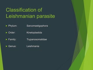
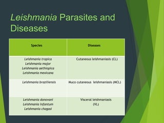
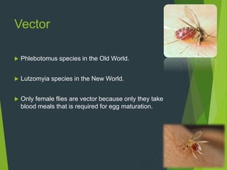
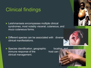
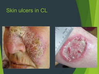
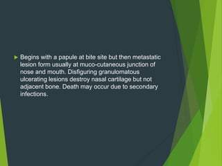
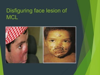
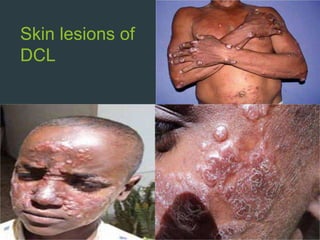
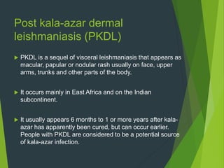
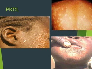
LEISHMANIASIS
By MUIB
Objectives
๏ต At the end of presentation, you will be able to
โ€ข Define leishmaniasis
โ€ข Typify leishmaniasis
โ€ข Manage leishmaniasis
Introduction
๏ต Leishmaniases are vector-borne diseases caused by
obligate intracellular protozoan parasites from more
than 20 Leishmania species.
๏ต There are three main forms of the disease: cutaneous
leishmaniasis (CL), visceral leishmaniasis (VL) or
kala-azar, and muco-cutaneous leishmaniasis (MCL).
๏ต Other forms include anthroponotic cutaneous
leishmaniasis, zoonotic cutaneous leishmaniasis &
post kala-azar dermal leishmaniasis (PKDL)
๏ต The Genus Leishmania includes four major
pathogens:
1. L.donovani complex with 3 species (L.
donovani, L. infantum, and L. chagasi)
2. L. mexicana complex with 3 main species (L.
mexicana, L. amazonensis, and L. venezuelensis)
3. L. tropica
4. L. braziliences
๏ต Majority of leishmaniasis are zoonoses while some
forms are considered to be non-zoonotic infections.
๏ต The different species are morphologically
indistinguishable, but they can be differentiated
by isoenzyme analysis, molecular methods, or
monoclonal antibodies.
๏ต Above is the False color SEM micrograph of
Promastigote form Leishmania mexicana as found in
the sand fly midgut. The cell body is shown in orange
and the flagellum is in red. 119 pixels/ฮผm.
Classification of
Leishmanian parasite
๏ต Phylum: Sarcomastigophora
๏ต Order: Kinetoplastida
๏ต Family: Trypanosomatidae
๏ต Genus: Leishmania
Leishmania Parasites and
Diseases
Species Diseases
Leishmania tropica
Leishmania major
Leishmania aethiopica
Leishmania mexicana
Cutaneous leishmaniasis (CL)
Leishmania braziliensis Muco cutaneous leishmaniasis (MCL)
Leishmania donovani
Leishmania infantum
Leishmania chagasi
Visceral leishmaniasis
(VL)
Vector
๏ต Phlebotomus species in the Old World.
๏ต Lutzomyia species in the New World.
๏ต Only female flies are vector because only they take
blood meals that is required for egg maturation.
๏ต The main way of parasite transmission is through the bite
of infected female Phlebotomus sand flies. Sand flies
become infected by sucking blood from an infected animal
or person. People might not realize that sand flies are
present because:
๏ต They do not make any noise;
๏ต They are small: they are only about one third the size of
typical mosquitoes or even smaller;
๏ต Their bites might not be noticed (the bites can be painless
or painful).
History
๏ต In 1903, Leishman identified the parasite and
Donovan described identical organisms in a splenic
puncture.
๏ต A devastating epidemic of visceral leishmaniasis
occurred in Sudan from 1984 to 1994
๏ต The recent epidemic in south Sudan has caused over
28 300 cases and nearly 900 deaths in the period
from 2009 to 2012.
๏ต In south America, Brazil has experienced a significant
increase in the number of cases of visceral
leishmaniasis since 1999.
๏ต Epidemics of anthroponotic cutaneous leishmaniasis
are of particular concern in Afghanistan, where
decades of war and civil unrest have created
conditions that favor the spread of the disease and
make its control especially difficult. The disease flared
up in 2002, with an estimated 100 000 cases in
Kabul. Because of their low resistance to the disease,
returning refugees and other displaced persons in
Kabul are at higher risk of infection.
Life cycle
๏ต Leishmaniasis is transmitted by the bite of infected female
Phlebotomus sand flies.
๏ต The sand flies inject the infective stage (i.e., promastigotes) from
their proboscis during blood meals.
๏ต Promastigotes that reach the puncture wound are phagocytized
by macrophages and other types of mononuclear phagocytic
cells.
๏ต Promastigotes transform in these cells into the tissue stage of
the parasite (i.e., amastigotes), which multiply by simple division
and proceed to infect other mononuclear phagocytic cells.
๏ต Parasite, host, and other factors affect whether the
infection becomes symptomatic and whether
cutaneous or visceral leishmaniasis results.
๏ต Sand flies become infected by ingesting infected cells
during blood meals.
๏ต In sand flies, amastigotes transform into
promastigotes, develop in the gut and migrate to the
proboscis.
Life cycle
Morphology
Digenetic Life Cycle
Promastigote
๏ต Insect stage
๏ต Motile
๏ต Gut
Amastigote
๏ต Mammalian stage
๏ต Non-motile
๏ต Intracellular
Promastigotes
๏ต Promastigotes are present in the digestive tract of sand fly
(vector) and in the culture media.
๏ต The fully developed promastigotes are long, slender and
spindle-shaped.
๏ต They measure 14.3 to 20 ฮผm in length and 1.5 to 1.8 ฮผm
in breadth.
๏ต A single nucleus lies in the center. The flagellum is single,
delicate and measures 15-28 ฮผm.
Promastigotes
Amastigotes
๏ต Amastigotes are round in shape & live inside
monocytes, polymorphonuclear leucocytes. They are
2.9-5.9 ฮผm in length.
๏ต They are stained well with Giemsa or Wright stain.
๏ต In the stained preparation, the cytoplasm appears
pale-blue and nucleus relatively is stained red.
Amastigotes
Transmission,
Pathogenesis &
Epidemiology
Transmission
๏ต Leishmania parasites are transmitted through the bite
of infected female sand flies (Phlebotomus species in
Old World & Lutzomyia in New World).
๏ต Transmission may occur by contamination of bite
wound or by contact when insect is crushed during
feeding.
๏ต Some 70 animal species, including humans, have
been found as natural reservoir hosts
of Leishmania parasites.
Mammalian Hosts
๏ต Rodents
๏ต Dogs
๏ต Foxes
๏ต Primates
๏ต Bats
๏ต Gerbils
๏ต Hyraxes
๏ต Sloths
๏ต Sand flies become infected by sucking blood from an
infected animal or person.
๏ต After infective meal, sand fly becomes infective in 6 to
9 days.
๏ต Transmission of kala-azar has also been recorded by
blood transfusion & through contaminated syringes
and needles.
๏ต Congenital transmission (spread from a pregnant
woman to her baby) has been reported.
Incubation Period
๏ต Incubation period is quite variable. It is generally 1 -3
months; but ranges from 2weeks to 2 years.
Pathogenesis
Visceral Leishmaniasis (VL)
๏ต Organs of reticuloendothelial system (liver, spleen &
bone marrow) most severely affected.
๏ต Reduced bone marrow activity coupled with cellular
destruction in spleen results in anemia, leucopenia &
thrombocytopenia.
๏ต This leads to secondary infections & a tendency to
bleed.
๏ต Striking splenomegaly is due to proliferation of
macrophages & sequestered RBCs.
Cutaneous Leishmaniasis
(CL) & Muco-cutaneous
Leishmaniasis (MCL)
๏ต Lesions are confined to skin in CL and to mucous
membranes, cartilages & skin in MCL.
๏ต A necrotic ulcer is formed at the bite site due to
granulomatous response.
๏ต The lesion has the tendency to become super
infected with bacteria.
Epidemiology
๏ต Leishmaniasis is found in people in focal areas of more
than 90 countries most of which are developing countries.
It occurs in the tropics, subtropics, and southern Europe.
The ecologic settings range from rain forests to deserts.
๏‚ง 90% of all VL: Bangladesh, Brazil, Ethiopia, India,
Nepal and Sudan.
๏‚ง 90% of all MCL: Bolivia, Brazil, Peru & Ethiopia.
๏‚ง 90% of all CL : Afghanistan, Brazil, Iran and Syria.
๏ต Annual incidence: 0.7-1.2 million cases of CL
: 0.2-0.4 million cases of VL and over
20,000 deaths annually
๏ต Prevalence: 12 million people
๏ต Population at risk: 310 million
๏ต Leishmaniasis is found on every continent except Australia
and Antarctica.
๏ต L. tropica & L. Mexicana both causes CL. Former organism
is found in the Old World whereas the latter is found in the
New World (American continents).
๏ต In the Old World (the Eastern Hemisphere), CL(Oriental
sore, Delhi boil, Bagdad sore) is found in some parts of
Asia, the Middle East, Africa (particularly in the tropical
region and North Africa, with some cases elsewhere), and
India. It is not found in Australia or the Pacific islands.
๏ต In the New World (the Western Hemisphere), CL (chicle
ulcer, bay sore) caused by L.mexicaca is found in Central &
South America. MCL (espundia) is found in Brazil & Central
America.
๏ต In many geographic areas where leishmaniasis is found in
people, infected people are not needed to maintain the
transmission cycle of the parasite in nature; infected
animals (such as rodents or dogs), along with sand flies,
maintain the cycle. However, in some parts of the world,
infected people are needed to maintain the cycle; this type
of transmission (humanโ€”sand flyโ€”human) is called
anthroponotic transmission. In areas with anthroponotic
transmission, effective treatment of individual patients can
help control the spread of the parasite.
๏ต Kala-azar occurs in three distinct epidemiological
patterns:
1. Mediterranean basin, Middle East, southern
Russia & parts of China, reservoir hosts are primarily
dogs & foxes.
2. Sub-Saharan Africa where rats and small
carnivores are main reservoirs
3. A third pattern is seen in India where humans
appear to be the only reservoir.
Leishmaniasis in Pakistan
๏ต It is especially present alongside regions bordering
the Afghanistan and cities that have had the
maximum influx of refugees. Moreover, the problem is
especially acute in Baluchistan and Sindh; mainly in
rural areas
๏ต Cross boarder movement of infected men is very
common and infected migrant carriers from
Afghanistan are probably the source of outbreak in
Pakistan.
๏ต In Afghanistan as well as in border areas of Khyber
Pakhtoon Khuwah especially, Waziristan, Kurram
Agency, Parah chinar, Swat, Bannu, Dera Ismail
Khan and Federally Administered area (FATA) several
new species of sand flies were recorded and are
known to be important endemic foci of leishmaniasis
in the region.
๏ต Complete data about sand fly fauna of both the
countries and specially vector species is still meager.
Pakistan map showing the endemic foci of leishmaniasis in blocks
๏ต Systemic leishmaniasis is rarer in Pakistan and
invariably fatal if not treated promptly.
๏ต Pakistan has a burden of cutaneous and visceral
leishmaniasis, the muco cutaneous form being almost
nonexistent.
๏ต In January 2002, a joint assessment mission of the Ministry
of Health and WHO identified 5,000 cases of cutaneous
leishmaniasis in Kurram Agency. The disease first appeared
in refugee camps but quickly reached neighboring villages
where an anthroponotic (person-to-person) transmission
occurred.
๏ต As leishmaniasis was previously unknown in the area the
local population had no immunity and there was a sharply
increased risk of a severe epidemic. The rapid response
team identified immediate needs for 15 000 vials of
pentavalent antimonials (first-line drugs) to treat the cases
which were donated by WHO mainly and by Iran.
๏ต The mission recommended a plan of action to involve
the Ministry of Health, WHO, United Nations High
Commissioner for Refugees (UNHCR) and
nongovernmental organizations in the area to prevent
further transmission by :
1. training of local health workers
2. translation of guidelines into the local languages
3. intensified surveillance
4. vector control activities to.
Map showing CL burden
Map showing VL burden
Major risk factors
๏ต Socioeconomic conditions: Poverty, poor housing and
domestic sanitary conditions (e.g. lack of waste
management, open sewerage) may increase sand fly
breeding. Human behavior, such as sleeping outside may
increase risk. The use of insecticide-treated bed nets
reduces risk.
๏ต Malnutrition: Diets lacking protein-energy, iron, vitamin A
and zinc increase the risk that an infection will progress to
kala-azar.
๏ต Population mobility: Often associated with migration and
the movement of non-immune people into areas with
existing transmission cycles.
๏ต Environmental changes: Leishmaniasis is affected
urbanization, deforestation and settlements into forested
areas.
๏ต Climate change: Leishmaniasis is affected by changes in
rainfall, temperature and humidity. Drought, famine and
flood resulting from climate change can lead to massive
displacement and migration of people to areas with
transmission of leishmaniasis, and poor nutrition could
compromise their immunity.
Clinical findings &
Diagnosis
Clinical findings
๏ต Leishmaniasis encompasses multiple clinical
syndromes, most notably visceral, cutaneous, and
muco cutaneous forms.
๏ต Different species can be associated with diverse
clinical manifestations.
๏ต Species identification, geographic location, and
immune response of the host can facilitate
clinical management.
Initial infections
๏ต Similar in all species.
๏ต Inoculation of promastigotes.
๏ต Inflammation & chemotaxis.
๏ต Receptor mediated phagocytosis.
๏ต The disease occurs in varying presentations, from the self-limited
and even self-healing cutaneous forms to fatal systemic disease.
๏ต Lesions of cutaneous leishmaniasis may occur anywhere on the
body but the most likely sites are the exposed parts. The initial
papule rapidly gives rise to an ulcer.
Cutaneous Leishmaniasis
(CL)
๏ต The most common form is cutaneous leishmaniasis, which
causes skin sores. The sores typically develop within a few
weeks or months of the sand fly bite. The sores can change
in size and appearance over time. The initial lesions may
start out as papules at bite site. This enlarges slowly to form
multiple satellite nodules. It may end up as ulcers (like a
volcano, with a raised edge and central crater); skin ulcers
might be covered by scab or crust. The sores usually are
painless but can be painful.
Skin ulcers in CL
Visceral Leishmaniasis (VL)
๏ต The other main form is visceral leishmaniasis, which
affects several internal organs (usually spleen, liver, and
bone marrow) and can be life threatening. The illness
typically develops within months (sometimes as long as
years) of the sand fly bite. Affected people usually have
intermittent fever, weakness & weight loss.
๏ต Hyper pigmentation of skin is seen in light skinned patients
(kala-azar means black sickness). Enlargement (swelling)
of the spleen and liver is present.
๏ต Certain blood tests are abnormal. For example,
patients usually have low blood counts, including a
low red blood cell count (anemia), low white blood
cell count, and low platelet count. Some patients
develop post kala-azar dermal leishmaniasis. Visceral
leishmaniasis is becoming an important opportunistic
infection in areas where it coexists with HIV.
๏ต Untreated severe disease is nearly always fatal as a
result of secondary infections.
Splenomegaly in VL
Teenage boy suffering from
visceral leishmaniasis.
The boy exhibits splenomegaly,
distended abdomen and
severe muscle wasting.
Jaundiced hands in VL
Muco cutaneous Leishmaniasis
(MCL)
๏ต Muco-cutaneous leishmaniasis is an example of
one of the less common forms of leishmaniasis. This
form can be a consequence of infection with some of
the species of the parasite that cause cutaneous
leishmaniasis in parts of Latin America: certain types
of the parasite might spread from the skin and cause
sores in the mucous membranes of the nose (most
common location), mouth, or throat.
๏ต Begins with a papule at bite site but then metastatic
lesion form usually at muco-cutaneous junction of
nose and mouth. Disfiguring granulomatous
ulcerating lesions destroy nasal cartilage but not
adjacent bone. Death may occur due to secondary
infections.
Disfiguring face lesion of
MCL
Diffuse cutaneous
leishmaniasis (DCL)
๏ต Both L. aethiopica (Old World) and L. amazonensis (New
World) are the causes of diffuse cutaneous leishmaniasis.
๏ต Skin lesions develop over a large area of the body.
๏ต The lesions on the eyebrows, nose and ears resemble those
of lepromatous leprosy.
๏ต At first, the lesions are smooth, and firm. Later they become
scaly and rough.
๏ต The nodules contain large numbers of amastigotes.
๏ต The lesions do not heal spontaneously and this is an
incurable condition characterized by the formation of
disfiguring nodules over the surface of the body.
Skin lesions of
DCL
Post kala-azar dermal
leishmaniasis (PKDL)
๏ต PKDL is a sequel of visceral leishmaniasis that appears as
macular, papular or nodular rash usually on face, upper
arms, trunks and other parts of the body.
๏ต It occurs mainly in East Africa and on the Indian
subcontinent.
๏ต It usually appears 6 months to 1 or more years after kala-
azar has apparently been cured, but can occur earlier.
People with PKDL are considered to be a potential source
of kala-azar infection.
PKDL
Leishmania-HIV co-infection
๏ต Leishmaniaโ€“HIV co-infection had been reported from 35
endemic countries with high rates reported from Brazil,
Ethiopia & India.
๏ต Leishmania-HIV co-infected people have high a chance of
developing the full-blown clinical disease, and high relapse
and mortality rates. Co-infection with HIV intensifies the
burden of visceral and cutaneous leishmaniasis by causing
severe forms that are more difficult to manage. HIV-infected
people are particularly vulnerable to VL, and VL accelerates
HIV replication and progression to AIDS.
๏ต Antiretroviral treatment reduces the development of the
disease, delays relapses and increases the survival of the
co-infected patients.
Diagnosis
๏ต Includes:
1. Clinical diagnosis based on clinical signs
& symptoms
2. Laboratory diagnosis
Lab diagnosis
Specimens that may be collected
๏ต Splenic aspirate and biopsy
๏ต Liver biopsy
๏ต Bone marrow (Sternum or iliac crest)
๏ต Blood
๏ต Dermal scrapings, sections from skin biopsy
Microscopy
๏ต In the human host, only the amastigotes stage is
seen upon microscopic examination of tissue
specimens. Amastigotes can be visualized with both
Giemsa and hematoxylin and eosin (H&E) stains.
๏ต The amastigotes of different Leishmania species are
morphologically indistinguishable. Amastigotes are
ovoid and measure 1-5 micrometers long by 1-2
micrometers wide and are present inside the
macrophages. They have a nucleus in center.
๏ต Amastigotes in an intact macrophage.
๏ต H&E stained skin tissue showing amastigotes.
Serology
๏ต A very high IgG titer is indicative of infection but not
diagnostic for leishmaniasis.
๏ต Various serological tests for leishmaniasis are
1. ELISA
2. Direct agglutination test
3. rk39 dipstick test (based on amastigote epitope
recombinant k 39 protein)
4. Indirect fluorescent antibody test
Aldehyde (formol-gel) test
of Napier
๏ต 1-2 ml serum and 1-2 drops of 40% formalin are added
together.
๏ต A positive test is indicated by milky white opacity.
๏ต If it occurs in 2-20 minutes, it is said to be strongly positive.
๏ต Reaction after 30 minutes is not significant.
๏ต Test becomes positive 2-3 months after onset of disease and
reverts to negative 6 months after cure.
๏ต Therefore test is good for surveillance, not for diagnosis.
๏ต Has low specificity as the test is positive in many other chronic
disease in which albumin to globulin ratio is reversed.
Leishmanin (Montenegro) test
๏ต Based on skin reaction.
๏ต Leishmanin is preparation of 106 per ml washed promastigotes
of leishmania, suspended in 0.5% phenol saline or merthiolate.
๏ต 0.1 ml leishmanin is injected intradermally on flexor surface of
forearm and examined after 48 to 72 hours.
๏ต Induration in noted. Induration of 5 mm or more is considered
positive.
๏ต Test is positive 4-6 weeks after onset of CL and MCL.
๏ต Usually negative in active phase of kala-azar, but becomes
positive in 75% of cases within 1 year of recovery.
๏ต Test is not species specific.
๏ต Helps to distinguish immune from non immune subjects
๏ต From this information, it may be possible to infer the endemicity
or epidemicity of the infection
Culture
Culture media for leishmaniasis are:
๏ต SOLID MEDIUM
๏‚ง NNN medium
๏‚ง Evanโ€™s modified Tobieโ€™s medium
๏ต LIQUID MEDIA
๏‚ง Schneiderโ€™s Drosophila medium
๏‚ง Graceโ€™s insect tissue culture medium
Promastigotes as seen in
artificial culture medium
Isoenzyme analysis
๏ต After isolation parasites can be characterized to the
complex and sometimes to the species level using
isoenzyme analysis, which is the conventional
diagnostic approach for Leishmania species
identification. Diagnostic identification
of Leishmania using this approach may take several
weeks.
Hematological findings
๏ต Anemia
๏ต Leucopenia
๏ต ESR is increased
๏ต Reversed albumin-globulin ratio
๏ต WBC:RBC ration is 1:1500 or even more (normal
ratio is 1:750)
Treatment, Control &
Prevention
Treatment
๏ต In general, all clinically manifest cases of VL and MCL
should be treated, whereas not all cases of
cutaneous leishmaniasis require treatment.
๏ต General points:
โ€ข Improvement of general health
โ€ข Balanced diet
โ€ข Avoidance of risk factors
CL
๏ต Therapy of cutaneous leishmaniasis may be indicated to:
1. decrease the risk for mucosal dissemination of disease.
2. accelerate healing of the skin lesions.
3. decrease the risk for relapse of the skin lesions.
4. decrease the local morbidity caused by large or
persistent skin lesions, particularly those on the face or ears
or near joints.
5. decrease the reservoir of infection in geographic areas
where infected persons serve as reservoir hosts (such as in
Kabul, Afghanistan, where transmission is anthroponotic)
CL & MCL
๏ต Antimony (Pentostamยฎ, Sodium stibogluconate) is
the drug of choice.
โ€ข 20 mg/kg body weight IM or IV for 20 days
VL
๏ต Liposomal amphotericin-B (AmBisomeยฎ) is the drug of
choice.
โ€ข 1 mg/kg body weight IV infusion daily or alternate day
for 15-20 infusions
๏ต Pentostamยฎ is an alternative therapy
โ€ข 28 days of therapy is required
๏ต Conventional amphotericin B deoxycholate is highly effective
therapy for visceral leishmaniasis but generally is more toxic than
liposomal amphotericin B. Immunocompetent patients typically
receive 0.5 to 1.0 mg per kgโ€”either daily or every other dayโ€”by
IV infusionโ€”for a total dose of approximately 15 to 20 mg per kg.
Longer courses of therapy may be indicated for some patients
๏ต Miltefosine (a phospholipid derivative) can be used for treatment
of cutaneous, mucosal, and visceral leishmaniasis caused by
particular Leishmania species in adults and adolescents at least
12 years of age who weigh at least 30 kg (66 pounds). 100 mg
daily in 2 divided doses for 4 weeks is required.
๏ต Miltefosine is contraindicated in pregnant women. Nursing
mothers should be advised not to breastfeed during the
treatment course or for 5 months thereafter.
๏ต Some medications that might have merit for treating
selected cases of leishmaniasis include
1. parenteral agents: amphotericin B
deoxycholate and pentamidine isethionate, parenteral
formulation of the aminoglycoside paromomycin (11
mg/kg for 21 days)
2. orally administered "azoles"
(ketoconazole, itraconazole, and fluconazole).
๏ต Recovery results in permanent immunity.
Control
๏ต It includes:
๏ต Early diagnosis and effective case management:
It reduces the prevalence of the disease and prevents
disabilities and death.
๏ต Vector control control: It helps to reduce or interrupt
transmission of disease. It can be done by the use of
various insecticides e.g. DDT.
๏ต Reservoir control: Potential reservoirs such as
dogs, rats, gerbils, other small mammals and rodents
should be controlled by methods such as incineration.
๏ต Education of the community: It should be done with
effective behavioral change interventions with locally
tailored communication strategies. Partnership and
collaboration with various stakeholders and other
vector-borne disease control programmes is critical.
Prevention
๏ต No vaccines or drugs to prevent infection are
available. The best way for travelers to prevent
infection is to protect themselves from sand fly bites.
To decrease the risk of being bitten, follow these
preventive measures:
๏ต Avoid outdoor activities, especially from dusk to dawn,
when sand flies generally are the most active.
๏ต When outdoors (or in unprotected quarters):
๏ต Minimize the amount of exposed (uncovered) skin. To
the extent that is tolerable in the climate, wear long-
sleeved shirts, long pants, and socks; and tuck your
shirt into your pants.
๏ต Apply insect repellent to exposed skin and under the
ends of sleeves and pant legs. Follow the instructions
on the label of the repellent.
๏ต When indoors:
๏ต Stay in well-screened or air-conditioned areas.
๏ต Keep in mind that sand flies are much smaller than
mosquitoes and therefore can get through smaller holes.
๏ต Spray living/sleeping areas with an insecticide to kill
insects.
๏ต If you are not sleeping in a well-screened or air-conditioned
area, use a bed net and tuck it under your mattress. If
possible, use a bed net that has been soaked in or sprayed
with an insecticide. The same treatment can be applied to
screens, curtains, sheets, and clothing (clothing should be
retreated after five washings).
Vaccine
๏ต There is as yet no effective vaccine for prevention of
any form of leishmaniasis.
๏ต Several potential vaccines are being developed, but
as of 2015 none are available.
๏ต In February 2012, the nonprofit Infectious Disease
Research Institute launched the worldโ€™s first
human clinical trial of the visceral leishmaniasis
vaccine in some endemic countries.
Leischmaniasis (Kala Azar)

Leischmaniasis (Kala Azar)

  • 1.
  • 2.
    Objectives ๏ต At theend of presentation, you will be able to โ€ข Define leishmaniasis โ€ข Typify leishmaniasis โ€ข Manage leishmaniasis
  • 3.
    Introduction ๏ต Leishmaniases arevector-borne diseases caused by obligate intracellular protozoan parasites from more than 20 Leishmania species. ๏ต There are three main forms of the disease: cutaneous leishmaniasis (CL), visceral leishmaniasis (VL) or kala-azar, and muco-cutaneous leishmaniasis (MCL). ๏ต Other forms include anthroponotic cutaneous leishmaniasis, zoonotic cutaneous leishmaniasis & post kala-azar dermal leishmaniasis (PKDL)
  • 4.
    ๏ต The GenusLeishmania includes four major pathogens: 1. L.donovani complex with 3 species (L. donovani, L. infantum, and L. chagasi) 2. L. mexicana complex with 3 main species (L. mexicana, L. amazonensis, and L. venezuelensis) 3. L. tropica 4. L. braziliences ๏ต Majority of leishmaniasis are zoonoses while some forms are considered to be non-zoonotic infections.
  • 5.
    ๏ต The differentspecies are morphologically indistinguishable, but they can be differentiated by isoenzyme analysis, molecular methods, or monoclonal antibodies. ๏ต Above is the False color SEM micrograph of Promastigote form Leishmania mexicana as found in the sand fly midgut. The cell body is shown in orange and the flagellum is in red. 119 pixels/ฮผm.
  • 6.
    Classification of Leishmanian parasite ๏ตPhylum: Sarcomastigophora ๏ต Order: Kinetoplastida ๏ต Family: Trypanosomatidae ๏ต Genus: Leishmania
  • 7.
    Leishmania Parasites and Diseases SpeciesDiseases Leishmania tropica Leishmania major Leishmania aethiopica Leishmania mexicana Cutaneous leishmaniasis (CL) Leishmania braziliensis Muco cutaneous leishmaniasis (MCL) Leishmania donovani Leishmania infantum Leishmania chagasi Visceral leishmaniasis (VL)
  • 8.
    Vector ๏ต Phlebotomus speciesin the Old World. ๏ต Lutzomyia species in the New World. ๏ต Only female flies are vector because only they take blood meals that is required for egg maturation.
  • 9.
    ๏ต The mainway of parasite transmission is through the bite of infected female Phlebotomus sand flies. Sand flies become infected by sucking blood from an infected animal or person. People might not realize that sand flies are present because: ๏ต They do not make any noise; ๏ต They are small: they are only about one third the size of typical mosquitoes or even smaller; ๏ต Their bites might not be noticed (the bites can be painless or painful).
  • 10.
    History ๏ต In 1903,Leishman identified the parasite and Donovan described identical organisms in a splenic puncture. ๏ต A devastating epidemic of visceral leishmaniasis occurred in Sudan from 1984 to 1994 ๏ต The recent epidemic in south Sudan has caused over 28 300 cases and nearly 900 deaths in the period from 2009 to 2012.
  • 11.
    ๏ต In southAmerica, Brazil has experienced a significant increase in the number of cases of visceral leishmaniasis since 1999. ๏ต Epidemics of anthroponotic cutaneous leishmaniasis are of particular concern in Afghanistan, where decades of war and civil unrest have created conditions that favor the spread of the disease and make its control especially difficult. The disease flared up in 2002, with an estimated 100 000 cases in Kabul. Because of their low resistance to the disease, returning refugees and other displaced persons in Kabul are at higher risk of infection.
  • 12.
    Life cycle ๏ต Leishmaniasisis transmitted by the bite of infected female Phlebotomus sand flies. ๏ต The sand flies inject the infective stage (i.e., promastigotes) from their proboscis during blood meals. ๏ต Promastigotes that reach the puncture wound are phagocytized by macrophages and other types of mononuclear phagocytic cells. ๏ต Promastigotes transform in these cells into the tissue stage of the parasite (i.e., amastigotes), which multiply by simple division and proceed to infect other mononuclear phagocytic cells.
  • 13.
    ๏ต Parasite, host,and other factors affect whether the infection becomes symptomatic and whether cutaneous or visceral leishmaniasis results. ๏ต Sand flies become infected by ingesting infected cells during blood meals. ๏ต In sand flies, amastigotes transform into promastigotes, develop in the gut and migrate to the proboscis.
  • 14.
  • 15.
    Morphology Digenetic Life Cycle Promastigote ๏ตInsect stage ๏ต Motile ๏ต Gut Amastigote ๏ต Mammalian stage ๏ต Non-motile ๏ต Intracellular
  • 16.
    Promastigotes ๏ต Promastigotes arepresent in the digestive tract of sand fly (vector) and in the culture media. ๏ต The fully developed promastigotes are long, slender and spindle-shaped. ๏ต They measure 14.3 to 20 ฮผm in length and 1.5 to 1.8 ฮผm in breadth. ๏ต A single nucleus lies in the center. The flagellum is single, delicate and measures 15-28 ฮผm.
  • 17.
  • 18.
    Amastigotes ๏ต Amastigotes areround in shape & live inside monocytes, polymorphonuclear leucocytes. They are 2.9-5.9 ฮผm in length. ๏ต They are stained well with Giemsa or Wright stain. ๏ต In the stained preparation, the cytoplasm appears pale-blue and nucleus relatively is stained red.
  • 19.
  • 20.
  • 21.
    Transmission ๏ต Leishmania parasitesare transmitted through the bite of infected female sand flies (Phlebotomus species in Old World & Lutzomyia in New World). ๏ต Transmission may occur by contamination of bite wound or by contact when insect is crushed during feeding. ๏ต Some 70 animal species, including humans, have been found as natural reservoir hosts of Leishmania parasites.
  • 22.
    Mammalian Hosts ๏ต Rodents ๏ตDogs ๏ต Foxes ๏ต Primates ๏ต Bats ๏ต Gerbils ๏ต Hyraxes ๏ต Sloths
  • 23.
    ๏ต Sand fliesbecome infected by sucking blood from an infected animal or person. ๏ต After infective meal, sand fly becomes infective in 6 to 9 days. ๏ต Transmission of kala-azar has also been recorded by blood transfusion & through contaminated syringes and needles. ๏ต Congenital transmission (spread from a pregnant woman to her baby) has been reported.
  • 24.
    Incubation Period ๏ต Incubationperiod is quite variable. It is generally 1 -3 months; but ranges from 2weeks to 2 years.
  • 25.
    Pathogenesis Visceral Leishmaniasis (VL) ๏ตOrgans of reticuloendothelial system (liver, spleen & bone marrow) most severely affected. ๏ต Reduced bone marrow activity coupled with cellular destruction in spleen results in anemia, leucopenia & thrombocytopenia. ๏ต This leads to secondary infections & a tendency to bleed. ๏ต Striking splenomegaly is due to proliferation of macrophages & sequestered RBCs.
  • 26.
    Cutaneous Leishmaniasis (CL) &Muco-cutaneous Leishmaniasis (MCL) ๏ต Lesions are confined to skin in CL and to mucous membranes, cartilages & skin in MCL. ๏ต A necrotic ulcer is formed at the bite site due to granulomatous response. ๏ต The lesion has the tendency to become super infected with bacteria.
  • 27.
    Epidemiology ๏ต Leishmaniasis isfound in people in focal areas of more than 90 countries most of which are developing countries. It occurs in the tropics, subtropics, and southern Europe. The ecologic settings range from rain forests to deserts. ๏‚ง 90% of all VL: Bangladesh, Brazil, Ethiopia, India, Nepal and Sudan. ๏‚ง 90% of all MCL: Bolivia, Brazil, Peru & Ethiopia. ๏‚ง 90% of all CL : Afghanistan, Brazil, Iran and Syria.
  • 28.
    ๏ต Annual incidence:0.7-1.2 million cases of CL : 0.2-0.4 million cases of VL and over 20,000 deaths annually ๏ต Prevalence: 12 million people ๏ต Population at risk: 310 million ๏ต Leishmaniasis is found on every continent except Australia and Antarctica.
  • 29.
    ๏ต L. tropica& L. Mexicana both causes CL. Former organism is found in the Old World whereas the latter is found in the New World (American continents). ๏ต In the Old World (the Eastern Hemisphere), CL(Oriental sore, Delhi boil, Bagdad sore) is found in some parts of Asia, the Middle East, Africa (particularly in the tropical region and North Africa, with some cases elsewhere), and India. It is not found in Australia or the Pacific islands. ๏ต In the New World (the Western Hemisphere), CL (chicle ulcer, bay sore) caused by L.mexicaca is found in Central & South America. MCL (espundia) is found in Brazil & Central America.
  • 30.
    ๏ต In manygeographic areas where leishmaniasis is found in people, infected people are not needed to maintain the transmission cycle of the parasite in nature; infected animals (such as rodents or dogs), along with sand flies, maintain the cycle. However, in some parts of the world, infected people are needed to maintain the cycle; this type of transmission (humanโ€”sand flyโ€”human) is called anthroponotic transmission. In areas with anthroponotic transmission, effective treatment of individual patients can help control the spread of the parasite.
  • 31.
    ๏ต Kala-azar occursin three distinct epidemiological patterns: 1. Mediterranean basin, Middle East, southern Russia & parts of China, reservoir hosts are primarily dogs & foxes. 2. Sub-Saharan Africa where rats and small carnivores are main reservoirs 3. A third pattern is seen in India where humans appear to be the only reservoir.
  • 32.
    Leishmaniasis in Pakistan ๏ตIt is especially present alongside regions bordering the Afghanistan and cities that have had the maximum influx of refugees. Moreover, the problem is especially acute in Baluchistan and Sindh; mainly in rural areas ๏ต Cross boarder movement of infected men is very common and infected migrant carriers from Afghanistan are probably the source of outbreak in Pakistan.
  • 33.
    ๏ต In Afghanistanas well as in border areas of Khyber Pakhtoon Khuwah especially, Waziristan, Kurram Agency, Parah chinar, Swat, Bannu, Dera Ismail Khan and Federally Administered area (FATA) several new species of sand flies were recorded and are known to be important endemic foci of leishmaniasis in the region. ๏ต Complete data about sand fly fauna of both the countries and specially vector species is still meager.
  • 34.
    Pakistan map showingthe endemic foci of leishmaniasis in blocks
  • 35.
    ๏ต Systemic leishmaniasisis rarer in Pakistan and invariably fatal if not treated promptly. ๏ต Pakistan has a burden of cutaneous and visceral leishmaniasis, the muco cutaneous form being almost nonexistent.
  • 36.
    ๏ต In January2002, a joint assessment mission of the Ministry of Health and WHO identified 5,000 cases of cutaneous leishmaniasis in Kurram Agency. The disease first appeared in refugee camps but quickly reached neighboring villages where an anthroponotic (person-to-person) transmission occurred. ๏ต As leishmaniasis was previously unknown in the area the local population had no immunity and there was a sharply increased risk of a severe epidemic. The rapid response team identified immediate needs for 15 000 vials of pentavalent antimonials (first-line drugs) to treat the cases which were donated by WHO mainly and by Iran.
  • 37.
    ๏ต The missionrecommended a plan of action to involve the Ministry of Health, WHO, United Nations High Commissioner for Refugees (UNHCR) and nongovernmental organizations in the area to prevent further transmission by : 1. training of local health workers 2. translation of guidelines into the local languages 3. intensified surveillance 4. vector control activities to.
  • 39.
  • 41.
  • 42.
    Major risk factors ๏ตSocioeconomic conditions: Poverty, poor housing and domestic sanitary conditions (e.g. lack of waste management, open sewerage) may increase sand fly breeding. Human behavior, such as sleeping outside may increase risk. The use of insecticide-treated bed nets reduces risk. ๏ต Malnutrition: Diets lacking protein-energy, iron, vitamin A and zinc increase the risk that an infection will progress to kala-azar. ๏ต Population mobility: Often associated with migration and the movement of non-immune people into areas with existing transmission cycles.
  • 43.
    ๏ต Environmental changes:Leishmaniasis is affected urbanization, deforestation and settlements into forested areas. ๏ต Climate change: Leishmaniasis is affected by changes in rainfall, temperature and humidity. Drought, famine and flood resulting from climate change can lead to massive displacement and migration of people to areas with transmission of leishmaniasis, and poor nutrition could compromise their immunity.
  • 44.
  • 45.
    Clinical findings ๏ต Leishmaniasisencompasses multiple clinical syndromes, most notably visceral, cutaneous, and muco cutaneous forms. ๏ต Different species can be associated with diverse clinical manifestations. ๏ต Species identification, geographic location, and immune response of the host can facilitate clinical management.
  • 46.
    Initial infections ๏ต Similarin all species. ๏ต Inoculation of promastigotes. ๏ต Inflammation & chemotaxis. ๏ต Receptor mediated phagocytosis. ๏ต The disease occurs in varying presentations, from the self-limited and even self-healing cutaneous forms to fatal systemic disease. ๏ต Lesions of cutaneous leishmaniasis may occur anywhere on the body but the most likely sites are the exposed parts. The initial papule rapidly gives rise to an ulcer.
  • 47.
    Cutaneous Leishmaniasis (CL) ๏ต Themost common form is cutaneous leishmaniasis, which causes skin sores. The sores typically develop within a few weeks or months of the sand fly bite. The sores can change in size and appearance over time. The initial lesions may start out as papules at bite site. This enlarges slowly to form multiple satellite nodules. It may end up as ulcers (like a volcano, with a raised edge and central crater); skin ulcers might be covered by scab or crust. The sores usually are painless but can be painful.
  • 48.
  • 49.
    Visceral Leishmaniasis (VL) ๏ตThe other main form is visceral leishmaniasis, which affects several internal organs (usually spleen, liver, and bone marrow) and can be life threatening. The illness typically develops within months (sometimes as long as years) of the sand fly bite. Affected people usually have intermittent fever, weakness & weight loss. ๏ต Hyper pigmentation of skin is seen in light skinned patients (kala-azar means black sickness). Enlargement (swelling) of the spleen and liver is present.
  • 50.
    ๏ต Certain bloodtests are abnormal. For example, patients usually have low blood counts, including a low red blood cell count (anemia), low white blood cell count, and low platelet count. Some patients develop post kala-azar dermal leishmaniasis. Visceral leishmaniasis is becoming an important opportunistic infection in areas where it coexists with HIV. ๏ต Untreated severe disease is nearly always fatal as a result of secondary infections.
  • 51.
  • 52.
    Teenage boy sufferingfrom visceral leishmaniasis. The boy exhibits splenomegaly, distended abdomen and severe muscle wasting.
  • 53.
  • 54.
    Muco cutaneous Leishmaniasis (MCL) ๏ตMuco-cutaneous leishmaniasis is an example of one of the less common forms of leishmaniasis. This form can be a consequence of infection with some of the species of the parasite that cause cutaneous leishmaniasis in parts of Latin America: certain types of the parasite might spread from the skin and cause sores in the mucous membranes of the nose (most common location), mouth, or throat.
  • 55.
    ๏ต Begins witha papule at bite site but then metastatic lesion form usually at muco-cutaneous junction of nose and mouth. Disfiguring granulomatous ulcerating lesions destroy nasal cartilage but not adjacent bone. Death may occur due to secondary infections.
  • 56.
  • 57.
    Diffuse cutaneous leishmaniasis (DCL) ๏ตBoth L. aethiopica (Old World) and L. amazonensis (New World) are the causes of diffuse cutaneous leishmaniasis. ๏ต Skin lesions develop over a large area of the body. ๏ต The lesions on the eyebrows, nose and ears resemble those of lepromatous leprosy. ๏ต At first, the lesions are smooth, and firm. Later they become scaly and rough. ๏ต The nodules contain large numbers of amastigotes. ๏ต The lesions do not heal spontaneously and this is an incurable condition characterized by the formation of disfiguring nodules over the surface of the body.
  • 58.
  • 59.
    Post kala-azar dermal leishmaniasis(PKDL) ๏ต PKDL is a sequel of visceral leishmaniasis that appears as macular, papular or nodular rash usually on face, upper arms, trunks and other parts of the body. ๏ต It occurs mainly in East Africa and on the Indian subcontinent. ๏ต It usually appears 6 months to 1 or more years after kala- azar has apparently been cured, but can occur earlier. People with PKDL are considered to be a potential source of kala-azar infection.
  • 60.
  • 61.
    Leishmania-HIV co-infection ๏ต Leishmaniaโ€“HIVco-infection had been reported from 35 endemic countries with high rates reported from Brazil, Ethiopia & India. ๏ต Leishmania-HIV co-infected people have high a chance of developing the full-blown clinical disease, and high relapse and mortality rates. Co-infection with HIV intensifies the burden of visceral and cutaneous leishmaniasis by causing severe forms that are more difficult to manage. HIV-infected people are particularly vulnerable to VL, and VL accelerates HIV replication and progression to AIDS. ๏ต Antiretroviral treatment reduces the development of the disease, delays relapses and increases the survival of the co-infected patients.
  • 62.
    Diagnosis ๏ต Includes: 1. Clinicaldiagnosis based on clinical signs & symptoms 2. Laboratory diagnosis
  • 63.
    Lab diagnosis Specimens thatmay be collected ๏ต Splenic aspirate and biopsy ๏ต Liver biopsy ๏ต Bone marrow (Sternum or iliac crest) ๏ต Blood ๏ต Dermal scrapings, sections from skin biopsy
  • 64.
    Microscopy ๏ต In thehuman host, only the amastigotes stage is seen upon microscopic examination of tissue specimens. Amastigotes can be visualized with both Giemsa and hematoxylin and eosin (H&E) stains. ๏ต The amastigotes of different Leishmania species are morphologically indistinguishable. Amastigotes are ovoid and measure 1-5 micrometers long by 1-2 micrometers wide and are present inside the macrophages. They have a nucleus in center.
  • 65.
    ๏ต Amastigotes inan intact macrophage. ๏ต H&E stained skin tissue showing amastigotes.
  • 66.
    Serology ๏ต A veryhigh IgG titer is indicative of infection but not diagnostic for leishmaniasis. ๏ต Various serological tests for leishmaniasis are 1. ELISA 2. Direct agglutination test 3. rk39 dipstick test (based on amastigote epitope recombinant k 39 protein) 4. Indirect fluorescent antibody test
  • 67.
    Aldehyde (formol-gel) test ofNapier ๏ต 1-2 ml serum and 1-2 drops of 40% formalin are added together. ๏ต A positive test is indicated by milky white opacity. ๏ต If it occurs in 2-20 minutes, it is said to be strongly positive. ๏ต Reaction after 30 minutes is not significant. ๏ต Test becomes positive 2-3 months after onset of disease and reverts to negative 6 months after cure. ๏ต Therefore test is good for surveillance, not for diagnosis. ๏ต Has low specificity as the test is positive in many other chronic disease in which albumin to globulin ratio is reversed.
  • 68.
    Leishmanin (Montenegro) test ๏ตBased on skin reaction. ๏ต Leishmanin is preparation of 106 per ml washed promastigotes of leishmania, suspended in 0.5% phenol saline or merthiolate. ๏ต 0.1 ml leishmanin is injected intradermally on flexor surface of forearm and examined after 48 to 72 hours. ๏ต Induration in noted. Induration of 5 mm or more is considered positive. ๏ต Test is positive 4-6 weeks after onset of CL and MCL. ๏ต Usually negative in active phase of kala-azar, but becomes positive in 75% of cases within 1 year of recovery. ๏ต Test is not species specific. ๏ต Helps to distinguish immune from non immune subjects ๏ต From this information, it may be possible to infer the endemicity or epidemicity of the infection
  • 69.
    Culture Culture media forleishmaniasis are: ๏ต SOLID MEDIUM ๏‚ง NNN medium ๏‚ง Evanโ€™s modified Tobieโ€™s medium ๏ต LIQUID MEDIA ๏‚ง Schneiderโ€™s Drosophila medium ๏‚ง Graceโ€™s insect tissue culture medium
  • 70.
    Promastigotes as seenin artificial culture medium
  • 71.
    Isoenzyme analysis ๏ต Afterisolation parasites can be characterized to the complex and sometimes to the species level using isoenzyme analysis, which is the conventional diagnostic approach for Leishmania species identification. Diagnostic identification of Leishmania using this approach may take several weeks.
  • 72.
    Hematological findings ๏ต Anemia ๏ตLeucopenia ๏ต ESR is increased ๏ต Reversed albumin-globulin ratio ๏ต WBC:RBC ration is 1:1500 or even more (normal ratio is 1:750)
  • 73.
  • 74.
    Treatment ๏ต In general,all clinically manifest cases of VL and MCL should be treated, whereas not all cases of cutaneous leishmaniasis require treatment. ๏ต General points: โ€ข Improvement of general health โ€ข Balanced diet โ€ข Avoidance of risk factors
  • 75.
    CL ๏ต Therapy ofcutaneous leishmaniasis may be indicated to: 1. decrease the risk for mucosal dissemination of disease. 2. accelerate healing of the skin lesions. 3. decrease the risk for relapse of the skin lesions. 4. decrease the local morbidity caused by large or persistent skin lesions, particularly those on the face or ears or near joints. 5. decrease the reservoir of infection in geographic areas where infected persons serve as reservoir hosts (such as in Kabul, Afghanistan, where transmission is anthroponotic)
  • 76.
    CL & MCL ๏ตAntimony (Pentostamยฎ, Sodium stibogluconate) is the drug of choice. โ€ข 20 mg/kg body weight IM or IV for 20 days
  • 77.
    VL ๏ต Liposomal amphotericin-B(AmBisomeยฎ) is the drug of choice. โ€ข 1 mg/kg body weight IV infusion daily or alternate day for 15-20 infusions ๏ต Pentostamยฎ is an alternative therapy โ€ข 28 days of therapy is required
  • 78.
    ๏ต Conventional amphotericinB deoxycholate is highly effective therapy for visceral leishmaniasis but generally is more toxic than liposomal amphotericin B. Immunocompetent patients typically receive 0.5 to 1.0 mg per kgโ€”either daily or every other dayโ€”by IV infusionโ€”for a total dose of approximately 15 to 20 mg per kg. Longer courses of therapy may be indicated for some patients ๏ต Miltefosine (a phospholipid derivative) can be used for treatment of cutaneous, mucosal, and visceral leishmaniasis caused by particular Leishmania species in adults and adolescents at least 12 years of age who weigh at least 30 kg (66 pounds). 100 mg daily in 2 divided doses for 4 weeks is required. ๏ต Miltefosine is contraindicated in pregnant women. Nursing mothers should be advised not to breastfeed during the treatment course or for 5 months thereafter.
  • 79.
    ๏ต Some medicationsthat might have merit for treating selected cases of leishmaniasis include 1. parenteral agents: amphotericin B deoxycholate and pentamidine isethionate, parenteral formulation of the aminoglycoside paromomycin (11 mg/kg for 21 days) 2. orally administered "azoles" (ketoconazole, itraconazole, and fluconazole). ๏ต Recovery results in permanent immunity.
  • 80.
    Control ๏ต It includes: ๏ตEarly diagnosis and effective case management: It reduces the prevalence of the disease and prevents disabilities and death. ๏ต Vector control control: It helps to reduce or interrupt transmission of disease. It can be done by the use of various insecticides e.g. DDT. ๏ต Reservoir control: Potential reservoirs such as dogs, rats, gerbils, other small mammals and rodents should be controlled by methods such as incineration.
  • 81.
    ๏ต Education ofthe community: It should be done with effective behavioral change interventions with locally tailored communication strategies. Partnership and collaboration with various stakeholders and other vector-borne disease control programmes is critical.
  • 82.
    Prevention ๏ต No vaccinesor drugs to prevent infection are available. The best way for travelers to prevent infection is to protect themselves from sand fly bites. To decrease the risk of being bitten, follow these preventive measures: ๏ต Avoid outdoor activities, especially from dusk to dawn, when sand flies generally are the most active.
  • 83.
    ๏ต When outdoors(or in unprotected quarters): ๏ต Minimize the amount of exposed (uncovered) skin. To the extent that is tolerable in the climate, wear long- sleeved shirts, long pants, and socks; and tuck your shirt into your pants. ๏ต Apply insect repellent to exposed skin and under the ends of sleeves and pant legs. Follow the instructions on the label of the repellent.
  • 84.
    ๏ต When indoors: ๏ตStay in well-screened or air-conditioned areas. ๏ต Keep in mind that sand flies are much smaller than mosquitoes and therefore can get through smaller holes. ๏ต Spray living/sleeping areas with an insecticide to kill insects. ๏ต If you are not sleeping in a well-screened or air-conditioned area, use a bed net and tuck it under your mattress. If possible, use a bed net that has been soaked in or sprayed with an insecticide. The same treatment can be applied to screens, curtains, sheets, and clothing (clothing should be retreated after five washings).
  • 85.
    Vaccine ๏ต There isas yet no effective vaccine for prevention of any form of leishmaniasis. ๏ต Several potential vaccines are being developed, but as of 2015 none are available. ๏ต In February 2012, the nonprofit Infectious Disease Research Institute launched the worldโ€™s first human clinical trial of the visceral leishmaniasis vaccine in some endemic countries.