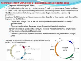
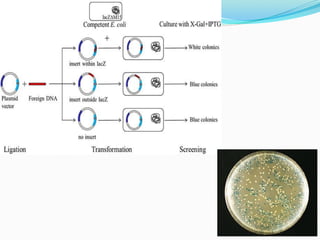
pUC plasmids
Dr. Manikandan Kathirvel M.Sc., Ph.D., (NET)
Assistant Professor,
Department of Life Sciences,
Kristu Jayanti College (Autonomous),
(Reaccredited with "A" Grade by NAAC)
Affiliated to Bengaluru North University,
K. Narayanapura, Kothanur (PO)
Bengaluru
Email: manikandan@kristujayanti.com
ORCID ID: 0000000270066334
pUC plasmids:
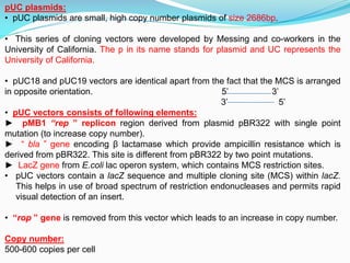
• pUC plasmids are small, high copy number plasmids of size 2686bp.
• This series of cloning vectors were developed by Messing and co-workers in the
University of California. The p in its name stands for plasmid and UC represents the
University of California.
• pUC18 and pUC19 vectors are identical apart from the fact that the MCS is arranged
in opposite orientation. 5’ 3’
3’ 5’
• pUC vectors consists of following elements:
► pMB1 “rep ” replicon region derived from plasmid pBR322 with single point
mutation (to increase copy number).
► “ bla ” gene encoding β lactamase which provide ampicillin resistance which is
derived from pBR322. This site is different from pBR322 by two point mutations.
► LacZ gene from E.coli lac operon system, which contains MCS restriction sites.
• pUC vectors contain a lacZ sequence and multiple cloning site (MCS) within lacZ.
This helps in use of broad spectrum of restriction endonucleases and permits rapid
visual detection of an insert.
• “rop ” gene is removed from this vector which leads to an increase in copy number.
Copy number:
500-600 copies per cell
 Ampicillin resistance gene (blaZ)
 Multiple cloning site inserted into the gene lacZ’ coding for the enzyme β-galactosidase
• An MCS is a short DNA sequence consisting of restriction sites for many different restriction endonucleases.
• The MCS is inserted into the lacZ sequence, which encodes the promoter and the α-peptide of β-
galactosidase.
• Insertion of the MCS into the lacZ fragment does not affect the ability of the α-peptide, while cloning DNA
fragments into the MCS does.
 Clones with foreign DNA in the MCS disrupt the ability of the cells to make β-
galactosidase
 Plate on media with a Substrate X-gal (β-galactosidase indicator) and
clones with intact β-galactosidase enzyme indicate that cells containing empty vector
without insert, will produce blue colonies
 Colorless (desirable) colonies indicates that cells contain the plasmid with foreign
DNA
• Therefore, recombinants can be detected by blue/white screening on growth medium containing X gal in
presence of IPTG as an inducer.
Cloning of insert DNA using β - galactosidase: as reporter gene
Cloning in pUC18/19:
Ω-fragment
Vector DNA
GOI
GOI- GENE OF INTEREST
β-galactosidase enzyme +
chromogenic substrate (X-gal)
Blue color colonies
MCS
 First generation plasmid cloning vectors include pBR322 and the pUC plasmids
 pUC has
 1 antibiotic resistance gene- Ampicillin resistance gene
 Variety of unique restriction sites in lacZ gene for inserting foreign DNA
 Most of these sites interrupt lacZ gene making screening straightforward
 Thus MCS in lacZ facilitates directional cloning.
Summary

Lecture on pUC18 vector

  • 1.
    pUC plasmids Dr. ManikandanKathirvel M.Sc., Ph.D., (NET) Assistant Professor, Department of Life Sciences, Kristu Jayanti College (Autonomous), (Reaccredited with "A" Grade by NAAC) Affiliated to Bengaluru North University, K. Narayanapura, Kothanur (PO) Bengaluru Email: manikandan@kristujayanti.com ORCID ID: 0000000270066334
  • 2.
    pUC plasmids: • pUCplasmids are small, high copy number plasmids of size 2686bp. • This series of cloning vectors were developed by Messing and co-workers in the University of California. The p in its name stands for plasmid and UC represents the University of California. • pUC18 and pUC19 vectors are identical apart from the fact that the MCS is arranged in opposite orientation. 5’ 3’ 3’ 5’ • pUC vectors consists of following elements: ► pMB1 “rep ” replicon region derived from plasmid pBR322 with single point mutation (to increase copy number). ► “ bla ” gene encoding β lactamase which provide ampicillin resistance which is derived from pBR322. This site is different from pBR322 by two point mutations. ► LacZ gene from E.coli lac operon system, which contains MCS restriction sites. • pUC vectors contain a lacZ sequence and multiple cloning site (MCS) within lacZ. This helps in use of broad spectrum of restriction endonucleases and permits rapid visual detection of an insert. • “rop ” gene is removed from this vector which leads to an increase in copy number. Copy number: 500-600 copies per cell
  • 3.
     Ampicillin resistancegene (blaZ)  Multiple cloning site inserted into the gene lacZ’ coding for the enzyme β-galactosidase • An MCS is a short DNA sequence consisting of restriction sites for many different restriction endonucleases. • The MCS is inserted into the lacZ sequence, which encodes the promoter and the α-peptide of β- galactosidase. • Insertion of the MCS into the lacZ fragment does not affect the ability of the α-peptide, while cloning DNA fragments into the MCS does.  Clones with foreign DNA in the MCS disrupt the ability of the cells to make β- galactosidase  Plate on media with a Substrate X-gal (β-galactosidase indicator) and clones with intact β-galactosidase enzyme indicate that cells containing empty vector without insert, will produce blue colonies  Colorless (desirable) colonies indicates that cells contain the plasmid with foreign DNA • Therefore, recombinants can be detected by blue/white screening on growth medium containing X gal in presence of IPTG as an inducer. Cloning of insert DNA using β - galactosidase: as reporter gene
  • 4.
    Cloning in pUC18/19: Ω-fragment VectorDNA GOI GOI- GENE OF INTEREST β-galactosidase enzyme + chromogenic substrate (X-gal) Blue color colonies MCS
  • 6.
     First generationplasmid cloning vectors include pBR322 and the pUC plasmids  pUC has  1 antibiotic resistance gene- Ampicillin resistance gene  Variety of unique restriction sites in lacZ gene for inserting foreign DNA  Most of these sites interrupt lacZ gene making screening straightforward  Thus MCS in lacZ facilitates directional cloning. Summary