The thymus is a two-lobed organ located in the upper chest that matures T cells from the bone marrow. T cells help fight infections. Tonsils trap and destroy bacteria in the throat and help the immune system. Lymphedema is swelling caused by damage to the lymphatic system from tumors, parasites, or surgery. Hypersensitivity refers to unwanted immune reactions and is classified into four types: Type I is immediate allergy responses, Type II involves antibodies attacking self cells, Type III occurs when immune complexes form, and Type IV is delayed hypersensitivity mediated by T cells. Allergies occur when IgE antibodies attach to mast cells and trigger histamine release in response to allergens.
Identify the organs of primary and secondary immune system- lymphoid organs, Know the functions of lymphoid organs, Understand the importance of lymphoid organs and Lymphatic circulatory system
• Gross anatomy:
– Components of the lymphatic system: lymphatic plexuses, lymphatics, lymphoid tissue
– Plan of the lymphatic system: Superficial lymphatic vessels, deep lymphatic vessels, lymph nodes, lymph trunks, cysterna chyli, lymph ducts: right lymph duct and thoracic duct.
– Lymphatic drainage of the lower limb
• Superficial inguinal lymph nodes: arrangement and drainage area.
• Deep inguinal lymph nodes: arrangement and drainage area. The popliteal lymph nodes
– Lymphatic drainage of the upper limb
• Superficial and deep lymphatics. Supratrochlear and infraclavicular lymph nodes.
• Axillary lymph nodes: arrangement and drainage area.
– Plan of the lymphatic drainage of the head and neck: deep cervical lymph nodes, inner and outer circle of lymph nodes.
• Deep cervical lymph nodes: location of the upper and lower groups, jugulodigastric node, jugulo-omohyoid, supraclavicular lymph nodes. Drainage area and efferent vessels.
• The outer circle of lymph nodes: submental, submandibular, buccal, mandibular, parotid, mastoid, occipital: location, drainage area and efferent vessels.
• The inner circle of lymph nodes: pretracheal, paratracheal and retropharyndeal.
• The tonsils and Waldeyer’s ring.
– Lymphatic drainage of the thorax:
• Lymph nodes of the chest wall: Parasternal, intercostal, and phrenic
• Lymph nodes of the mediastinum: Nodes around the division of the trachea and the main bronchi, anterior and posterior mediastinal nodes.
– Plan of lymphatic drainage of the abdomen: lumbar and intestinal lymph trunks.
• Pre-aortic lymph nodes: mesenteric, celiac, superior and inferior mesenteric lymph nodes.
• Para-aortic lymph nodes.
• MALT & Peyer’s patches.
– Lymphatic drainage in the pelvis: External and internal iliac lymph nodes, lymph nodes in fascial sheaths, sacral and common iliac lymph nodes.
• Applied anatomy
• Functional and clinical importance of the lymphatic system; Virchow’s lymph nodes; Retropharyngeal abscess; Clinical applications of enlarged thoracic lymph nodes: involvement of left recurrent laryngeal nerve and phrenic nerve. Pressure on the esophagus. Carinal lymph nodes and bronchoscopy; Communications of lymphatics between thorax and abdomen.
• Radiographic anatomy:
– Lymphangiogrms.
• Surface anatomy of palpable lymph node groups: superficial inguinal, axillary, infraclavicular, outer circle of crevical lymph nodes, deep cervical lymph nodes.
Introduction
Hypersensitivity is increased reactivity or increased sensitivity by the animal body to an antigen to
which it has been previously exposed.
The term is often used as a synonym for allergy, which describes a state of altered reactivity to an
antigen.
Hypersensitivity has been divided into categories based upon whether it can be passively transferred
by antibodies or by specifically immune lymphoid cells.
The most widely adopted current classification is that of Coombs and Gell that designates
immunoglobulin-mediated (immediate) hypersensitivity reactions as types I, II, and III, and
lymphoid cell-mediated (delayed-type) hypersensitivity/cell-mediated immunity as a type IV
reaction.
“Hypersensitivity” generally represents the “dark side,” signifying the undesirable aspects of an
immune reaction, whereas the term “immunity” implies a desirable effect.
A hypersensitive response (HR) is an anti-pathogen response in plants produced by avr-R system
activation that leads to alterations in Ca+ flux, MAPK activation, and NO and ROI formation.
There is rapid necrosis of plant cells in contact with the pathogen.
This process prevents spread of the pathogen and releases hydrolytic enzymes that facilitate injury to
the pathogen’s structural integrity.
Causes of Hypersensitivity
Immune responses that are the cause of hypersensitivity diseases may be specific for antigens from different
sources:
Autoimmunity: reactions against self antigens.
Reactions against microbes.
Reactions against non-microbial environmental antigens.
Mechanism of Hypersensitivity
Hypersensitivity diseases are commonly classified according to the type of immune response and the
effector mechanism responsible for cell and tissue injury. These mechanisms include some that are
predominantly dependent on antibodies and others predominantly dependent on T cells, although a role for
both humoral and cell-mediated immunity is often found in many hypersensitivity diseases.
Screening for the immunological diseasesduresameen24
Immunodeficiency disorders usually result from use of a medication or from a long-lasting serious disorder (such as cancer) but occasionally are inherited.
People usually have frequent, unusual, or unusually severe or prolonged infections and may develop an autoimmune disorder or cancer.
Doctors suspect immunodeficiency based on symptoms and do blood tests to identify the particular disorder.
People may be given antimicrobial drugs (such as antibiotics) to prevent and treat infections.
Immune globulin may be given if there are too few antibodies (immunoglobulins) or they are not functioning normally.
For some severe immunodeficiency disorders, stem cell transplantation is sometimes done.
Immunodeficiency disorders impair the immune system’s ability to defend the body against foreign or abnormal cells that invade or attack it (such as bacteria, viruses, fungi, and cancer cells). As a result, unusual bacterial, viral, or fungal infections or lymphomas or other cancers may develop.
Another problem is that up to 25% of people who have an immunodeficiency disorder also have an autoimmune disorder (such as immune thrombocytopenia). In an autoimmune disorder, the immune system attacks the body's own tissues. Sometimes the autoimmune disorder develops before the immunodeficiency causes any symptoms.
There are two types of immunodeficiency disorders:
Primary: These disorders are usually present at birth and are genetic disorders that are usually hereditary. They typically become evident during infancy or childhood. However, some primary immunodeficiency disorders (such as common variable immunodeficiency) are not recognized until adulthood. There are more than 100 primary immunodeficiency disorders. All are relatively rare.
Secondary: These disorders generally develop later in life and often result from use of certain medications or from another disorder, such as diabetes or human immunodeficiency virus (HIV) infection. They are more common than primary immunodeficiency disorders.
Autoimmunity and autoimmune diseases dr. ihsan alsaimarydr.Ihsan alsaimary
Dr. ihsan edan abdulkareem alsaimary
PROFESSOR IN MEDICAL MICROBIOLOGY AND MOLECULAR IMMUNOLOGY
ihsanalsaimary@gmail.com
mobile : 009647801410838
university of basrah - college of medicine - basrah -IRAQ
Ozempic: Preoperative Management of Patients on GLP-1 Receptor Agonists Saeid Safari
Preoperative Management of Patients on GLP-1 Receptor Agonists like Ozempic and Semiglutide
ASA GUIDELINE
NYSORA Guideline
2 Case Reports of Gastric Ultrasound
TEST BANK for Operations Management, 14th Edition by William J. Stevenson, Ve...kevinkariuki227
TEST BANK for Operations Management, 14th Edition by William J. Stevenson, Verified Chapters 1 - 19, Complete Newest Version.pdf
Ethanol (CH3CH2OH), or beverage alcohol, is a two-carbon alcohol
that is rapidly distributed in the body and brain. Ethanol alters many
neurochemical systems and has rewarding and addictive properties. It
is the oldest recreational drug and likely contributes to more morbidity,
mortality, and public health costs than all illicit drugs combined. The
5th edition of the Diagnostic and Statistical Manual of Mental Disorders
(DSM-5) integrates alcohol abuse and alcohol dependence into a single
disorder called alcohol use disorder (AUD), with mild, moderate,
and severe subclassifications (American Psychiatric Association, 2013).
In the DSM-5, all types of substance abuse and dependence have been
combined into a single substance use disorder (SUD) on a continuum
from mild to severe. A diagnosis of AUD requires that at least two of
the 11 DSM-5 behaviors be present within a 12-month period (mild
AUD: 2–3 criteria; moderate AUD: 4–5 criteria; severe AUD: 6–11 criteria).
The four main behavioral effects of AUD are impaired control over
drinking, negative social consequences, risky use, and altered physiological
effects (tolerance, withdrawal). This chapter presents an overview
of the prevalence and harmful consequences of AUD in the U.S.,
the systemic nature of the disease, neurocircuitry and stages of AUD,
comorbidities, fetal alcohol spectrum disorders, genetic risk factors, and
pharmacotherapies for AUD.
Tom Selleck Health: A Comprehensive Look at the Iconic Actor’s Wellness Journeygreendigital
Tom Selleck, an enduring figure in Hollywood. has captivated audiences for decades with his rugged charm, iconic moustache. and memorable roles in television and film. From his breakout role as Thomas Magnum in Magnum P.I. to his current portrayal of Frank Reagan in Blue Bloods. Selleck's career has spanned over 50 years. But beyond his professional achievements. fans have often been curious about Tom Selleck Health. especially as he has aged in the public eye.
Follow us on: Pinterest
Introduction
Many have been interested in Tom Selleck health. not only because of his enduring presence on screen but also because of the challenges. and lifestyle choices he has faced and made over the years. This article delves into the various aspects of Tom Selleck health. exploring his fitness regimen, diet, mental health. and the challenges he has encountered as he ages. We'll look at how he maintains his well-being. the health issues he has faced, and his approach to ageing .
Early Life and Career
Childhood and Athletic Beginnings
Tom Selleck was born on January 29, 1945, in Detroit, Michigan, and grew up in Sherman Oaks, California. From an early age, he was involved in sports, particularly basketball. which played a significant role in his physical development. His athletic pursuits continued into college. where he attended the University of Southern California (USC) on a basketball scholarship. This early involvement in sports laid a strong foundation for his physical health and disciplined lifestyle.
Transition to Acting
Selleck's transition from an athlete to an actor came with its physical demands. His first significant role in "Magnum P.I." required him to perform various stunts and maintain a fit appearance. This role, which he played from 1980 to 1988. necessitated a rigorous fitness routine to meet the show's demands. setting the stage for his long-term commitment to health and wellness.
Fitness Regimen
Workout Routine
Tom Selleck health and fitness regimen has evolved. adapting to his changing roles and age. During his "Magnum, P.I." days. Selleck's workouts were intense and focused on building and maintaining muscle mass. His routine included weightlifting, cardiovascular exercises. and specific training for the stunts he performed on the show.
Selleck adjusted his fitness routine as he aged to suit his body's needs. Today, his workouts focus on maintaining flexibility, strength, and cardiovascular health. He incorporates low-impact exercises such as swimming, walking, and light weightlifting. This balanced approach helps him stay fit without putting undue strain on his joints and muscles.
Importance of Flexibility and Mobility
In recent years, Selleck has emphasized the importance of flexibility and mobility in his fitness regimen. Understanding the natural decline in muscle mass and joint flexibility with age. he includes stretching and yoga in his routine. These practices help prevent injuries, improve posture, and maintain mobilit
ARTIFICIAL INTELLIGENCE IN HEALTHCARE.pdfAnujkumaranit
Artificial intelligence (AI) refers to the simulation of human intelligence processes by machines, especially computer systems. It encompasses tasks such as learning, reasoning, problem-solving, perception, and language understanding. AI technologies are revolutionizing various fields, from healthcare to finance, by enabling machines to perform tasks that typically require human intelligence.
Report Back from SGO 2024: What’s the Latest in Cervical Cancer?bkling
Are you curious about what’s new in cervical cancer research or unsure what the findings mean? Join Dr. Emily Ko, a gynecologic oncologist at Penn Medicine, to learn about the latest updates from the Society of Gynecologic Oncology (SGO) 2024 Annual Meeting on Women’s Cancer. Dr. Ko will discuss what the research presented at the conference means for you and answer your questions about the new developments.
MANAGEMENT OF ATRIOVENTRICULAR CONDUCTION BLOCK.pdfJim Jacob Roy
Cardiac conduction defects can occur due to various causes.
Atrioventricular conduction blocks ( AV blocks ) are classified into 3 types.
This document describes the acute management of AV block.
The prostate is an exocrine gland of the male mammalian reproductive system
It is a walnut-sized gland that forms part of the male reproductive system and is located in front of the rectum and just below the urinary bladder
Function is to store and secrete a clear, slightly alkaline fluid that constitutes 10-30% of the volume of the seminal fluid that along with the spermatozoa, constitutes semen
A healthy human prostate measures (4cm-vertical, by 3cm-horizontal, 2cm ant-post ).
It surrounds the urethra just below the urinary bladder. It has anterior, median, posterior and two lateral lobes
It’s work is regulated by androgens which are responsible for male sex characteristics
Generalised disease of the prostate due to hormonal derangement which leads to non malignant enlargement of the gland (increase in the number of epithelial cells and stromal tissue)to cause compression of the urethra leading to symptoms (LUTS
Couples presenting to the infertility clinic- Do they really have infertility...Sujoy Dasgupta
Dr Sujoy Dasgupta presented the study on "Couples presenting to the infertility clinic- Do they really have infertility? – The unexplored stories of non-consummation" in the 13th Congress of the Asia Pacific Initiative on Reproduction (ASPIRE 2024) at Manila on 24 May, 2024.
Flu Vaccine Alert in Bangalore Karnatakaaddon Scans
As flu season approaches, health officials in Bangalore, Karnataka, are urging residents to get their flu vaccinations. The seasonal flu, while common, can lead to severe health complications, particularly for vulnerable populations such as young children, the elderly, and those with underlying health conditions.
Dr. Vidisha Kumari, a leading epidemiologist in Bangalore, emphasizes the importance of getting vaccinated. "The flu vaccine is our best defense against the influenza virus. It not only protects individuals but also helps prevent the spread of the virus in our communities," he says.
This year, the flu season is expected to coincide with a potential increase in other respiratory illnesses. The Karnataka Health Department has launched an awareness campaign highlighting the significance of flu vaccinations. They have set up multiple vaccination centers across Bangalore, making it convenient for residents to receive their shots.
To encourage widespread vaccination, the government is also collaborating with local schools, workplaces, and community centers to facilitate vaccination drives. Special attention is being given to ensuring that the vaccine is accessible to all, including marginalized communities who may have limited access to healthcare.
Residents are reminded that the flu vaccine is safe and effective. Common side effects are mild and may include soreness at the injection site, mild fever, or muscle aches. These side effects are generally short-lived and far less severe than the flu itself.
Healthcare providers are also stressing the importance of continuing COVID-19 precautions. Wearing masks, practicing good hand hygiene, and maintaining social distancing are still crucial, especially in crowded places.
Protect yourself and your loved ones by getting vaccinated. Together, we can help keep Bangalore healthy and safe this flu season. For more information on vaccination centers and schedules, residents can visit the Karnataka Health Department’s official website or follow their social media pages.
Stay informed, stay safe, and get your flu shot today!
These lecture slides, by Dr Sidra Arshad, offer a quick overview of physiological basis of a normal electrocardiogram.
Learning objectives:
1. Define an electrocardiogram (ECG) and electrocardiography
2. Describe how dipoles generated by the heart produce the waveforms of the ECG
3. Describe the components of a normal electrocardiogram of a typical bipolar leads (limb II)
4. Differentiate between intervals and segments
5. Enlist some common indications for obtaining an ECG
Study Resources:
1. Chapter 11, Guyton and Hall Textbook of Medical Physiology, 14th edition
2. Chapter 9, Human Physiology - From Cells to Systems, Lauralee Sherwood, 9th edition
3. Chapter 29, Ganong’s Review of Medical Physiology, 26th edition
4. Electrocardiogram, StatPearls - https://www.ncbi.nlm.nih.gov/books/NBK549803/
5. ECG in Medical Practice by ABM Abdullah, 4th edition
6. ECG Basics, http://www.nataliescasebook.com/tag/e-c-g-basics
Prix Galien International 2024 Forum ProgramLevi Shapiro
June 20, 2024, Prix Galien International and Jerusalem Ethics Forum in ROME. Detailed agenda including panels:
- ADVANCES IN CARDIOLOGY: A NEW PARADIGM IS COMING
- WOMEN’S HEALTH: FERTILITY PRESERVATION
- WHAT’S NEW IN THE TREATMENT OF INFECTIOUS,
ONCOLOGICAL AND INFLAMMATORY SKIN DISEASES?
- ARTIFICIAL INTELLIGENCE AND ETHICS
- GENE THERAPY
- BEYOND BORDERS: GLOBAL INITIATIVES FOR DEMOCRATIZING LIFE SCIENCE TECHNOLOGIES AND PROMOTING ACCESS TO HEALTHCARE
- ETHICAL CHALLENGES IN LIFE SCIENCES
- Prix Galien International Awards Ceremony
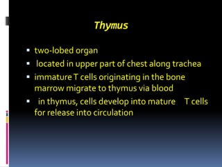
1. Thymus two-lobed organ located in upper part of chest along trachea immature T cells originating in the bone marrow migrate to thymus via blood in thymus, cells develop into mature T cells for release into circulation
4. Tonsils Two masses of tissue on either side of the back of the throat Assist the body in its defense against incoming bacteria and viruses Three types: pharyngeal tonsils Palatine tonsil lingual tonsils
7. blockage of lymph drainage Lymphedema Is a condition of localized fluid retention and tissue swelling due to damage of lymphatic system - due to tumor pressure, parasites, or surgery
8. Edema lowers colloid osmotic pressure Increase hydrostatic pressure poor lymph drainage increased capillary permeability as in inflammation
9. Hypersensitivity Refers to undesirable reactions produced by the normal immune system There are four groups classification: Type I (allergy) Type II (Antibody dependent) Type III (Immune complex diseases Type IV (Delayed type hypersensitivity)
10. Classification of Hypersensitivity Diseases Type Immunologic Mechanisms Examples Type I: Immediate hypersensitivity Type II: Antibody Mediated Type III: Immune complex Mediated Type IV: Delayed type hypersensitivity T-cell mediated IgE antibody mediated-mast cell activation and degranulation Antibodies (IgM, IgG) formed Against cell surface or matrix Ags. Complement is usually involved Immune complexes of circulating antigens.Complement and Leukocytes (neutrophils, macrophages) are often involved. Mononuclear cells (T lymphocytes, macrophages) involved. Th1 diseases Allergies (“Hay fever”), asthma,anaphylaxis Autoimmune hemolytic anemias,Myasthenia gravis, Rheumatic fever, Graves disease Serum sickness, Lupus, glomerulonephritis Diabetes, Rheumatoid Arthritis, Inflammatory Bowel Disease; Multiple sclerosis
11. Allergy Allergy is one of four forms of hypersensitivity Allergic person who has excess IgE The allergic tendency is genetically passed from parent to child Characterized by presence of large quantities if IgE (reagins) in the blood IgE attached to mast cells and basophils Antigen (allergen) react with IgE that attached to mast cell and basophil Some of mast cell produce chemical substance Attract neutrophils to the reactive site
12. Anaphylaxis Specific allergen enters vascular system Reaction occurs of allergen –IgE that attached to basophil Histamine released into the circulation Treated with epinephrine
13. Urticaria Resulting from antigen entering skin area Histamine released and causes vasodilatin and increase permeability of the capillaries
14. Hay fever The allergen- reagin reaction occurs in the nose Histamine released in response to the reaction leads to increase capillary pressure and permeability Using antihistamine prevent swelling reaction
15. Asthma The allergen- reagin reaction occurs in the bronchioles of the lungs Product released from mast cell called slow reacting substance of anaphylaxis which caused spasm of smooth muscles
16. Type II (Antibody Mediated) Antibodies produced by the immune response bind to antigen on the patients own cell surface Autoimmune hemolytic anemia: where the body immune system attacks its own RBCs leading to their destruction Drugs bind to RBC causing them to be recognized different. IgM and IgG antibodies bind to these antigens that cause cell lysis and death