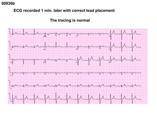
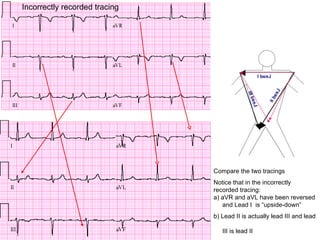
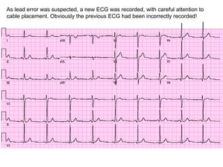
This document discusses common errors involving limb leads in electrocardiograms (ECGs), including:
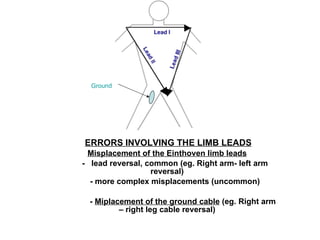
1) Lead reversal is a common error, such as switching the right and left arm leads. This can cause leads like aVR and aVL to be reversed.
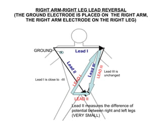
2) Misplacing the ground cable, such as attaching it to the right arm instead of the right leg, is another error.
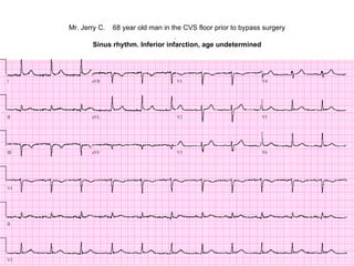
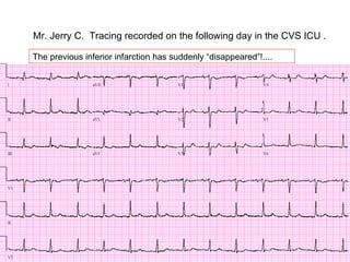
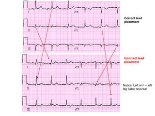
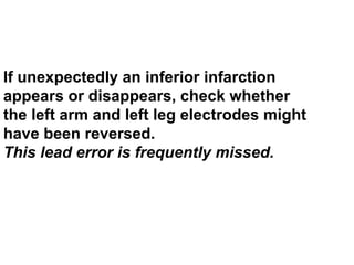
3) Complex misplacements of the limb leads are less common. Examples provided demonstrate how specific lead errors can cause abnormalities to appear or disappear on ECG tracings.