This document introduces the 2018 revision of the AO/OTA Fracture and Dislocation Classification Compendium. The revision was undertaken by the International Comprehensive Classification of Fractures and Dislocations Committee (ICCFC) to address issues, criticisms, and simplify the classification system based on fracture patterns and complexity. The revision streamlines the classification while maintaining the original principles. It standardizes terminology, separates radius and ulna classifications, integrates other validated classifications, adds a fibula classification, and places common modifiers in a universal list to simplify usage. The goal is to provide a more concise and clinically relevant compendium for classifying fractures and dislocations.


![Fundamentals of fracture classification
Classification is the process by which related groups are organized
based on similarities and differences.5
It provides the language neces-
sary to convey information among individuals to ensure standardiza-
tion. This classification process may be looked upon as the systematic
methodology of describing a fracture or dislocation. It is critical to note
that a fracture should be coded only after all the information is ob-
tained. It must be remembered that if there is doubt, then waiting until
the complete information is available is mandatory before determining
the final classification.23–28
The final classification may be delayed until
the operative procedure is completed and the fracture fully visualized.
This system provides the clinician with standardized definitions so
the verbal fracture description is precise and consistent from bone
to bone and fracture to fracture. These standard definitions and
guidelines for application assure consistency in the classification
process.16,24–37
With the improved consistency of fracture descriptions,
future investigations assessing treatment guidelines, prognosis, and
risk of complications will be more reliable and meaningful. The system
also provides a mechanism to convert the verbal description into an
alphanumeric code to allow for data storage and future recall. The use
of this alphanumeric coding scheme is absolutely necessary for multi-
center collaboration, retrospective comparison of results, international
communication, and to standardize recording information about all
fractures in a trauma database.
The classification offers several other benefits. It provides a hierarchy
of severity as the descriptions generally proceed from simple to mul-
tifragmentary fractures. This hierarchy is based on the energy of injury
or potential complexity of treatment. Ease of use is also an important
aspect for a classification. This system allows the clinician to be as
general or detailed as necessary according to their clinical or research
needs. The classification is logical, comprehensible, and does not
contain an unmanageable number of categories,
a problem that ensures poor reliability.
Principles of fracture and dislocation
classification
The principles of classification2
are based on understanding and apply-
ing standardized definitions. These definitions are universal and allow
consistency in classification and communication. Although clinical
decisions are sometimes made on incomplete information, this should
be avoided as much as possible when classifying a fracture—the more
precise the description the better the data recorded. Attention should
be paid to upper-case versus lower-case letters and ( ) versus [ ] as
this will aid in accurate fracture pattern retrieval from databases.
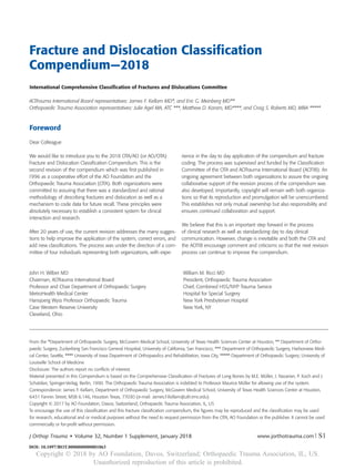
Fracture localization—bones and segments
The bone is identified (Fig 1).
Humerus 1
Thorax 16
Femur 3
Fibula 4F_
Tibia 4
Hand 7
Foot 8
Talus 81
Calcaneus 82
Navicular 83
Cuboid 84
Cuneiforms 85
Metatarsals 87
Phalanges 88
Lunate 71
Scaphoid 72
Capitate 73
Hamate 74
Trapezium 75
Other carpal bones 76
Metacarpals 77
Phalanges 78
Craniomaxillofacial bones 9
Clavicle 15
Scapula14
Acetabulum 62
32
33
Patella 34
43
Malleolus 44
4F3
42 4F2
41 4F1
31
11
12
13
14
15
2R1
2U1
2R2 2U2
2R3 2U3
Spine 5
Pelvis 6Radius 2R_
Ulna 2U_
Pelvic ring 61
Fig 1 Designation of bone location.
J Orthop Trauma • Volume 32, Number 1 Supplement, January 2018Introduction
Copyright © 2017 by AO Foundation, Davos, Switzerland; Orthopaedic Trauma Association, IL, USS4 | www.jorthotrauma.com
Copyright Ó 2018 by AO Foundation, Davos, Switzerland; Orthopaedic Trauma Association, IL, US.
Unauthorized reproduction of this article is prohibited.](https://image.slidesharecdn.com/ao2018-191013152516/85/Ao-2018-4-320.jpg)


![Universal modifiers
The universal modifiers are descriptive terms of fracture morphology,
displacement, associated injury, or location that are generalizable to
most fractures. They provide detail that are optional for users.
Universal modifiers may be added to the end of the fracture code
within square brackets, eg, [1].
Multiple universal modifiers may be contained within the same set of
squared brackets and separated by a comma.
Example: A proximal humerus fracture-dislocation with
displacement, anterior dislocation, cartilage injury, and
osteopenia = 11A1.2[2,5a,8e,9]
Example: Humerus, proximal end segment, articular or 4-part frac-
ture, with multifragmentary metaphyseal fracture and articular fracture
with an anterior dislocation = 11C3.2[5a]
List of universal modifiers
1 Nondisplaced
2 Displaced
3 Impaction
3a Articular
3b Metaphyseal
4 No impaction
5 Dislocation
5a Anterior (volar, palmar, plantar)
5b Posterior (dorsal)
5c Medial (ulnar)
5d Lateral (radial)
5e Inferior (with hip is also obturator)
5f Multidirectional
6 Subluxation/ligamentous instability
6a Anterior (volar, palmar, plantar)
6b Posterior (dorsal)
6c Medial (ulnar)
6d Lateral (radial)
6e Inferior (with hip is also obturator)
6f Multidirectional
7 Diaphyseal extension
8 Articular cartilage injury#
8a ICRS Grade 0 Normal
8b ICRS Grade 1 Superficial indentation (A) and /or superficial fissures and cracks (B)
8c ICRS Grade 2 Abnormal lesions extending down to 50% of cartilage depth
8d ICRS Grade 3 (A) Severely abnormal with defects extending down >50% of cartilage
depth; (B) down to calcified layer; (C) down to subchondral bone but not
through; (D) blisters included
8e ICRS Grade 4 Severely Abnormal Cartilage loss through subchondral bone
9 Poor bone quality
10 Replantation
11 Amputation associated with a fracture
12 Associated with a nonarthroplasty implant
13 Spiral type fracture
14 Bending type fracture
#
This grading system is used with the permission of the International Cartilage Repair Society.38
J Orthop Trauma • Volume 32, Number 1 Supplement, January 2018 Introduction
www.jorthotrauma.com | S7Copyright © 2017 by AO Foundation, Davos, Switzerland; Orthopaedic Trauma Association, IL, US
Copyright Ó 2018 by AO Foundation, Davos, Switzerland; Orthopaedic Trauma Association, IL, US.
Unauthorized reproduction of this article is prohibited.](https://image.slidesharecdn.com/ao2018-191013152516/85/Ao-2018-7-320.jpg)
![Qualifications
The fracture qualifications are descriptive terms of fracture morphology
or location that are specific to each fracture.
• All fracture classification qualifications are lower-case letters to
differentiate them from the fracture type, which is always an
upper-case letter.
• All fracture qualifications are inserted in place of the asterisk in
the fracture code as a lower-case letter within a round bracket,
eg, (a).
• Where appropriate in the classification sections, the qualification
that corresponds to an the image is bolded.
Example: Humerus, proximal end segment, articular or 4-part fracture,
with multifragmentary metaphyseal fracture and simple articular
fracture with an anterior dislocation 11C3.2(x)[5a]
The process of classification and coding a diaphyseal fracture
The process of classification and coding an end-segment fracture
Step Question Answer
1 What is the bone? Specific bone (see Fig 1 for bone number)
2 At which end is the fracture located? Proximal (1) or distal (3)
3 Type: Does the fracture enter the joint surface (type)? No—extraarticular (A), go to 5
Yes—articular (B or C), go to 4(a,b)
4a Type: If articular, is it partial (part of joint attached to
metaphysis)?
Yes (type B), go to 6
4b Type: If articular, is it complete (no part of joint attached to
metaphysis)?
Yes (type C), go to 7
5 Group: If extraarticular (A) what is the fracture pattern? Avulsion (1), simple (2), wedge or multifragmentary (3)
6 Group: If partial articular (B) what is the fracture pattern? Simple (1), split and/or depression (2), fragmentary (3)
7 Group: If complete articular (C) what is the articular fracture
pattern?
Simple (1), multifragmentary (2)
8 Subgroup: If complete articular (C) what is the metaphyseal
fracture pattern?
Simple articular with simple metaphyseal (1),
simple articular fracture with multifragmentary metaphyseal (2),
multifragmentary articular with multifragmentary metaphyseal (3)
9 Add qualifications and/or universal modifiers
Step Question Answer
1 What is the bone? Specific bone (see Fig 1 for bone number)
2 Is the fracture at the end or middle segment? Middle—diaphyseal segment (2)
3 What is the type? Simple (A), wedge (B), multifragmentary (C)
4a Group: If simple (A): What is the fracture pattern (group)? Spiral (1), oblique (2), transverse (3)
4b Group: If wedge (B): What is the fracture pattern (group)? Intact (2) or fragmentary (3)
4c Group: If multifragmentary (C): What is the fracture pattern
(group)?
Intact segmental (2) or fragmentary segmental (3)
5 Add qualifications and/or universal modifiers
J Orthop Trauma • Volume 32, Number 1 Supplement, January 2018Introduction
Copyright © 2017 by AO Foundation, Davos, Switzerland; Orthopaedic Trauma Association, IL, USS8 | www.jorthotrauma.com
Copyright Ó 2018 by AO Foundation, Davos, Switzerland; Orthopaedic Trauma Association, IL, US.
Unauthorized reproduction of this article is prohibited.](https://image.slidesharecdn.com/ao2018-191013152516/85/Ao-2018-8-320.jpg)
![Summary
Since the original publication of the AO/OTA Fracture Classification in
the 1996 Journal of Orthopaedic Trauma Compendium, there has
been important progress in fracture classification toward the goal of a
universally accepted comprehensive fracture language. The 21 years
of use of the AO/OTA compendium has demonstrated its strengths
and shortcomings. Although admirable, the process of classification
validation has been time consuming and expensive and generally
not practical in a retrospective manner for accepted classifications.
With the increased use of validated patient outcomes, a standardized
comprehensive classification of injury is necessary. The AOTIB and OTA
Classification Committee through the International Comprehensive
Classification of Fractures and Dislocations Committee realized the
need to make the compendium as comprehensive and standardized as
possible. This third compendium addresses many of the prior criticisms
as well as updating the prior editions and adding new published
classifications. These changes in content and presentation should
make the compendium more universal and simpler to use. These
standardized classification systems should make injury description
more standardized and so improve research and fracture outcomes
assessments.
The collaboration of the AOTIB and the OTA through their classification
committees has resulted in the return of the compendium copyright
to both organizations so it is available for any clinician to use without
charge. This collaboration has allowed its worldwide dissemination.
The organizations are committed to working together to continually
evaluate the compendium and revise as necessary.
Alphanumeric structure of the AO/OTA classification
Acknowledgments
The committee would like to acknowledge and gratefully thank
Jessica Schisel, Jecca Reichmuth, Marcel Erismann, and Kathleen
Caswell, all the staff of the OTA office, and the AO Education
Institute for their tremendous support and encouragement.
Bone Location Type Group Subgroup Qualifications Universal modifiers
Localization Morphology
Diagnosis
X-rays, CT scans, MRIs as required, operative findings
Qualifications are applied at asterisk as a lower-case
letter in rounded brackets after the fracture code ( ).
Universal modifiers added in square brackets after
the fracture code [ ].
Universal modifiers and qualifications are applied
when appropriate.
The committee would also like to acknowledge the important
contributions to the prior compendiums from Larry Marsh, MD,
Theddy Slongo MD, and Laurent Audige PhD.
J Orthop Trauma • Volume 32, Number 1 Supplement, January 2018 Introduction
www.jorthotrauma.com | S9Copyright © 2017 by AO Foundation, Davos, Switzerland; Orthopaedic Trauma Association, IL, US
Copyright Ó 2018 by AO Foundation, Davos, Switzerland; Orthopaedic Trauma Association, IL, US.
Unauthorized reproduction of this article is prohibited.](https://image.slidesharecdn.com/ao2018-191013152516/85/Ao-2018-9-320.jpg)


![Avulsion of ligamentum teres fracture
31C1.1
Chondral lesion
31C2.1
Depression impaction fracture
31C2.2
Split depression fracture
31C2.3
Split, infrafoveal fracture
31C1.2
Split, suprafoveal fracture
31C1.3
31C
Type: Femur, proximal end segment, femoral head fracture 31C
Group: Femur, proximal end segment, femoral head, split fracture 31C1
Subgroups:
Group: Femur, proximal end segment, femoral head, depression fracture 31C2
Subgroups:
→ Associated dislocations are coded using the dislocation direction universal modifier in square brackets [5_].
J Orthop Trauma • Volume 32, Number 1 Supplement, January 2018 Femur
Copyright © 2017 by AO Foundation, Davos, Switzerland; Orthopaedic Trauma Association, IL, USS36 | www.jorthotrauma.com
Copyright Ó 2018 by AO Foundation, Davos, Switzerland; Orthopaedic Trauma Association, IL, US.
Unauthorized reproduction of this article is prohibited.](https://image.slidesharecdn.com/ao2018-191013152516/85/Ao-2018-36-320.jpg)


![Infratectal fracture
62B1.1*
Juxtatectal fracture
62B1.2*
Transtectal fracture
62B1.3*
Anterior wall fracture
62A3.1*
High anterior column fracture
(exits along iliac crest)
62A3.2*
Low anterior column fracture
(exits below anterior superior iIiac spine
[ASIS])
62A3.3*
62B
Type: Pelvis, acetabulum, partial articular, transverse type fracture 62B
Group: Pelvis, acetabulum, partial articular, transverse type, transverse fracture 62B1
Subgroups:
Group: Pelvis, acetabulum, partial articular, isolated column or wall, anterior column or wall fracture 62A3
Subgroups:
*Qualification:
a With marginal impaction
*Qualifications:
b Associated posterior wall fracture
c Associated posterior wall fracture with marginal impaction
J Orthop Trauma • Volume 32, Number 1 Supplement, January 2018Acetabulum
www.jorthotrauma.com | S79Copyright © 2017 by AO Foundation, Davos, Switzerland; Orthopaedic Trauma Association, IL, US
Copyright Ó 2018 by AO Foundation, Davos, Switzerland; Orthopaedic Trauma Association, IL, US.
Unauthorized reproduction of this article is prohibited.](https://image.slidesharecdn.com/ao2018-191013152516/85/Ao-2018-79-320.jpg)
![With infratectal transverse component
62B2.1*
Associated with anterior wall
62B3.1
With juxtatectal transverse component
62B2.2*
High anterior column fracture
(exits along iliac crest)
62B3.2
With transtectal transverse component
62B2.3*
Low anterior column fracture
(exits below anterior superior iIiac spine
[ASIS])
62B3.3
Group: Pelvis, acetabulum, partial articular, transverse type, T fracture 62B2
Subgroups:
Group: Pelvis, acetabulum, partial articular, transverse type, with anterior column, posterior hemitransverse fracture 62B3
Subgroups:
*Qualifications:
b Associated posterior wall fracture
c Associated posterior wall fracture with marginal impaction
J Orthop Trauma • Volume 32, Number 1 Supplement, January 2018 Acetabulum
Copyright © 2017 by AO Foundation, Davos, Switzerland; Orthopaedic Trauma Association, IL, USS80 | www.jorthotrauma.com
Copyright Ó 2018 by AO Foundation, Davos, Switzerland; Orthopaedic Trauma Association, IL, US.
Unauthorized reproduction of this article is prohibited.](https://image.slidesharecdn.com/ao2018-191013152516/85/Ao-2018-80-320.jpg)
![Pelvis, acetabulum, complete articular, both
columns, high anterior column fracture
(exits along iliac crest)
62C1*
Pelvis, acetabulum, complete articular, both
columns, low anterior column fracture
(exits below anterior superior iIiac spine
[ASIS])
62C2*
Pelvis, acetabulum, complete articular, both
columns, involving the sacroiliac (SI) joint
62C3*
62C
Type: Pelvis, acetabulum, complete articular, associated both column fracture
Groups:
*Qualifications:
d Both columns simple
e Multifragmentary anterior column
f Multifragmentary posterior column
g Both columns multifragmentary
*Qualifications:
d Both columns simple
e Multifragmentary anterior column
f Multifragmentary posterior column
g Both columns multifragmentary
*Qualifications:
d Both columns simple
e Multifragmentary anterior column
f Multifragmentary posterior column
g Both columns multifragmentary
Qualifications are optional and applied to the fracture code where the asterisk is located as a lower-case letter within rounded brackets. More than one
qualification can be applied for a given fracture classification, separated by a comma. For a more detailed explanation, see the compendium introduction.
Based on the Letournel Classification of Acetabular Fractures:
Letournel E, Judet R. Fractures of the Acetabulum. Berlin: Springer–Verlag; 1993.
J Orthop Trauma • Volume 32, Number 1 Supplement, January 2018Acetabulum
www.jorthotrauma.com | S81Copyright © 2017 by AO Foundation, Davos, Switzerland; Orthopaedic Trauma Association, IL, US
Copyright Ó 2018 by AO Foundation, Davos, Switzerland; Orthopaedic Trauma Association, IL, US.
Unauthorized reproduction of this article is prohibited.](https://image.slidesharecdn.com/ao2018-191013152516/85/Ao-2018-81-320.jpg)


![Type: Scapula, glenoid fossa, multifragmentary (three or more fracture lines) 14F2
Groups:
Scapula, glenoid fossa, multifragmentary
(3 or more articular fragments), glenoid
fossa fracture
14F2.1
Scapula, glenoid fossa, multifragmentary
(3 or more articular fragments with rim exits),
central fracture dislocation
14F2.2
Qualifications are optional and applied to the fracture code where the asterisk is located as a lower-case letter within rounded brackets. More than one
qualification can be applied for a given fracture classification, separated by a comma. For a more detailed explanation, see the compendium introduction.
NOTE: Glenoid fractures with extension into the body are classified as a glenoid fracture, with the body
fracture code added to the end of the code in square brackets [ ].
Audige L, Kellam JF, Lambert S, et al. The AO Foundation and Orthopaedic
Trauma Association (AO/OTA) scapula fracture classification system: focus on body
involvement. J Shoulder Elbow Surg. 2014 Feb;23(2):189–196.
Harvey E, Audige L, Herscovici D, Jr, et al. Development and validation of the
new international classification for scapula fractures. J Orthop Trauma. 2012
Jun;26(6):364–369.
Jaeger M, Lambert S, Sudkamp NP, et al. The AO Foundation and Orthopaedic
Trauma Association (AO/OTA) scapula fracture classification system: focus on
glenoid fossa involvement. J Shoulder Elbow Surg. 2013 Apr;22(4):512–520.
References
J Orthop Trauma • Volume 32, Number 1 Supplement, January 2018 Scapula
Copyright © 2017 by AO Foundation, Davos, Switzerland; Orthopaedic Trauma Association, IL, USS104 | www.jorthotrauma.com
Copyright Ó 2018 by AO Foundation, Davos, Switzerland; Orthopaedic Trauma Association, IL, US.
Unauthorized reproduction of this article is prohibited.](https://image.slidesharecdn.com/ao2018-191013152516/85/Ao-2018-104-320.jpg)


![Dislocations
The coding is as follows:
• The first number represents the distal bone of the dislocated joint.
• The second number is 0 which represents the dislocation (with the exception of the shoulder girdle where all dislocations are 10).
• The third character (A, B, C, D, and E) is used when there are more than two bone articulations in the anatomical region.
• The direction of the dislocation is coded using the universal modifier for dislocation direction, within square brackets [5_].
Anatomical region: Shoulder girdle 10
Locations:
10
Shoulder girdle, glenohumeral
10A[5_]
Shoulder girdle, sternoclavicular
10C[5_]
Shoulder girdle, acromioclavicular
10B[5_]
Shoulder girdle, scapulothoracic
10D[5_]
Qualifications are optional and applied to the fracture code where the asterisk is located as a lower-case letter within rounded brackets. More than one
qualification can be applied for a given fracture classification, separated by a comma. For a more detailed explanation, see the compendium introduction.
www.jorthotrauma.com | S107J Orthop Trauma • Volume 32, Number 1 Supplement, January 2018
DOI: 10.1097/BOT.0000000000001057
Copyright Ó 2018 by AO Foundation, Davos, Switzerland; Orthopaedic Trauma Association, IL, US.
Unauthorized reproduction of this article is prohibited.](https://image.slidesharecdn.com/ao2018-191013152516/85/Ao-2018-107-320.jpg)
![Elbow, ulnohumeral with radiohumeral
20A[5_]
Elbow, radiohumeral
20B[5_]
Elbow, ulnohumeral
20C[5_]
Anatomical region: Elbow 20
Locations:
20
J Orthop Trauma • Volume 32, Number 1 Supplement, January 2018 Dislocations
Copyright © 2017 by AO Foundation, Davos, Switzerland; Orthopaedic Trauma Association, IL, USS108 | www.jorthotrauma.com
Copyright Ó 2018 by AO Foundation, Davos, Switzerland; Orthopaedic Trauma Association, IL, US.
Unauthorized reproduction of this article is prohibited.](https://image.slidesharecdn.com/ao2018-191013152516/85/Ao-2018-108-320.jpg)
![Anatomical region: Hip joint 30[5_]
30
J Orthop Trauma • Volume 32, Number 1 Supplement, January 2018Dislocations
www.jorthotrauma.com | S109Copyright © 2017 by AO Foundation, Davos, Switzerland; Orthopaedic Trauma Association, IL, US
Copyright Ó 2018 by AO Foundation, Davos, Switzerland; Orthopaedic Trauma Association, IL, US.
Unauthorized reproduction of this article is prohibited.](https://image.slidesharecdn.com/ao2018-191013152516/85/Ao-2018-109-320.jpg)
![Knee, tibiofemoral
40A*[5_]
Knee, patellofemoral
40B[5_]
Knee, tibiofibular proximal
40C[5_]
Anatomical region: Knee 40
Locations:
*Qualifications:
a KD1—Multiligamentous rupture with either cruciate intact
b KDII—Bicruciate rupture with collateral ligaments intact
c KDIIIM—Bicruciate rupture with medial collateral ligament rupture
d KDIIIL—Bicruciate rupture with lateral collateral ligament rupture
e KDIV—MCL, LCL, ACL, PCL rupture
f KDV— Fracture dislocation
g associated arterial injury
h associated nerve injury
More specific coding would use the fracture code and universal modifier for dislocation
and direction.1,2
40
J Orthop Trauma • Volume 32, Number 1 Supplement, January 2018 Dislocations
Copyright © 2017 by AO Foundation, Davos, Switzerland; Orthopaedic Trauma Association, IL, USS110 | www.jorthotrauma.com
Copyright Ó 2018 by AO Foundation, Davos, Switzerland; Orthopaedic Trauma Association, IL, US.
Unauthorized reproduction of this article is prohibited.](https://image.slidesharecdn.com/ao2018-191013152516/85/Ao-2018-110-320.jpg)
![Anatomical region: Hand and wrist 70
Locations:
70
Hand and wrist, distal radioulnar joint
70A[5_]
Hand and wrist, intercarpal joint
70C[5_]
Hand and wrist, phalangeal joint
70E[5_]
Hand and wrist, radiocarpal joint
70B[5_]
Hand and wrist, carpal-metacarpal joint
70D[5_]
J Orthop Trauma • Volume 32, Number 1 Supplement, January 2018Dislocations
www.jorthotrauma.com | S111Copyright © 2017 by AO Foundation, Davos, Switzerland; Orthopaedic Trauma Association, IL, US
Copyright Ó 2018 by AO Foundation, Davos, Switzerland; Orthopaedic Trauma Association, IL, US.
Unauthorized reproduction of this article is prohibited.](https://image.slidesharecdn.com/ao2018-191013152516/85/Ao-2018-111-320.jpg)
![Location: Hand and wrist, carpal-metacarpal joint 70D
Types:
70D
Hand and wrist, carpal-metacarpal joint, 1st metacarpal-trapezial joint 70D1[5_]
Hand and wrist, carpal-metacarpal joint, 2nd metacarpal-trapezoid joint 70D2[5_]
Hand and wrist, carpal-metacarpal joint, 3rd metacarpal capitate joint 70D3[5_]
Hand and wrist, carpal-metacarpal joint, 4th metacarpal hamate joint 70D4[5_]
Hand and wrist, carpal-metacarpal joint, 5th metacarpal triquetrum joint 70D5[5_]
Hand and wrist, carpal-metacarpal joint, multiple carpal-metacarpal joint 70D6[5_]
1
234
5
J Orthop Trauma • Volume 32, Number 1 Supplement, January 2018 Dislocations
Copyright © 2017 by AO Foundation, Davos, Switzerland; Orthopaedic Trauma Association, IL, USS112 | www.jorthotrauma.com
Copyright Ó 2018 by AO Foundation, Davos, Switzerland; Orthopaedic Trauma Association, IL, US.
Unauthorized reproduction of this article is prohibited.](https://image.slidesharecdn.com/ao2018-191013152516/85/Ao-2018-112-320.jpg)
![Location: Hand and wrist, phalangeal joint 70E
Type: Hand and wrist, phalangeal joint, metacarpal phalangeal joint 70E1
Groups:
Type: Hand and wrist, phalangeal joint, proximal interphalangeal joint 70E2
Groups:
Type: Hand and wrist, phalangeal joint, distal interphalangeal joint 70E3
Groups:
Type: Hand and wrist, sesamoid joint dislocation 70E4[5_ ]
Type: Hand and wrist, multiple phalangeal joint dislocations 70E5
70E
Hand and wrist, phalangeal joint, 1st metacarpal phalangeal joint 70E1.1.[5_]
Hand and wrist, phalangeal joint, 2nd metacarpal phalangeal joint 70E1.2.[5_]
Hand and wrist, phalangeal joint, 3rd metacarpal phalangeal joint 70E1.3.[5_]
Hand and wrist, phalangeal joint, 4th metacarpal phalangeal joint 70E1.4.[5_]
Hand and wrist, phalangeal joint, 5th metacarpal phalangeal joint 70E1.5.[5_]
Hand and wrist, phalangeal joint, proximal interphalangeal joint, thumb (1st) 70E2.1[5_]
Hand and wrist, phalangeal joint, proximal interphalangeal joint, index (2nd) 70E2.2[5_]
Hand and wrist, phalangeal joint, proximal interphalangeal joint, long (3rd) 70E2.3[5_]
Hand and wrist, phalangeal joint, proximal interphalangeal joint, ring (4th) 70E2.4[5_]
Hand and wrist, phalangeal joint, proximal interphalangeal joint, little (5th) 70E2.5[5_]
Hand and wrist, phalangeal joint, distal interphalangeal joint, index (2nd) 70E3.2[5_]
Hand and wrist, phalangeal joint, distal interphalangeal joint, long (3rd) 70E3.3[5_]
Hand and wrist, phalangeal joint, distal interphalangeal joint, ring (4th) 70E3.4[5_]
Hand and wrist, phalangeal joint, distal interphalangeal joint, little (5th) 70E3.5[5_]
1
70E3.__.
(distal interphalangeal joints 2−5)
70E2.__.
(proximal interphalangeal joints 1−5)
70E1.__.
(metacarpal phalangeal joints 1−5)
2
3
4
5
→ The interphanangeal joints are identified as follows: thumb = 1, index = 2, long or middle = 3, ring = 4, and little = 5.
→ The identifier is added to the code after the type code.
J Orthop Trauma • Volume 32, Number 1 Supplement, January 2018Dislocations
www.jorthotrauma.com | S113Copyright © 2017 by AO Foundation, Davos, Switzerland; Orthopaedic Trauma Association, IL, US
Copyright Ó 2018 by AO Foundation, Davos, Switzerland; Orthopaedic Trauma Association, IL, US.
Unauthorized reproduction of this article is prohibited.](https://image.slidesharecdn.com/ao2018-191013152516/85/Ao-2018-113-320.jpg)
![Foot and ankle, syndesmosis
80A[5_]
Foot and ankle, hindfoot (subtalar joint)
80C[5_]
Foot and ankle, forefoot
80E
Foot and ankle, ankle joint (tibiotalar/talocrural)
80B[5_]
Foot and ankle, midfoot
80D
Anatomical region: Foot and ankle 80D
Locations:
80
J Orthop Trauma • Volume 32, Number 1 Supplement, January 2018 Dislocations
Copyright © 2017 by AO Foundation, Davos, Switzerland; Orthopaedic Trauma Association, IL, USS114 | www.jorthotrauma.com
Copyright Ó 2018 by AO Foundation, Davos, Switzerland; Orthopaedic Trauma Association, IL, US.
Unauthorized reproduction of this article is prohibited.](https://image.slidesharecdn.com/ao2018-191013152516/85/Ao-2018-114-320.jpg)
![Location: Foot and ankle, midfoot 80D
Types:
Type: Foot and ankle, midfoot, multiple joint dislocations 80D6
Groups:
80D
Foot and ankle, midfoot, tarsal-metatarsal joint, 1st metatarsal medial cuneiform 80D5.1[5_]
Foot and ankle, midfoot, tarsal-metatarsal joint, 2nd metatarsal middle cuneiform 80D5.2[5_]
Foot and ankle, midfoot, tarsal-metatarsal joint, 3rd metatarsal lateral cuneiform 80D5.3[5_]
Foot and ankle, midfoot, tarsal-metatarsal joint, 4th metatarsal cuboid 80D5.4[5_]
Foot and ankle, midfoot, tarsal-metatarsal joint, 5th metatarsal cuboid 80D5.5[5_]
Foot and ankle, midfoot, tarsal-metatarsal joint, multiple metatarsal-tarsal 80D5.6[5_]
Foot and ankle, midfoot, multiple joint dislocations 80D6
Foot and ankle, midfoot, talonavicular joint 80D1[5_]
Foot and ankle, midfoot, calcaneocuboid joint 80D2[5_]
Foot and ankle, midfoot, navicular-cuneiform joint 80D3[5_]
Foot and ankle, midfoot, intercuneiform joint 80D4[5_]
Foot and ankle, midfoot, tarsal-metatarsal joint 80D5
Metatarsal
identifiers
1 2
3
4
5
J Orthop Trauma • Volume 32, Number 1 Supplement, January 2018Dislocations
www.jorthotrauma.com | S115Copyright © 2017 by AO Foundation, Davos, Switzerland; Orthopaedic Trauma Association, IL, US
Copyright Ó 2018 by AO Foundation, Davos, Switzerland; Orthopaedic Trauma Association, IL, US.
Unauthorized reproduction of this article is prohibited.](https://image.slidesharecdn.com/ao2018-191013152516/85/Ao-2018-115-320.jpg)
![Location: Foot and ankle, forefoot 80E
Type: Foot and ankle, forefoot, phalangeal joint 80E1
Type: Forefoot, phalangeal joint, proximal interphalangeal joint 80E2
Type: Forefoot, phalangeal joint, distal interphalangeal joint 80E3
Type: Foot and ankle, forefoot, sesamoid dislocation (any) 80E4[5_]
Type: Foot and ankle, forefoot, multiple dislocations 80E5
1. Schenck RC, Jr. The dislocated knee. Instr Course Lect. 1994;43:127–136.
2. Wascher DC. High-velocity knee dislocation with vascular injury. Treatment principles. Clin Sports Med. 2000 Jul;19(3):457–477.
Groups (by joint medial to lateral):
Groups (by joint medial to lateral):
Groups (by joint medial to lateral):
80E
Foot and ankle, forefoot, phalangeal joint, 1st metatarsal phalangeal joint 80E1.1.[5_]
Foot and ankle, forefoot, phalangeal joint, 2nd metatarsal phalangeal joint 80E1.2.[5_]
Foot and ankle, forefoot, phalangeal joint, 3rd metatarsal phalangeal joint 80E1.3.[5_]
Foot and ankle, forefoot, phalangeal joint, 4th metatarsal phalangeal joint 80E1.4.[5_]
Foot and ankle, forefoot, phalangeal joint, 5th metatarsal phalangeal joint 80E1.5.[5_]
Forefoot, phalangeal joint, proximal interphalangeal joint, 1st toe (IP joint as there is no DIP in
great toe) 80E2.1[5_]
Forefoot, phalangeal joint, proximal interphalangeal joint, 2nd toe 80E2.2.[5_]
Forefoot, phalangeal joint, proximal interphalangeal joint, 3rd toe 80E2.3.[5_]
Forefoot, phalangeal joint, proximal interphalangeal joint, 4th toe 80E2.4.[5_]
Forefoot, phalangeal joint, proximal interphalangeal joint, 5th toe 80E2.5.[5_]
Forefoot, phalangeal joint, distal interphalangeal joint, 2nd toe 80E3.2.[5_]
Forefoot, phalangeal joint, distal interphalangeal joint, 3rd toe 80E3.3.[5_]
Forefoot, phalangeal joint, distal interphalangeal joint, 4th toe 80DE3.4.[5_]
Forefoot, phalangeal joint, distal interphalangeal joint, 5th toe 80DE3.5.[5_]
Interphalangeal
joints
80E3.__.
(distal interphalangeal joints 2−5)
80E2.__.
(proximal interphalangeal joints 1−5)
80E1.__.
(metacarpal phalangeal joints 1−5)
1 2
Toe identifiers
3
4
5
References
Qualifications are optional and applied to the fracture code where the asterisk is located as a lower-case letter within rounded brackets. More than one
qualification can be applied for a given fracture classification, separated by a comma. For a more detailed explanation, see the compendium introduction.
J Orthop Trauma • Volume 32, Number 1 Supplement, January 2018 Dislocations
Copyright © 2017 by AO Foundation, Davos, Switzerland; Orthopaedic Trauma Association, IL, USS116 | www.jorthotrauma.com
Copyright Ó 2018 by AO Foundation, Davos, Switzerland; Orthopaedic Trauma Association, IL, US.
Unauthorized reproduction of this article is prohibited.](https://image.slidesharecdn.com/ao2018-191013152516/85/Ao-2018-116-320.jpg)


![Unified Classification System for
Periprosthetic Fractures (UCPF)
The UCPF is based upon the following factors:
1. The fracture location may involve either the bone supporting the implant or distant to it.
2. The stability of the components must be assessed to determine if the bone implant surface is stable prior to fracture and after fracture.
3. The adequacy of the bone stock and bone strength supporting the implant must be sufficient to support internal fixation or a revision with-
out additional major reconstruction.
4. For clinical use, the definitions and terminology of the UCPF are used. In order to maintain consistency in coding and allow easy data retriev-
al for data collection, the UCPF has been modified so that the AO/OTA bone code appears first.
5. The UCPF code follows as a qualification in square brackets.
6. Fractures about or in a bone with a nonarthroplasty implant are coded using the universal modifier [12] following the AO/OTA fracture code.
1. The bone is identified by the AO/OTA code
(see Fig 1). The fracture morphology maybe
classified in as much detail as needed.
2. The UCPF for the joint involved is added as a
modifier in square brackets [_] after the bone
code (see Fig 1).
3. The fracture type is based on the location of
the fracture in relation to the implant as follows:
• Apophysis adjacent implant with no
effect on implant stability—Type A
– Tuberosities of the humerus
– Epicondyles or olecranon of distal humerus
– Trochanters and epicondyles of femur
– Spines of the pelvis
– Poles or tips of the patella
– Tibial tuberosity and malleoli
• Bed of the implant or around the im-
plant—Type B
– Good bone no implant loosening—Type B1
– Good bone but implant loose—Type B2
– Poor bone or defect, implant loose—Type
B3
• Clear of the implant—Type C
• Dividing the bone between
two implants—Type D
• Each of the two bones supporting the
implant—Type E
• Facing and articulating with a hemiar-
throplasty—Type F
The table provides the unified codes that follow
the fracture classification.
Example: A spiral fracture about a femoral
prosthesis of a total hip, which on x-rays shows
loosening of the implant but good bone stock =
32A1[IVB2]
AO/OTA codes:
Humerus 1
Radius 2R
Ulna 2U
Carpus and hand 7
Scapula 14
Pelvis 61
Acetabulum 62
Femur 3
Patella 34
Tibia 4
Fibula 4F
Ankle 44
Foot 8
UCPF codes:
Shoulder I
Elbow II
Wrist III
Hip IV
Knee V
Ankle VI
Joint Bone
I
1
14
6
3
34
4
8
2
7
II
III
IV
V
VI
Principles
Classification and coding process
Fig 1 AO/OTA bone codes and UCPF joint codes.
www.jorthotrauma.com | S141J Orthop Trauma • Volume 32, Number 1 Supplement, January 2018
DOI: 10.1097/BOT.0000000000001068
Copyright Ó 2018 by AO Foundation, Davos, Switzerland; Orthopaedic Trauma Association, IL, US.
Unauthorized reproduction of this article is prohibited.](https://image.slidesharecdn.com/ao2018-191013152516/85/Ao-2018-141-320.jpg)


![Universal fracture modifiers for the thorax section only
• Universal modifiers may be added to the end of the fracture code
within squared brackets [1]
• Multiple universal modifiers may be contained within the same
set of squared brackets and separated by a comma and no space
[1,2,3,etc]
Lung contusion
Pneumothorax
Hemothorax
Cardiac injury
Great vessel injury
Intercostal artery injury
Soft tissue injury
1
2
3
4
5
6
7
Acknowledgements
The Thoracic segment classification has been developed with the
collaboration of the AO Foundation TK Thoracic Surgery Expert
Group. The members are:
Michael Bemelman, MD; St Elisabeth Hospital, Department of
Surgery and Trauma, Hilvarenbeekseweg 60, 5022 GC, Tilburg,
Netherlands.
Edward A Black, FRCS; Cardiothoracic Department, PO Box 1006,
Al Ain, United Arab Emirates.
Mario Gasparri, MD; SSM Health, St Mary’s Madison, Division of
Thoracic Surgery; 700 S Park Street, Madison, WI 53715, USA.
Arthur T Martella, MD; University of Pennsylvania, School of Medi-
cine, Department of Cardiothoracic Surgery; MOB 824 Main Street,
Suite 302, Phoenixville, PA 19460, USA.
Fredric M Pieracci, MD/MPH, FACS; Associate Professor of Surgery,
University of Colorado Denver SOM, 777 Bannock Street, Denver,
CO 80204, USA.
Stefan Schulz-Drost, MD, PhD, FEBS.EmSurg; Senior Surgeon,
Unfallkrankenhaus Berlin, Department of Trauma and Orthopedic
Surgery, Warener Strasse 7, 12683 Berlin, and University Hospital
Erlangen, Department of Trauma Surgery, Krankenhausstrasse 12,
91054 Erlangen, Germany.
Qualifications are optional and applied to the fracture code where the asterisk is located as a lower-case letter within rounded brackets. More than one
qualification can be applied for a given fracture classification, separated by a comma. For a more detailed explanation, see the compendium introduction.
J Orthop Trauma • Volume 32, Number 1 Supplement, January 2018Thorax
www.jorthotrauma.com | S165Copyright © 2017 by AO Foundation, Davos, Switzerland; Orthopaedic Trauma Association, IL, US
Copyright Ó 2018 by AO Foundation, Davos, Switzerland; Orthopaedic Trauma Association, IL, US.
Unauthorized reproduction of this article is prohibited.](https://image.slidesharecdn.com/ao2018-191013152516/85/Ao-2018-149-320.jpg)


![In this section, guides to help the coder classify fractures are provided.
Within each bone segment, references are made to this section if specif-
ic definitions or suggestions for coding are required.
Radius and Ulna
To facilitate the coding of radius and ulna fractures, they are coded by
the individual bone. The following guidelines are suggested:
• The location of the end segment requires that the square has as its
side dimension the widest part of the end segment, which includes
both the radius and ulna (Fig 1).
Galeazzi
Radial shaft, distal diaphysis, intact
wedge fracture = 2R2B2(c) with
dislocation of distal radio-ulnar joint
2R2B2(c,g)
Monteggia
Ulna, proximal diapyhsis, intact
wedge fracture = 2U2B2(a) with
anterior dislocation of proximal radio-
ulnar joint [5a] = 2U2B2(a,m)[5a]
Fig 1 Determine the location of the end segment.
• Each fracture is coded, stored, and searched for separately ie, as two
codes.
• Galeazzi and Monteggia fracture codes:
–Galeazzi and Monteggia fracture patterns consist of a shaft fracture
with associated joint dislocation or injury. The code for the injury
complex is the radius or ulna fracture code with a qualifier of g for
Galeazzi representing disruption of the distal radioulnar joint (DRUJ)
and m for Monteggia representing disruption of the proximal radio-
ulnar joint (PRUJ). This qualification is placed at the end of the code
in round brackets (__).
• If the coder feels that it is necessary to code for joint dislocation and
its direction, the dislocation code from the universal modifiers is
added within square brackets [5_] following the round brackets
(Fig 2).
Fig 2 Example of a Galeazzi and a Monteggia fracture.
Appendix
www.jorthotrauma.com | S167J Orthop Trauma • Volume 32, Number 1 Supplement, January 2018
DOI: 10.1097/BOT.0000000000001055
Copyright Ó 2018 by AO Foundation, Davos, Switzerland; Orthopaedic Trauma Association, IL, US.
Unauthorized reproduction of this article is prohibited.](https://image.slidesharecdn.com/ao2018-191013152516/85/Ao-2018-167-320.jpg)

![Malleolar segment
An isolated medial malleolar fracture is classified as a tibial distal end
segment partial articular fracture, 43B1.2 or 43B2.2.
If the medial malleolar fracture is associated with a lateral side ankle
injury, it is classified as a malleolar fracture, 44.
A fracture of the posterior articular margin (Volkmann) without a lesion
of the fibula is considered a fracture of the distal end segment of the
tibia ie, 43B1.1 or 43B2.1.
If a fibula fracture is part of ankle fracture it is coded as a 44. The
fibula code is used only for fibula fractures not associated with ankle
fractures.
Scapula
The four quadrants (Fig 5) are defined by the equatorial line and the
intertubercular line (maximum glenoid meridian) running from the
supraglenoid tubercle to the infraglenoid tubercle.
Dislocations
The coding is as follows:
• The first number represents the distal bone of the dislocated joint
• The second number is 0 for dislocation (with the exception of the
shoulder girdle where all dislocations are 10.
• The third letter (A, B, C, D, or E) is utilized when there are more than
two bone articulations in the anatomical region.
• The direction of the dislocation is coded using the universal mod-
ifiers for dislocation direction [5_]. By convention, the direction of the
dislocation is defined as the position of the distal bone relative to its
anatomical position.
17 Periprosthetic fracture—arthroplasty related
The importance of the Unified Classification of Periprosthetic Fractures
(UCPF) is its descriptive nature of the prosthesis-bone interface and
relationship of the fracture to the prosthesis. Consequently, the use of
the classification demands that the UCPF be used as the description
of the fracture in the clinical scenario.
To standardize the coding process for the compendium, a modification
of the UCPF was required. In collaboration with Duncan and Haddad,
an agreement was reached to have the bone fracture code described
first followed by the UCPF code enclosed in square brackets, thereby
utilizing it as a universal modifier.
References
1. Stimson LA. A Practical Treatise on Fractures and Dislocations. 8th ed.
New York and Philadelphia: Lea & Febiger; 1917:394.
2. Watson-Jones, R. Fractures and Other Bone and Joint Injuries: Second
Edition. Edinburgh: E&S Livingstone; 1941:4934.
3. Bohler, L. The Treatment of Fractures. Vol 2. New York: Grune and
Stratton; 1957:1370–1376.
4. Wilson PD, Cochrane WA. Fractures and Dislocations. Philadelphia and
London: JB Lippincott; 1925: 513–519.
5. Tang HC, Chen IJ, Yeh YC, et al. Correlation of parameters on
preoperative CT images with intra-articular soft-tissue injuries in acute
tibial plateau fractures: A review of 132 patients receiving ARIF. Injury.
2017 Mar;48(3):745–750.
6. Palm H, Jacobsen S, Sonne-Holm S, et al. Hip Fracture Study
Group. Integrity of the lateral femoral wall in intertrochanteric hip
fractures: An important predictor of a reoperation. J Bone Joint Surg Am.
2007;89:470–475.
7. Hsu CE, Shih CM, Wang CC, et al. Lateral femoral wall thickness.
A reliable predictor of post-operative lateral wall fracture in inte-
rtrochanteric fractures. Bone Joint J. 2013 Aug;95-b(8):1134–1138.
Posterior
Equatorial
line
Intertubercular
line
Anterior
Fig 5 Quadrants of the proximal glenoid fossa.
J Orthop Trauma • Volume 32, Number 1 Supplement, January 2018Appendix
www.jorthotrauma.com | S169Copyright © 2017 by AO Foundation, Davos, Switzerland; Orthopaedic Trauma Association, IL, US
Copyright Ó 2018 by AO Foundation, Davos, Switzerland; Orthopaedic Trauma Association, IL, US.
Unauthorized reproduction of this article is prohibited.](https://image.slidesharecdn.com/ao2018-191013152516/85/Ao-2018-169-320.jpg)


