INTRODUCTION
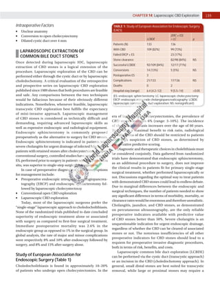
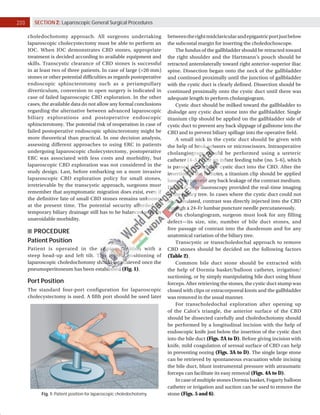
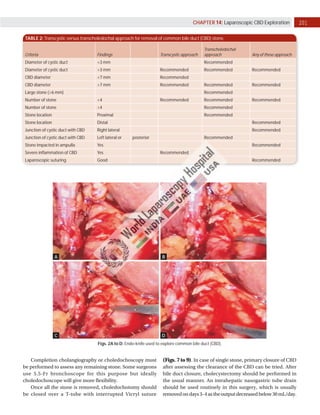
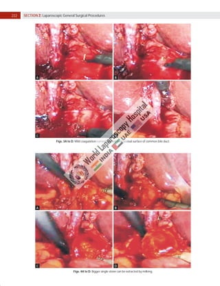
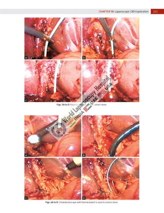
Laparoscopic exploration of the common bile duct (CBD)
is performed either for the diagnosis or the treatment of
CBD stones. CBD stones demonstrated by laparoscopic
intraoperative cholangiography (IOC) or laparoscopic
ultrasonography (LUS) are extracted either through the
cystic duct or through choledochotomy. An alternative for
the treatment of CBD stones is to perform an endoscopic
sphincterotomy either before, during, or after laparoscopic
cholecystectomy.
The main advantages of IOC during cholecystectomy are:
	
■ Detection of CBD stone
	
■ Reduction of the incidence of residual CBD stone
	
■ Delineation of the biliary anatomical variations at risk for
bile duct injury
Intraoperative cholangiogram is a highly sensitive tool
for detecting choledocholithiasis, with an overall accuracy
of 95%. Routine IOC can diagnose unsuspected CBD stone
in 1–14% (average 5%) of patients without indications for
ductal exploration.
INTRAOPERATIVE CHOLANGIOGRAPHY
Techniques of Cholangiography
Cholangiograms obtained during laparoscopy are usually
performed after catheterization of the cystic duct through
a cholangioclamp (Storz Endoscopy, USA), or inserting a
catheter through a hollow gasketed needle pinned through
the abdominal wall along the right subcostal margin.
Difficulties in catheterization of the small cystic duct have
led to consider cholecystocholangiography by direct
puncture of the gallbladder as an alternative to cystic duct
cholangiography. Cystic duct cholangiography is clearly
better than cholecystocholangiography, and fluoroscopic
imagingshouldbethestandardforIOC.Untilnow,nospecific
clinically significant complications directly attributable to
laparoscopic IOC have been reported.
Expected success rates for laparoscopic IOC are in a
90–100% range. Inability to cannulate a narrow cystic duct
is the main cause of failure. When performed after clipping
(but not cutting), the anatomical structures identified by
Laparoscopic CBD Exploration
careful dissection such as the cystic artery and the cystic
duct, a correctly interpreted IOC allows the detection of the
most frequently reported cause of CBD injury, i.e., mistaken
identification of a narrow main bile duct in place of the cystic
duct.
LAPAROSCOPIC ULTRASONOGRAPHY
Several studies on LUS have been published and
conclusions of these studies favor LUS as compared to IOC.
LUS is performed with a higher success rate, in less time,
with better specificity, but with less precision with regard
to the delineation of biliary tree anatomy. LUS is of little,
if any, helps in the diagnosis or prevention of bile duct
injuries. While detection of smaller stones by LUS should
increase its sensitivity, most of these stones are reputed
to be flushed out through the sphincter and therefore the
question arises if such small stones require any treatment
at all. Specificity of LUS is higher (less false positives) than
of IOC. When IOC and LUS were combined, there is chance
of less than 1% of false positives. The question that comes
to mind is whether LUS should be a screening test, and
IOC performed only in case of doubt or should IOC be the
screening test, and LUS used only when IOC is of doubtful
value? IOC performs better than LUS to delineate the entire
biliary tree, from the intrahepatic tree to the pancreatic
portion of the CBD. Injection of saline into the biliary tree
enhanced the images obtained by LUS, especially in the
distal portion of the bile duct.
Criteria for Routine Intraoperative
Cholangiography
Preoperative Factors
	
■ Failed endoscopic retrograde cholangiography ±
sphincterotomy
	
■ Ultrasonographic findings
	
■ CBD size (>6 mm)
	
■ Choledocholithiasis
	
■ History of jaundice or pancreatitis
	
■ Elevated bilirubin, alkaline phosphatase, and
transaminases
Prof. Dr. R. K. Mishra
199
CHAPTER 14: Laparoscopic CBD Exploration
TABLE 1: Study of European Association for Endoscopic Surgery
(EAES).
LCBDE
(ERC ± ES)
+ LC p
Patients (N) 133 136
With CBD 109 (82%) 99 (73%)
Failed ERCP ± ES 23 (17%)
Stone clearance 82/98 (84%) NS
Successful LCBDE 92/109 (84%) 12/17 (71%)
Conversions 14 (13%) 5 (5%) NS
Postoperative ES 3
Complications 21/133 17/136 NS
Deaths 0 2
Hospital stay (range) 6.4 (4.2–12) 9 (5.5–14) <0.05
(ES: endoscopic sphincterotomy; LC: laparoscopic cholecystectomy;
ERCP: endoscopic retrograde cholangiopancreaticography; LCBDE:
laparoscopic common bile duct exploration; NS: nonsignificant)
Intraoperative Factors
	
■ Unclear anatomy
	
■ Conversion to open cholecystectomy
	
■ Dilated cystic duct over 4 mm
LAPAROSCOPIC EXTRACTION OF
COMMON BILE DUCT STONES
Once detected during laparoscopic IOC, laparoscopic
extraction of CBD stones is a logical extension of the
procedure. Laparoscopic exploration of the CBD can be
performed either through the cystic duct or by laparoscopic
choledochotomy. A critical evaluation of the retrospective
and prospective series on laparoscopic CBD exploration
published since 1989 shows that both procedures are feasible
and safe. Any comparisons between the two techniques
would be fallacious because of their obviously different
indications. Nonetheless, whenever feasible, laparoscopic
transcystic CBD exploration best fulfills the expectancy
of mini-invasive approach. Laparoscopic management
of CBD stones is considered as technically difficult and
demanding, requiring advanced laparoscopic skills as
well as expensive endoscopic and radiological equipment.
Endoscopic sphincterotomy is commonly proposed
preoperatively as the alternative to surgery for CBD stones.
Endoscopic sphincterotomy is indicated in patients with
severe cholangitis for urgent drainage of infected bile, and in
patients with retained stones after cholecystectomy. In open
conventional surgery, controlled studies have not shown that
ES, performed prior to surgery in patients with CBD stones in
situ, was superior to single-step surgical management.
In case of preoperative diagnosis of CBD stone, options
for management include:
	
■ Preoperative endoscopic retrograde cholangiopancrea-
tography (ERCP) and endoscopic sphincterotomy fol-
lowed by laparoscopic cholecystectomy
	
■ Conventional open CBD exploration
	
■ Laparoscopic CBD exploration
Today, most of the laparoscopic surgeons prefer the
‘‘single-stage’’ laparoscopic approach to choledocholithiasis.
None of the randomized trials published to date concluded
superiority of endoscopic treatment alone or associated
with surgery as compared to first-line surgical treatment.
Immediate postoperative mortality was 2.6% in the
endoscopic group as opposed to 1% in the surgical group. In
global analysis, the rate of major and minor complications
were respectively 8% and 10% after endoscopy followed by
surgery, and 8% and 15% after surgery alone.
Study of European Association for
Endoscopic Surgery (Table 1)
Choledocholithiasis is found in approximately 10–20%
of patients who undergo open cholecystectomies. In the
era of laparoscopic cholecystectomies, the prevalence of
CBD stones averages 6% (range: 3–10%). The incidence
of choledocholithiasis increases over the age of 60 years.
To achieve a maximal benefit to risk ratio, radiological
investigations of the CBD should be restricted to patients
with high suspicion of CBD stones, as determined by
preoperative predictive scoring.
Diagnostic and therapeutic choices in cholelithiasis must
be considered conjointly. Data gathered from randomized
trials have demonstrated that endoscopic sphincterotomy,
as an additional procedure to surgery, does not improve
the clinical results in patients fit for primary single-stage
surgical treatment, whether performed laparoscopically or
not. Discussions regarding the optimal way to treat patients
with demonstrated CBD stones could lead to endless debate.
Due to marginal differences between the endoscopic and
surgical techniques, the number of patients needed to show
any significant difference in terms of morbidity, mortality, or
clearancerateswouldbeenormousandthereforeunrealistic.
Cholangitis, jaundice, and CBD stones, as demonstrated
on percutaneous ultrasonography, are the only reliable
preoperative indicators available with predictive value
of CBD stones better than 50%. Severe cholangitis is an
unquestionable indication for urgent endoscopic drainage,
regardless of whether the CBD can be cleared of associated
stones or not. The notorious insufficiencies of all other
preoperative indicators for CBD stones should lead to a
requiem for preoperative invasive diagnostic procedures,
both in terms of risk, benefits, and costs.
Laparoscopic common bile duct exploration (LCBDE)
can be performed via the cystic duct (transcystic approach)
or an incision in the CBD (choledochotomy approach). In
general, small distal stones are best suited for transcystic
removal, while large or proximal stones may require a
200 SECTION 2: Laparoscopic General Surgical Procedures
Fig. 1: Patient position for laparoscopic choledochotomy.
choledochotomy approach. All surgeons undertaking
laparoscopic cholecystectomy must be able to perform an
IOC. When IOC demonstrates CBD stones, appropriate
treatment is decided according to available equipment and
skills. Transcystic clearance of CBD stones is successful
in at least two of three patients. In case of large (>20 mm)
stones or other potential difficulties as regards postoperative
endoscopic sphincterotomy such as a periampullary
diverticulum, conversion to open surgery is indicated in
case of failed laparoscopic CBD exploration. In the other
cases, the available data do not allow any formal conclusions
regarding the alternative between advanced laparoscopic
biliary explorations and postoperative endoscopic
sphincterotomy. The potential risk of reoperation in case of
failed postoperative endoscopic sphincterotomy might be
more theoretical than practical. In one decision analysis,
assessing different approaches to using ERC in patients
undergoing laparoscopic cholecystectomy, postoperative
ERC was associated with less costs and morbidity, but
laparoscopic CBD exploration was not considered in the
study design. Last, before embarking on a more invasive
laparoscopic CBD exploration policy for small stones,
irretrievable by the transcystic approach, surgeons must
remember that asymptomatic migration does exist, even if
the definitive fate of small CBD stones remains unknown
at the present time. The potential security afforded by
temporary biliary drainage still has to be balanced with its
unavoidable morbidity.
PROCEDURE
Patient Position
Patient is operated in the supine position with a
steep head-up and left tilt. This typical positioning of
laparoscopic choledochotomy should be achieved once the
pneumoperitoneum has been established (Fig. 1).
Port Position
The standard four-port configuration for laparoscopic
cholecystectomy is used. A fifth port should be used later
betweentherightmidclavicularandepigastricportjustbelow
the subcostal margin for inserting the choledochoscope.
The fundus of the gallbladder should be retracted toward
the right shoulder and the Hartmann’s pouch should be
retracted anterolaterally toward right anterior–superior iliac
spine. Dissection began onto the neck of the gallbladder
and continued proximally until the junction of gallbladder
with the cystic duct is clearly defined. Dissection should be
continued proximally onto the cystic duct until there was
adequate length to perform cholangiogram.
Cystic duct should be milked toward the gallbladder to
dislodge any cystic duct stone into the gallbladder. Single
titanium clip should be applied on the gallbladder side of
cystic duct to prevent any back slippage of gallstone into the
CBD and to prevent biliary spillage into the operative field.
A small nick in the cystic duct should be given with
the help of hook scissors or microscissors. Intraoperative
cholangiogram should be performed using a ureteric
catheter (4–5 Fr) or an infant feeding tube (no. 5–6), which
is passed through the cystic duct into the CBD. After the
insertion of the catheter, a titanium clip should be applied
loosely to prevent any back leakage of the contrast medium.
Digital C-arm fluoroscopy provided the real-time imaging
of the biliary tree. In cases where the cystic duct could not
be cannulated, contrast was directly injected into the CBD
through a 24-Fr lumbar puncture needle percutaneously.
On cholangiogram, surgeon must look for any filling
defect—its size, site, number of bile duct stones, and
free passage of contrast into the duodenum and for any
anatomical variation of the biliary tree.
Transcystic or transcholedochal approach to remove
CBD stones should be decided on the following factors
(Table 2).
Common bile duct stone should be extracted with
the help of Dormia basket/balloon catheter, irrigation/
suctioning, or by simply manipulating bile duct using blunt
forceps. After retrieving the stones, the cystic duct stump was
closed with clips or extracorporeal knots and the gallbladder
was removed in the usual manner.
For transcholedochal exploration after opening up
of the Calot’s triangle, the anterior surface of the CBD
should be dissected carefully and choledochotomy should
be performed by a longitudinal incision with the help of
endoscopic knife just below the insertion of the cystic duct
into the bile duct (Figs. 2A to D). Before giving incision with
knife, mild coagulation of serosal surface of CBD can help
in preventing oozing (Figs. 3A to D). The single large stone
can be retrieved by spontaneous evacuation while incising
the bile duct, blunt instrumental pressure with atraumatic
forceps can facilitate its easy removal (Figs. 4A to D).
In case of multiple stones Dormia basket, Fogarty balloon
catheter or irrigation and suction can be used to remove the
stone (Figs. 5 and 6).
201
CHAPTER 14: Laparoscopic CBD Exploration
TABLE 2: Transcystic versus transcholedochal approach for removal of common bile duct (CBD) stone.
Criteria Findings Transcystic approach
Transcholedochal
approach Any of these approach
Diameter of cystic duct <3 mm Recommended
Diameter of cystic duct >3 mm Recommended Recommended Recommended
CBD diameter <7 mm Recommended
CBD diameter >7 mm Recommended Recommended Recommended
Large stone (>6 mm) Recommended
Number of stone <4 Recommended Recommended Recommended
Number of stone >4 Recommended
Stone location Proximal Recommended
Stone location Distal Recommended
Junction of cystic duct with CBD Right lateral Recommended
Junction of cystic duct with CBD Left lateral or posterior Recommended
Stone impacted in ampulla Yes Recommended
Severe inflammation of CBD Yes Recommended
Laparoscopic suturing Good Recommended
A
C
B
D
Figs. 2A to D: Endo-knife used to explore common bile duct (CBD).
Completion cholangiography or choledochoscopy must
be performed to assess any remaining stone. Some surgeons
use 5.5-Fr bronchoscope for this purpose but ideally
choledochoscope will give more flexibility.
Once all the stone is removed, choledochotomy should
be closed over a T-tube with interrupted Vicryl suture
(Figs. 7 to 9). In case of single stone, primary closure of CBD
after assessing the clearance of the CBD can be tried. After
bile duct closure, cholecystectomy should be performed in
the usual manner. An intrahepatic nasogastric tube drain
should be used routinely in this surgery, which is usually
removedondays3–4astheoutputdecreasedbelow30mL/day.
202 SECTION 2: Laparoscopic General Surgical Procedures
A B
C D
Figs. 3A to D: Mild coagulation current applied over serosal surface of common bile duct.
A
C
B
D
Figs. 4A to D: Bigger single stone can be extracted by milking.
203
CHAPTER 14: Laparoscopic CBD Exploration
A
C
B
D
Figs. 5A to D: Fogarty catheter used to extract stone.
A
C
B
D
Figs. 6A to D: Choledochoscope with Dormia basket is used to extract stone.
204 SECTION 2: Laparoscopic General Surgical Procedures
C
A
D
B
Figs. 7A to D: Introduction of T-tube.
T-tube Management
If a T-tube has been placed, a T-tube cholangiogram is
performed 24–48 hours postoperatively. If the initial T-tube
cholangiogram shows biliary obstruction or retained stones,
the T-tube is left open for 1–2 weeks. If a repeat T-tube
cholangiogram shows persistent stone or biliary obstruction,
ERCP or interventional radiologic procedure via the T-tube
is required to clear the duct. A normal T-tube cholangiogram
and liver function tests (LFTs) should be ascertained prior to
T-tube removal. In patients with a T-tube, the drain should
be kept until a T-tube cholangiogram shows no biliary leak
or stenosis. In patients without a T-tube, the drain may be
removed after LFTs normalize without bilious drain output;
alternatively, an HIDA scan can also be obtained to access
the biliary anatomy prior to drain removal.
COMPLICATIONS
Common complications after laparoscopic exploration are:
	
■ Bile duct leak
	
■ Persistent stones
	
■ Impacted stones
	
■ Bile duct stricture
	
■ Pancreatitis
Most of the complications are same as found in open
CBD exploration. Bile may leak from the cystic duct orifice,
cystic duct-CBD junction, or the CBD itself. It occurs in
2–16% of cases. Stones persist in the CBD in 0–5% of patients
after surgical CBD exploration. Rarely, severely impacted
stones are encountered. Strictures of the bile duct are a long-
term complication of CBD exploration that occurs in 0–0.8%
of cases. Impacted stones are difficult to remove with Fogarty
balloon catheters or wire baskets. Pancreatitis occurs in
0–3% of patients following CBD exploration due to reflux of
contrast into the pancreatic duct or ampullary obstruction
by stones, edema, or blood clots. In the latter case, ERCP
may be required to relieve the obstruction. According
to data from the American College of Surgeons National
Surgical Quality Improvement Program (ACS NSQIP)
database, laparoscopic CBD exploration has significantly
lower mortality (0.25 vs. 5.5%), surgical site infection
rate (1.2 vs. 10%), and overall morbidity rate (3.7 vs.
22%) compared with open CBD exploration. However,
the increased morbidity and mortality associated with
open exploration may be a reflection of underlying
comorbidities or more complicated disease requiring an
open approach.
205
CHAPTER 14: Laparoscopic CBD Exploration
A
C
E
B
D
F
Figs. 8A to F: T-tube is fixed with intracorporeal interrupted surgeons knot.
Fig. 9: Diagrammatic representation of T-tube fixation with intracorporeal interrupted surgeons knot.
206 SECTION 2: Laparoscopic General Surgical Procedures
BIBLIOGRAPHY
1. Altman DG. Practical Statistics for Medical Research. London,
England: Chapman and Hall; 1992.
2. Anciaux ML, Pelletier G, Attali P, Meduri B, Liguory C, Etienne
JP. Prospective study of clinical and biochemical features of
symptomatic choledocholithiasis. Dig Dis Sci. 1986;31:449-53.
3. Bansal VK, Misra MC, Rajan K, Kilambi R, Kumar S, Krishna A, et
al. Single-stage laparoscopic common bile duct exploration and
cholecystectomy versus two-stage endoscopic stone extraction
followed by laparoscopic cholecystectomy for patients with
concomitant gallbladder stones and common bile duct stones: a
randomized controlled trial. Surg Endosc. 2014;28:875.
4. Barkun AN, Barkun JS, Fried GM, Ghitulescu G, Steinmetz O,
Pham C, et al. Useful predictors of bile duct stones in patients
undergoing laparoscopic cholecystectomy. Ann Surg. 1994;
220:32-9.
5. Bates T, Ebbs SR, Harrison M, A’Hern RP. Influence of
cholecystectomy on symptoms. Br J Surg. 1991;78:964-7.
6. Behan M, Kazam E. Sonography of the common bile duct: value of
the right anterior oblique view. Am J Roentgenol. 1978;130:701-9.
7. Bhargava S, Vashisht S, Kakaria A, Tandon RK, Berry M.
Choledocholithiasis: an ultrasonic study with comparative
evaluation with ERCP/PTC. Australas Radiol. 1988;32:220-6.
8. Clemets D, Aslan S, Wilkins WE. Common bile duct gallstones,
anicteric presentation in the elderly: under-recognized but
important. Postgrad Med J. 1990;66:878-9.
9. Cooperberg PL, Li D, Wong P, Cohen MM, Burhenne HJ. Accuracy
of common hepatic duct size in the evaluation of extrahepatic
biliary obstruction. Radiology. 1980;135:141-4.
10. Cotton PB. Endoscopic retrograde cholangiopancreatography
and laparoscopic cholecystectomy. Am J Surg. 1993;165:474-8.
11. Cuscheri A, Croce E, Faggioni A, Jakimowicz J, Lacy A, Lezoche E,
et al. EAES ductal stone study. Surg Endosc. 1996;10:1130-5.
12. DePaula AL, Hashiba K, Bafatto M. Laparoscopic management of
choledocholithiasis. Surg Endosc. 1994;8:1399-1403.
13. Diehl AK, Sugarek NJ, Todd KH. Clinical evaluation for gallstone
disease: usefulness of symptoms and signs in diagnosis. Am J
Med. 1990;89:29-33.
14. Dorman JP, Franklin ME Jr, Glass JL. Laparoscopic common bile
duct exploration by choledochotomy: an effective and efficient
method of treatment of choledocholithiasis. Surg Endosc.
1998;12:926-8.
15. Edwin B, Rosseland ARR, Trondsen E. Prophylactic endoscopic
sphincterotomy as treatment. Hepatogastroenterology. 1993;
40:550-5.
16. Ferzli GS, Hurwitz JB, Massaad AA, Piperno B. Laparoscopic
common bile duct exploration: a review. J Laparoendosc Surg.
1996;6:413-19.
17. Gigot JF, Navez B, Etienne J, Cambier E, Jadoul P, Guiot P, et al.
A stratified intraoperative surgical strategy is mandatory
during laparoscopic common bile duct exploration for common
bile duct stones. Surg Endosc. 1997;11:722-8.
18. Gilliland TM, Traverso LW. Modern standards for comparison
of cholecystectomy with alternative treatments for symptomatic
cholelithiasis with emphasis on long-term relief of symptoms.
Surg Gynecol Obstet. 1990;170:39-44.
19. Graham SM, Flowers JL, Scott TR. Laparoscopic cholecystectomy
and common bile duct stones. Ann Surg. 1993;1:61-7.
20. Gross BH, Harter LP, Gore RM, Callen PW, Filly RA, Shapiro HA,
et al. Ultrasonic evaluation of common bile duct stones:
prospective comparison with endoscopic retrograde cholangio-
graphy. Radiology. 1983;146:471-4.
21. Hand DJ. Discrimination and Classification. New York, NY: John
Wiley and Sons; 1976.
22. Hauer-Jensen M, Karesen R, Nygaard K, Solheim K, Amlie E,
Havig O, et al. Predictive ability of choledocholithiasis indicators:
a prospective evaluation. Ann Surg. 1985;202:64-8.
23. Hauer-Jensen M, Karesen R, Nygaard K, Solheim K, Amlie E,
Havig O, et al. Consequences of routine preoperative
cholangiography during cholecystectomy for gallstone disease:
a prospective, randomized study. World J Surg. 1986;10:
996-1002.
24. Houdart R, Perniceni T, Darne B, Salmeron M, Simon JF.
Predicting common bile duct lithiasis: determination and
prospective validation of a model predicting low risk. Am J Surg.
1995;170:38-43.
25. HuntDR,ReiterL,ScottAJ.Preoperativeultrasoundmeasurement
of bile duct diameter: basis for selective cholangiography Aust N
Z J Surg. 1990;60:189-92.
26. Hunt DR, Scott AJ. Changes in bile duct diameter after
cholecystectomy: a 5-year prospective study. Gastroenterology.
1989;97:1485-8.
27. Hunter JG. Laparoscopic transcystic common bile duct
exploration. Am J Surg. 1992;163:53-8.
28. Jennrich RI. Stepwise discriminant analysis. In: Enslein K, Ralston
A, Wilf H (Eds). Statistical Methods for Digital Computers. New
York, NY: John Wiley and Sons; 1979.
29. Jorgensen T. Abdominal symptoms and gallstone disease: an
epidemiological investigation. Hepatology. 1989;9:856-60.
30. Kelly TR. Gallstone pancreatitis: the timing of surgery. Surgery.
1980;88:345-50.
31. Lacaine F, Corlette MB, Bismuth H. Preoperative evaluation
of the risk of common bile duct stones. Arch Surg. 1980;115:
1114-6.
32. Larson GM, Vitale GC, Casey J, Evans JS, Gilliam G, Heuser L,
et al. Multi-practice analysis of laparoscopic cholecystectomy in
1,983 patients. Am J Surg. 1991;163:221-6.
33. Liberman MA, Phillips EH, Carroll BJ, Fallas MJ, Rosenthal R,
Hiatt J. Cost-effective management of complicated choledo-
cholithiasis: laparoscopic transcystic duct exploration or
endoscopic sphincterotomy. J Am Coll Surg. 1996;182:488-94.
34. MjaalandO,RaederJ,AaseboeV,TrondsenE,BuanesT.Outpatient
laparoscopic cholecystectomy, patient satisfaction and safety:
prospective study of 200 patients. Br J Surg. 2007;31:1010-5.
35. Naude GP, Stabile BE, Bongard FS. Antegrade laparoscopic
common bile duct stone removal using a balloon-tipped
embolectomy catheter. J Am Coll Surg. 1997;184:655-7.
36. Neoptolemos JP, Carr-Locke DL, Fossard DP. Prospective
randomized study of preoperative endoscopic sphincterotomy
versus surgery alone for common bile duct stones. BMJ.
1987;294:470-4.
37. Neuhaus H, Feussner H, Ungeheuer A, Hoffmann W,
Siewert JR, Classen M. Prospective evaluation of the use of
endoscopic retrograde cholangiography prior to laparoscopic
cholecystography. Endoscopy. 1992;24:745-9.
38. Niederau C, Sonnenberg A, Mueller J. Comparison of the
extrahepaticbileductsizemeasuredbyultrasoundandbydifferent
radiographic methods. Gastroenterology. 1984;87:615-21.
39. Noble H, Tranter S, Chesworth T, Norton S, Thompson M.
A randomized, clinical trial to compare endoscopic sphin-
cterotomy and subsequent laparoscopic cholecystectomy
with primary laparoscopic bile duct exploration during
cholecystectomy in higher risk patients with choledocholithiasis.
J Laparoendosc Adv Surg Tech A. 2009;19:713.
40. O’Connor HJ, Bartlett RJ, Hamilton I, Ellis WR, Watters JK,
Lintott DJ, et al. Bile duct calibre: the value of ultrasonic and
cholangiographic measurement in the postcholecystectomy
patient. Gut. 1984;25:A576.
207
CHAPTER 14: Laparoscopic CBD Exploration
41. Onken J, Brazer S, Eisen G, et al. Accurate prediction of choledo-
cholithiasis.In:ProgramandabstractsoftheAmericanAssociation
for the Study of Liver Disease, American Gastroenterological
Association, American Society for Gastrointestinal Endoscopy,
and Society for Surgery of the Alimentary Tract group conference;
May 15–18, 1994; New Orleans, La. Abstract 699.
42. Pan L, Chen M, Ji L, Zheng L, Yan P, Fang J, et al. The safety and
efficacy of laparoscopic common bile duct exploration combined
with cholecystectomy for the management of cholecysto-
choledocholithiasis: an up-to-date meta-analysis. Ann Surg.
2018;268:247.
43. Pasanen P, Partanen K, Pikkarainen P, Alhava E, Pirinen A,
Janatuinen E. Ultrasonography, CT, and ERCP in the diagnosis of
choledochal stones. Acta Radiol. 1992;33:53-6.
44. Patwardhan RV, Smith OJ, Farmelant MH. Serum transaminase
levels and cholescintigraphic anomalies in acute biliary tract
obstruction. Arch Intern Med. 1987;147:1249-53.
45. Petelin JB. Clinical results of common bile duct exploration.
Endosc Surg Allied Technol. 1993;1:125-9.
46. Petelin JB. Laparoscopic approach to common duct pathology.
Am J Surg. 1993;165:487-91.
47. Phillips E, Daykhovsky L, Carroll B, Gershman A, Grundfest WS.
Laparoscopic cholecystectomy: instrumentation and technique.
J Laparoendosc Surg. 1990;1:3-15.
48. Phillips EH, Berci G, Carroll B, Daykhovsky L, Sackier J, Paz-
Partlow M. The importance of intraoperative cholangiography
during laparoscopic cholecystectomy. Am Surg. 1990;56:
792-5.
49. Phillips EH, Rosenthal RJ, Carroll BJ, Fallas MJ. Laparoscopic
transcystic-duct common bile duct exploration. Surg Endosc.
1994;8:1389-94.
50. Poh BR, Ho SP, Sritharan M, Yeong CC, Swan MP,
Devonshire DA, et al. Randomized clinical trial of intraoperative
endoscopic retrograde cholangiopancreatography versus
laparoscopic bile duct exploration in patients with choledo-
cholithiasis. Br J Surg. 2016;103:1117.
51. Poulose BK, Arbogast PG, Holzman MD. National analysis
of in-hospital resource utilization in choledocholithiasis
management using propensity scores. Surg Endosc. 2006;20:186.
52. ReissR,DeutschAA,NudelmanI,KottI.Statisticalvalueofvarious
clinical parameters in predicting the presence of choledochal
stones. Surg Gynecol Obstet. 1984;159:273-6.
53. Robertson GSM, Jagger C, Johnson PRV, Rathbone BJ,
Wicks AC, Lloyd DM, et al. Selection criteria for preoperative
endoscopic retrograde cholangiography in the laparoscopic era.
Arch Surg. 1996;131:89-94.
54. Ros E, Zambon D. Postcholecystectomy symptoms: a prospective
study of gallstone patients before and two years after surgery. Gut.
1987;28:1500-4.
55. Roslyn JJ, Binns GS, Hughes EF, Saunders-Kirkwood K,
Zinner MJ, Cates JA. Open cholecystectomy: a contemporary
analysis of 42,474 patients. Ann Surg. 1993;218:129-37.
56. Rosseland AR, Osnes M. Biliary concrements: the endoscopic
approach. World J Surg. 1989;13:178-85.
57. Rosseland AR, Solhaug JH. Early or delayed endoscopic
papillotomy (EPT) in gallstone pancreatitis. Ann Surg. 1984;
199:165-7.
58. Roush TS, Traverso LW. Management and long-term follow-up
of patients with positive cholangiograms during laparoscopic
cholecystectomy. Am J Surg. 1995;169:484-7.
59. Saltzstein EC, Peacock JB, Thomas MD. Preoperative bilirubin,
alkalinephosphataseandamylaselevelsaspredictorsofcommon
duct stones. Surg Gynecol Obstet. 1982;154:381-4.
60. Santo PD, Kazarian KK, Rogers JF, Bevins PA, Hall JR. Prediction
of operative cholangiography in patients undergoing elective
cholecystectomy with routine liver function chemistry. Surgery.
1985;98:7-11.
61. Sauerbrei EE, Cooperberg PL, Gordon P, Li D, Cohen MM,
Burhenne HJ. The discrepancy between radiographic and
sonographic bile duct measurements. Radiology. 1980;137:751-5.
62. Schultz LS, Kamel M, Graber JN, Hickok DF. Four-year outcome
data for 400 laparoscopic cholecystectomy patients: recognition
of persistent symptoms. Int Surg. 1994;79:205-8.
63. Sgourakis G, Karaliotas K. Laparoscopic common bile duct explo-
ration and cholecystectomy versus endoscopic stone extraction
and laparoscopic cholecystectomy for choledocholithiasis.
A prospective randomized study. Minerva Chir. 2002;57:467.
64. Stefanini P, Carboni M, Patrassi N, L origa P, De Bernardinis
G, Negro P. Factors influencing the long-term results of
cholecystectomy. Surg Gynecol Obstet. 1974;139:734-8.
65. Stiegman GV, Goff JS, Mansour A, Pearlman N, Reveille RM,
Norton L. Precholecystectomy endoscopic cholangiography
and stone removal is not superior to cholecystectomy,
cholangiography, and common bile duct exploration. Am J Surg.
1992;163:227-30.
66. Stoker ME. Common bile duct exploration in the era of
laparoscopic surgery. Arch Surg. 1995;130:265-9.
67. Taylor TV, Armstrong CP, Rimmer S, Lucas SB, Jeacock J, Gunn AA.
Prediction of choledocholithiasis using a pocket microcomputer.
Br J Surg. 1988;75:138-40.
68. Trondsen E, Edwin B, Reiertsen O, Fagertun H, Rosseland AR.
Selection criteria for endoscopic retrograde cholangiography
(ERCP) in patients with gallstone disease. World J Surg.
1995;19:852-7.
69. Vander Velpen GC, Shimi SM, Cuschieri A. Outcome after
cholecystectomy for symptomatic gallstone disease and
effect of surgical access: laparoscopic vs open approach. Gut.
1993;34:1448-51.
70. Voyles CR, Petro AB, Meena AL, Haick AJ, Koury AM. A
practical approach to laparoscopic cholecystectomy. Am J Surg.
1991;161:365-70.
71. Wegge C, Kjaergaard J. Evaluation of symptoms and signs of
gallstone disease in patients admitted with upper abdominal
pain. Scand J Gastroenterol. 1985;20:933-6.
72. Welbourn CRB, Mehta D, Armstrong CP, Gear MWL, Eyre-Brook IA.
Selective preoperative endoscopic retrograde cholangiography
with sphincterotomy avoids bile duct exploration during
laparoscopic cholecystectomy. Gut. 1995;37:576-9.

Laparoscopic CBD Exploration

  • 1.
    INTRODUCTION Laparoscopic exploration ofthe common bile duct (CBD) is performed either for the diagnosis or the treatment of CBD stones. CBD stones demonstrated by laparoscopic intraoperative cholangiography (IOC) or laparoscopic ultrasonography (LUS) are extracted either through the cystic duct or through choledochotomy. An alternative for the treatment of CBD stones is to perform an endoscopic sphincterotomy either before, during, or after laparoscopic cholecystectomy. The main advantages of IOC during cholecystectomy are: ■ Detection of CBD stone ■ Reduction of the incidence of residual CBD stone ■ Delineation of the biliary anatomical variations at risk for bile duct injury Intraoperative cholangiogram is a highly sensitive tool for detecting choledocholithiasis, with an overall accuracy of 95%. Routine IOC can diagnose unsuspected CBD stone in 1–14% (average 5%) of patients without indications for ductal exploration. INTRAOPERATIVE CHOLANGIOGRAPHY Techniques of Cholangiography Cholangiograms obtained during laparoscopy are usually performed after catheterization of the cystic duct through a cholangioclamp (Storz Endoscopy, USA), or inserting a catheter through a hollow gasketed needle pinned through the abdominal wall along the right subcostal margin. Difficulties in catheterization of the small cystic duct have led to consider cholecystocholangiography by direct puncture of the gallbladder as an alternative to cystic duct cholangiography. Cystic duct cholangiography is clearly better than cholecystocholangiography, and fluoroscopic imagingshouldbethestandardforIOC.Untilnow,nospecific clinically significant complications directly attributable to laparoscopic IOC have been reported. Expected success rates for laparoscopic IOC are in a 90–100% range. Inability to cannulate a narrow cystic duct is the main cause of failure. When performed after clipping (but not cutting), the anatomical structures identified by Laparoscopic CBD Exploration careful dissection such as the cystic artery and the cystic duct, a correctly interpreted IOC allows the detection of the most frequently reported cause of CBD injury, i.e., mistaken identification of a narrow main bile duct in place of the cystic duct. LAPAROSCOPIC ULTRASONOGRAPHY Several studies on LUS have been published and conclusions of these studies favor LUS as compared to IOC. LUS is performed with a higher success rate, in less time, with better specificity, but with less precision with regard to the delineation of biliary tree anatomy. LUS is of little, if any, helps in the diagnosis or prevention of bile duct injuries. While detection of smaller stones by LUS should increase its sensitivity, most of these stones are reputed to be flushed out through the sphincter and therefore the question arises if such small stones require any treatment at all. Specificity of LUS is higher (less false positives) than of IOC. When IOC and LUS were combined, there is chance of less than 1% of false positives. The question that comes to mind is whether LUS should be a screening test, and IOC performed only in case of doubt or should IOC be the screening test, and LUS used only when IOC is of doubtful value? IOC performs better than LUS to delineate the entire biliary tree, from the intrahepatic tree to the pancreatic portion of the CBD. Injection of saline into the biliary tree enhanced the images obtained by LUS, especially in the distal portion of the bile duct. Criteria for Routine Intraoperative Cholangiography Preoperative Factors ■ Failed endoscopic retrograde cholangiography ± sphincterotomy ■ Ultrasonographic findings ■ CBD size (>6 mm) ■ Choledocholithiasis ■ History of jaundice or pancreatitis ■ Elevated bilirubin, alkaline phosphatase, and transaminases Prof. Dr. R. K. Mishra
  • 2.
    199 CHAPTER 14: LaparoscopicCBD Exploration TABLE 1: Study of European Association for Endoscopic Surgery (EAES). LCBDE (ERC ± ES) + LC p Patients (N) 133 136 With CBD 109 (82%) 99 (73%) Failed ERCP ± ES 23 (17%) Stone clearance 82/98 (84%) NS Successful LCBDE 92/109 (84%) 12/17 (71%) Conversions 14 (13%) 5 (5%) NS Postoperative ES 3 Complications 21/133 17/136 NS Deaths 0 2 Hospital stay (range) 6.4 (4.2–12) 9 (5.5–14) <0.05 (ES: endoscopic sphincterotomy; LC: laparoscopic cholecystectomy; ERCP: endoscopic retrograde cholangiopancreaticography; LCBDE: laparoscopic common bile duct exploration; NS: nonsignificant) Intraoperative Factors ■ Unclear anatomy ■ Conversion to open cholecystectomy ■ Dilated cystic duct over 4 mm LAPAROSCOPIC EXTRACTION OF COMMON BILE DUCT STONES Once detected during laparoscopic IOC, laparoscopic extraction of CBD stones is a logical extension of the procedure. Laparoscopic exploration of the CBD can be performed either through the cystic duct or by laparoscopic choledochotomy. A critical evaluation of the retrospective and prospective series on laparoscopic CBD exploration published since 1989 shows that both procedures are feasible and safe. Any comparisons between the two techniques would be fallacious because of their obviously different indications. Nonetheless, whenever feasible, laparoscopic transcystic CBD exploration best fulfills the expectancy of mini-invasive approach. Laparoscopic management of CBD stones is considered as technically difficult and demanding, requiring advanced laparoscopic skills as well as expensive endoscopic and radiological equipment. Endoscopic sphincterotomy is commonly proposed preoperatively as the alternative to surgery for CBD stones. Endoscopic sphincterotomy is indicated in patients with severe cholangitis for urgent drainage of infected bile, and in patients with retained stones after cholecystectomy. In open conventional surgery, controlled studies have not shown that ES, performed prior to surgery in patients with CBD stones in situ, was superior to single-step surgical management. In case of preoperative diagnosis of CBD stone, options for management include: ■ Preoperative endoscopic retrograde cholangiopancrea- tography (ERCP) and endoscopic sphincterotomy fol- lowed by laparoscopic cholecystectomy ■ Conventional open CBD exploration ■ Laparoscopic CBD exploration Today, most of the laparoscopic surgeons prefer the ‘‘single-stage’’ laparoscopic approach to choledocholithiasis. None of the randomized trials published to date concluded superiority of endoscopic treatment alone or associated with surgery as compared to first-line surgical treatment. Immediate postoperative mortality was 2.6% in the endoscopic group as opposed to 1% in the surgical group. In global analysis, the rate of major and minor complications were respectively 8% and 10% after endoscopy followed by surgery, and 8% and 15% after surgery alone. Study of European Association for Endoscopic Surgery (Table 1) Choledocholithiasis is found in approximately 10–20% of patients who undergo open cholecystectomies. In the era of laparoscopic cholecystectomies, the prevalence of CBD stones averages 6% (range: 3–10%). The incidence of choledocholithiasis increases over the age of 60 years. To achieve a maximal benefit to risk ratio, radiological investigations of the CBD should be restricted to patients with high suspicion of CBD stones, as determined by preoperative predictive scoring. Diagnostic and therapeutic choices in cholelithiasis must be considered conjointly. Data gathered from randomized trials have demonstrated that endoscopic sphincterotomy, as an additional procedure to surgery, does not improve the clinical results in patients fit for primary single-stage surgical treatment, whether performed laparoscopically or not. Discussions regarding the optimal way to treat patients with demonstrated CBD stones could lead to endless debate. Due to marginal differences between the endoscopic and surgical techniques, the number of patients needed to show any significant difference in terms of morbidity, mortality, or clearancerateswouldbeenormousandthereforeunrealistic. Cholangitis, jaundice, and CBD stones, as demonstrated on percutaneous ultrasonography, are the only reliable preoperative indicators available with predictive value of CBD stones better than 50%. Severe cholangitis is an unquestionable indication for urgent endoscopic drainage, regardless of whether the CBD can be cleared of associated stones or not. The notorious insufficiencies of all other preoperative indicators for CBD stones should lead to a requiem for preoperative invasive diagnostic procedures, both in terms of risk, benefits, and costs. Laparoscopic common bile duct exploration (LCBDE) can be performed via the cystic duct (transcystic approach) or an incision in the CBD (choledochotomy approach). In general, small distal stones are best suited for transcystic removal, while large or proximal stones may require a
  • 3.
    200 SECTION 2:Laparoscopic General Surgical Procedures Fig. 1: Patient position for laparoscopic choledochotomy. choledochotomy approach. All surgeons undertaking laparoscopic cholecystectomy must be able to perform an IOC. When IOC demonstrates CBD stones, appropriate treatment is decided according to available equipment and skills. Transcystic clearance of CBD stones is successful in at least two of three patients. In case of large (>20 mm) stones or other potential difficulties as regards postoperative endoscopic sphincterotomy such as a periampullary diverticulum, conversion to open surgery is indicated in case of failed laparoscopic CBD exploration. In the other cases, the available data do not allow any formal conclusions regarding the alternative between advanced laparoscopic biliary explorations and postoperative endoscopic sphincterotomy. The potential risk of reoperation in case of failed postoperative endoscopic sphincterotomy might be more theoretical than practical. In one decision analysis, assessing different approaches to using ERC in patients undergoing laparoscopic cholecystectomy, postoperative ERC was associated with less costs and morbidity, but laparoscopic CBD exploration was not considered in the study design. Last, before embarking on a more invasive laparoscopic CBD exploration policy for small stones, irretrievable by the transcystic approach, surgeons must remember that asymptomatic migration does exist, even if the definitive fate of small CBD stones remains unknown at the present time. The potential security afforded by temporary biliary drainage still has to be balanced with its unavoidable morbidity. PROCEDURE Patient Position Patient is operated in the supine position with a steep head-up and left tilt. This typical positioning of laparoscopic choledochotomy should be achieved once the pneumoperitoneum has been established (Fig. 1). Port Position The standard four-port configuration for laparoscopic cholecystectomy is used. A fifth port should be used later betweentherightmidclavicularandepigastricportjustbelow the subcostal margin for inserting the choledochoscope. The fundus of the gallbladder should be retracted toward the right shoulder and the Hartmann’s pouch should be retracted anterolaterally toward right anterior–superior iliac spine. Dissection began onto the neck of the gallbladder and continued proximally until the junction of gallbladder with the cystic duct is clearly defined. Dissection should be continued proximally onto the cystic duct until there was adequate length to perform cholangiogram. Cystic duct should be milked toward the gallbladder to dislodge any cystic duct stone into the gallbladder. Single titanium clip should be applied on the gallbladder side of cystic duct to prevent any back slippage of gallstone into the CBD and to prevent biliary spillage into the operative field. A small nick in the cystic duct should be given with the help of hook scissors or microscissors. Intraoperative cholangiogram should be performed using a ureteric catheter (4–5 Fr) or an infant feeding tube (no. 5–6), which is passed through the cystic duct into the CBD. After the insertion of the catheter, a titanium clip should be applied loosely to prevent any back leakage of the contrast medium. Digital C-arm fluoroscopy provided the real-time imaging of the biliary tree. In cases where the cystic duct could not be cannulated, contrast was directly injected into the CBD through a 24-Fr lumbar puncture needle percutaneously. On cholangiogram, surgeon must look for any filling defect—its size, site, number of bile duct stones, and free passage of contrast into the duodenum and for any anatomical variation of the biliary tree. Transcystic or transcholedochal approach to remove CBD stones should be decided on the following factors (Table 2). Common bile duct stone should be extracted with the help of Dormia basket/balloon catheter, irrigation/ suctioning, or by simply manipulating bile duct using blunt forceps. After retrieving the stones, the cystic duct stump was closed with clips or extracorporeal knots and the gallbladder was removed in the usual manner. For transcholedochal exploration after opening up of the Calot’s triangle, the anterior surface of the CBD should be dissected carefully and choledochotomy should be performed by a longitudinal incision with the help of endoscopic knife just below the insertion of the cystic duct into the bile duct (Figs. 2A to D). Before giving incision with knife, mild coagulation of serosal surface of CBD can help in preventing oozing (Figs. 3A to D). The single large stone can be retrieved by spontaneous evacuation while incising the bile duct, blunt instrumental pressure with atraumatic forceps can facilitate its easy removal (Figs. 4A to D). In case of multiple stones Dormia basket, Fogarty balloon catheter or irrigation and suction can be used to remove the stone (Figs. 5 and 6).
  • 4.
    201 CHAPTER 14: LaparoscopicCBD Exploration TABLE 2: Transcystic versus transcholedochal approach for removal of common bile duct (CBD) stone. Criteria Findings Transcystic approach Transcholedochal approach Any of these approach Diameter of cystic duct <3 mm Recommended Diameter of cystic duct >3 mm Recommended Recommended Recommended CBD diameter <7 mm Recommended CBD diameter >7 mm Recommended Recommended Recommended Large stone (>6 mm) Recommended Number of stone <4 Recommended Recommended Recommended Number of stone >4 Recommended Stone location Proximal Recommended Stone location Distal Recommended Junction of cystic duct with CBD Right lateral Recommended Junction of cystic duct with CBD Left lateral or posterior Recommended Stone impacted in ampulla Yes Recommended Severe inflammation of CBD Yes Recommended Laparoscopic suturing Good Recommended A C B D Figs. 2A to D: Endo-knife used to explore common bile duct (CBD). Completion cholangiography or choledochoscopy must be performed to assess any remaining stone. Some surgeons use 5.5-Fr bronchoscope for this purpose but ideally choledochoscope will give more flexibility. Once all the stone is removed, choledochotomy should be closed over a T-tube with interrupted Vicryl suture (Figs. 7 to 9). In case of single stone, primary closure of CBD after assessing the clearance of the CBD can be tried. After bile duct closure, cholecystectomy should be performed in the usual manner. An intrahepatic nasogastric tube drain should be used routinely in this surgery, which is usually removedondays3–4astheoutputdecreasedbelow30mL/day.
  • 5.
    202 SECTION 2:Laparoscopic General Surgical Procedures A B C D Figs. 3A to D: Mild coagulation current applied over serosal surface of common bile duct. A C B D Figs. 4A to D: Bigger single stone can be extracted by milking.
  • 6.
    203 CHAPTER 14: LaparoscopicCBD Exploration A C B D Figs. 5A to D: Fogarty catheter used to extract stone. A C B D Figs. 6A to D: Choledochoscope with Dormia basket is used to extract stone.
  • 7.
    204 SECTION 2:Laparoscopic General Surgical Procedures C A D B Figs. 7A to D: Introduction of T-tube. T-tube Management If a T-tube has been placed, a T-tube cholangiogram is performed 24–48 hours postoperatively. If the initial T-tube cholangiogram shows biliary obstruction or retained stones, the T-tube is left open for 1–2 weeks. If a repeat T-tube cholangiogram shows persistent stone or biliary obstruction, ERCP or interventional radiologic procedure via the T-tube is required to clear the duct. A normal T-tube cholangiogram and liver function tests (LFTs) should be ascertained prior to T-tube removal. In patients with a T-tube, the drain should be kept until a T-tube cholangiogram shows no biliary leak or stenosis. In patients without a T-tube, the drain may be removed after LFTs normalize without bilious drain output; alternatively, an HIDA scan can also be obtained to access the biliary anatomy prior to drain removal. COMPLICATIONS Common complications after laparoscopic exploration are: ■ Bile duct leak ■ Persistent stones ■ Impacted stones ■ Bile duct stricture ■ Pancreatitis Most of the complications are same as found in open CBD exploration. Bile may leak from the cystic duct orifice, cystic duct-CBD junction, or the CBD itself. It occurs in 2–16% of cases. Stones persist in the CBD in 0–5% of patients after surgical CBD exploration. Rarely, severely impacted stones are encountered. Strictures of the bile duct are a long- term complication of CBD exploration that occurs in 0–0.8% of cases. Impacted stones are difficult to remove with Fogarty balloon catheters or wire baskets. Pancreatitis occurs in 0–3% of patients following CBD exploration due to reflux of contrast into the pancreatic duct or ampullary obstruction by stones, edema, or blood clots. In the latter case, ERCP may be required to relieve the obstruction. According to data from the American College of Surgeons National Surgical Quality Improvement Program (ACS NSQIP) database, laparoscopic CBD exploration has significantly lower mortality (0.25 vs. 5.5%), surgical site infection rate (1.2 vs. 10%), and overall morbidity rate (3.7 vs. 22%) compared with open CBD exploration. However, the increased morbidity and mortality associated with open exploration may be a reflection of underlying comorbidities or more complicated disease requiring an open approach.
  • 8.
    205 CHAPTER 14: LaparoscopicCBD Exploration A C E B D F Figs. 8A to F: T-tube is fixed with intracorporeal interrupted surgeons knot. Fig. 9: Diagrammatic representation of T-tube fixation with intracorporeal interrupted surgeons knot.
  • 9.
    206 SECTION 2:Laparoscopic General Surgical Procedures BIBLIOGRAPHY 1. Altman DG. Practical Statistics for Medical Research. London, England: Chapman and Hall; 1992. 2. Anciaux ML, Pelletier G, Attali P, Meduri B, Liguory C, Etienne JP. Prospective study of clinical and biochemical features of symptomatic choledocholithiasis. Dig Dis Sci. 1986;31:449-53. 3. Bansal VK, Misra MC, Rajan K, Kilambi R, Kumar S, Krishna A, et al. Single-stage laparoscopic common bile duct exploration and cholecystectomy versus two-stage endoscopic stone extraction followed by laparoscopic cholecystectomy for patients with concomitant gallbladder stones and common bile duct stones: a randomized controlled trial. Surg Endosc. 2014;28:875. 4. Barkun AN, Barkun JS, Fried GM, Ghitulescu G, Steinmetz O, Pham C, et al. Useful predictors of bile duct stones in patients undergoing laparoscopic cholecystectomy. Ann Surg. 1994; 220:32-9. 5. Bates T, Ebbs SR, Harrison M, A’Hern RP. Influence of cholecystectomy on symptoms. Br J Surg. 1991;78:964-7. 6. Behan M, Kazam E. Sonography of the common bile duct: value of the right anterior oblique view. Am J Roentgenol. 1978;130:701-9. 7. Bhargava S, Vashisht S, Kakaria A, Tandon RK, Berry M. Choledocholithiasis: an ultrasonic study with comparative evaluation with ERCP/PTC. Australas Radiol. 1988;32:220-6. 8. Clemets D, Aslan S, Wilkins WE. Common bile duct gallstones, anicteric presentation in the elderly: under-recognized but important. Postgrad Med J. 1990;66:878-9. 9. Cooperberg PL, Li D, Wong P, Cohen MM, Burhenne HJ. Accuracy of common hepatic duct size in the evaluation of extrahepatic biliary obstruction. Radiology. 1980;135:141-4. 10. Cotton PB. Endoscopic retrograde cholangiopancreatography and laparoscopic cholecystectomy. Am J Surg. 1993;165:474-8. 11. Cuscheri A, Croce E, Faggioni A, Jakimowicz J, Lacy A, Lezoche E, et al. EAES ductal stone study. Surg Endosc. 1996;10:1130-5. 12. DePaula AL, Hashiba K, Bafatto M. Laparoscopic management of choledocholithiasis. Surg Endosc. 1994;8:1399-1403. 13. Diehl AK, Sugarek NJ, Todd KH. Clinical evaluation for gallstone disease: usefulness of symptoms and signs in diagnosis. Am J Med. 1990;89:29-33. 14. Dorman JP, Franklin ME Jr, Glass JL. Laparoscopic common bile duct exploration by choledochotomy: an effective and efficient method of treatment of choledocholithiasis. Surg Endosc. 1998;12:926-8. 15. Edwin B, Rosseland ARR, Trondsen E. Prophylactic endoscopic sphincterotomy as treatment. Hepatogastroenterology. 1993; 40:550-5. 16. Ferzli GS, Hurwitz JB, Massaad AA, Piperno B. Laparoscopic common bile duct exploration: a review. J Laparoendosc Surg. 1996;6:413-19. 17. Gigot JF, Navez B, Etienne J, Cambier E, Jadoul P, Guiot P, et al. A stratified intraoperative surgical strategy is mandatory during laparoscopic common bile duct exploration for common bile duct stones. Surg Endosc. 1997;11:722-8. 18. Gilliland TM, Traverso LW. Modern standards for comparison of cholecystectomy with alternative treatments for symptomatic cholelithiasis with emphasis on long-term relief of symptoms. Surg Gynecol Obstet. 1990;170:39-44. 19. Graham SM, Flowers JL, Scott TR. Laparoscopic cholecystectomy and common bile duct stones. Ann Surg. 1993;1:61-7. 20. Gross BH, Harter LP, Gore RM, Callen PW, Filly RA, Shapiro HA, et al. Ultrasonic evaluation of common bile duct stones: prospective comparison with endoscopic retrograde cholangio- graphy. Radiology. 1983;146:471-4. 21. Hand DJ. Discrimination and Classification. New York, NY: John Wiley and Sons; 1976. 22. Hauer-Jensen M, Karesen R, Nygaard K, Solheim K, Amlie E, Havig O, et al. Predictive ability of choledocholithiasis indicators: a prospective evaluation. Ann Surg. 1985;202:64-8. 23. Hauer-Jensen M, Karesen R, Nygaard K, Solheim K, Amlie E, Havig O, et al. Consequences of routine preoperative cholangiography during cholecystectomy for gallstone disease: a prospective, randomized study. World J Surg. 1986;10: 996-1002. 24. Houdart R, Perniceni T, Darne B, Salmeron M, Simon JF. Predicting common bile duct lithiasis: determination and prospective validation of a model predicting low risk. Am J Surg. 1995;170:38-43. 25. HuntDR,ReiterL,ScottAJ.Preoperativeultrasoundmeasurement of bile duct diameter: basis for selective cholangiography Aust N Z J Surg. 1990;60:189-92. 26. Hunt DR, Scott AJ. Changes in bile duct diameter after cholecystectomy: a 5-year prospective study. Gastroenterology. 1989;97:1485-8. 27. Hunter JG. Laparoscopic transcystic common bile duct exploration. Am J Surg. 1992;163:53-8. 28. Jennrich RI. Stepwise discriminant analysis. In: Enslein K, Ralston A, Wilf H (Eds). Statistical Methods for Digital Computers. New York, NY: John Wiley and Sons; 1979. 29. Jorgensen T. Abdominal symptoms and gallstone disease: an epidemiological investigation. Hepatology. 1989;9:856-60. 30. Kelly TR. Gallstone pancreatitis: the timing of surgery. Surgery. 1980;88:345-50. 31. Lacaine F, Corlette MB, Bismuth H. Preoperative evaluation of the risk of common bile duct stones. Arch Surg. 1980;115: 1114-6. 32. Larson GM, Vitale GC, Casey J, Evans JS, Gilliam G, Heuser L, et al. Multi-practice analysis of laparoscopic cholecystectomy in 1,983 patients. Am J Surg. 1991;163:221-6. 33. Liberman MA, Phillips EH, Carroll BJ, Fallas MJ, Rosenthal R, Hiatt J. Cost-effective management of complicated choledo- cholithiasis: laparoscopic transcystic duct exploration or endoscopic sphincterotomy. J Am Coll Surg. 1996;182:488-94. 34. MjaalandO,RaederJ,AaseboeV,TrondsenE,BuanesT.Outpatient laparoscopic cholecystectomy, patient satisfaction and safety: prospective study of 200 patients. Br J Surg. 2007;31:1010-5. 35. Naude GP, Stabile BE, Bongard FS. Antegrade laparoscopic common bile duct stone removal using a balloon-tipped embolectomy catheter. J Am Coll Surg. 1997;184:655-7. 36. Neoptolemos JP, Carr-Locke DL, Fossard DP. Prospective randomized study of preoperative endoscopic sphincterotomy versus surgery alone for common bile duct stones. BMJ. 1987;294:470-4. 37. Neuhaus H, Feussner H, Ungeheuer A, Hoffmann W, Siewert JR, Classen M. Prospective evaluation of the use of endoscopic retrograde cholangiography prior to laparoscopic cholecystography. Endoscopy. 1992;24:745-9. 38. Niederau C, Sonnenberg A, Mueller J. Comparison of the extrahepaticbileductsizemeasuredbyultrasoundandbydifferent radiographic methods. Gastroenterology. 1984;87:615-21. 39. Noble H, Tranter S, Chesworth T, Norton S, Thompson M. A randomized, clinical trial to compare endoscopic sphin- cterotomy and subsequent laparoscopic cholecystectomy with primary laparoscopic bile duct exploration during cholecystectomy in higher risk patients with choledocholithiasis. J Laparoendosc Adv Surg Tech A. 2009;19:713. 40. O’Connor HJ, Bartlett RJ, Hamilton I, Ellis WR, Watters JK, Lintott DJ, et al. Bile duct calibre: the value of ultrasonic and cholangiographic measurement in the postcholecystectomy patient. Gut. 1984;25:A576.
  • 10.
    207 CHAPTER 14: LaparoscopicCBD Exploration 41. Onken J, Brazer S, Eisen G, et al. Accurate prediction of choledo- cholithiasis.In:ProgramandabstractsoftheAmericanAssociation for the Study of Liver Disease, American Gastroenterological Association, American Society for Gastrointestinal Endoscopy, and Society for Surgery of the Alimentary Tract group conference; May 15–18, 1994; New Orleans, La. Abstract 699. 42. Pan L, Chen M, Ji L, Zheng L, Yan P, Fang J, et al. The safety and efficacy of laparoscopic common bile duct exploration combined with cholecystectomy for the management of cholecysto- choledocholithiasis: an up-to-date meta-analysis. Ann Surg. 2018;268:247. 43. Pasanen P, Partanen K, Pikkarainen P, Alhava E, Pirinen A, Janatuinen E. Ultrasonography, CT, and ERCP in the diagnosis of choledochal stones. Acta Radiol. 1992;33:53-6. 44. Patwardhan RV, Smith OJ, Farmelant MH. Serum transaminase levels and cholescintigraphic anomalies in acute biliary tract obstruction. Arch Intern Med. 1987;147:1249-53. 45. Petelin JB. Clinical results of common bile duct exploration. Endosc Surg Allied Technol. 1993;1:125-9. 46. Petelin JB. Laparoscopic approach to common duct pathology. Am J Surg. 1993;165:487-91. 47. Phillips E, Daykhovsky L, Carroll B, Gershman A, Grundfest WS. Laparoscopic cholecystectomy: instrumentation and technique. J Laparoendosc Surg. 1990;1:3-15. 48. Phillips EH, Berci G, Carroll B, Daykhovsky L, Sackier J, Paz- Partlow M. The importance of intraoperative cholangiography during laparoscopic cholecystectomy. Am Surg. 1990;56: 792-5. 49. Phillips EH, Rosenthal RJ, Carroll BJ, Fallas MJ. Laparoscopic transcystic-duct common bile duct exploration. Surg Endosc. 1994;8:1389-94. 50. Poh BR, Ho SP, Sritharan M, Yeong CC, Swan MP, Devonshire DA, et al. Randomized clinical trial of intraoperative endoscopic retrograde cholangiopancreatography versus laparoscopic bile duct exploration in patients with choledo- cholithiasis. Br J Surg. 2016;103:1117. 51. Poulose BK, Arbogast PG, Holzman MD. National analysis of in-hospital resource utilization in choledocholithiasis management using propensity scores. Surg Endosc. 2006;20:186. 52. ReissR,DeutschAA,NudelmanI,KottI.Statisticalvalueofvarious clinical parameters in predicting the presence of choledochal stones. Surg Gynecol Obstet. 1984;159:273-6. 53. Robertson GSM, Jagger C, Johnson PRV, Rathbone BJ, Wicks AC, Lloyd DM, et al. Selection criteria for preoperative endoscopic retrograde cholangiography in the laparoscopic era. Arch Surg. 1996;131:89-94. 54. Ros E, Zambon D. Postcholecystectomy symptoms: a prospective study of gallstone patients before and two years after surgery. Gut. 1987;28:1500-4. 55. Roslyn JJ, Binns GS, Hughes EF, Saunders-Kirkwood K, Zinner MJ, Cates JA. Open cholecystectomy: a contemporary analysis of 42,474 patients. Ann Surg. 1993;218:129-37. 56. Rosseland AR, Osnes M. Biliary concrements: the endoscopic approach. World J Surg. 1989;13:178-85. 57. Rosseland AR, Solhaug JH. Early or delayed endoscopic papillotomy (EPT) in gallstone pancreatitis. Ann Surg. 1984; 199:165-7. 58. Roush TS, Traverso LW. Management and long-term follow-up of patients with positive cholangiograms during laparoscopic cholecystectomy. Am J Surg. 1995;169:484-7. 59. Saltzstein EC, Peacock JB, Thomas MD. Preoperative bilirubin, alkalinephosphataseandamylaselevelsaspredictorsofcommon duct stones. Surg Gynecol Obstet. 1982;154:381-4. 60. Santo PD, Kazarian KK, Rogers JF, Bevins PA, Hall JR. Prediction of operative cholangiography in patients undergoing elective cholecystectomy with routine liver function chemistry. Surgery. 1985;98:7-11. 61. Sauerbrei EE, Cooperberg PL, Gordon P, Li D, Cohen MM, Burhenne HJ. The discrepancy between radiographic and sonographic bile duct measurements. Radiology. 1980;137:751-5. 62. Schultz LS, Kamel M, Graber JN, Hickok DF. Four-year outcome data for 400 laparoscopic cholecystectomy patients: recognition of persistent symptoms. Int Surg. 1994;79:205-8. 63. Sgourakis G, Karaliotas K. Laparoscopic common bile duct explo- ration and cholecystectomy versus endoscopic stone extraction and laparoscopic cholecystectomy for choledocholithiasis. A prospective randomized study. Minerva Chir. 2002;57:467. 64. Stefanini P, Carboni M, Patrassi N, L origa P, De Bernardinis G, Negro P. Factors influencing the long-term results of cholecystectomy. Surg Gynecol Obstet. 1974;139:734-8. 65. Stiegman GV, Goff JS, Mansour A, Pearlman N, Reveille RM, Norton L. Precholecystectomy endoscopic cholangiography and stone removal is not superior to cholecystectomy, cholangiography, and common bile duct exploration. Am J Surg. 1992;163:227-30. 66. Stoker ME. Common bile duct exploration in the era of laparoscopic surgery. Arch Surg. 1995;130:265-9. 67. Taylor TV, Armstrong CP, Rimmer S, Lucas SB, Jeacock J, Gunn AA. Prediction of choledocholithiasis using a pocket microcomputer. Br J Surg. 1988;75:138-40. 68. Trondsen E, Edwin B, Reiertsen O, Fagertun H, Rosseland AR. Selection criteria for endoscopic retrograde cholangiography (ERCP) in patients with gallstone disease. World J Surg. 1995;19:852-7. 69. Vander Velpen GC, Shimi SM, Cuschieri A. Outcome after cholecystectomy for symptomatic gallstone disease and effect of surgical access: laparoscopic vs open approach. Gut. 1993;34:1448-51. 70. Voyles CR, Petro AB, Meena AL, Haick AJ, Koury AM. A practical approach to laparoscopic cholecystectomy. Am J Surg. 1991;161:365-70. 71. Wegge C, Kjaergaard J. Evaluation of symptoms and signs of gallstone disease in patients admitted with upper abdominal pain. Scand J Gastroenterol. 1985;20:933-6. 72. Welbourn CRB, Mehta D, Armstrong CP, Gear MWL, Eyre-Brook IA. Selective preoperative endoscopic retrograde cholangiography with sphincterotomy avoids bile duct exploration during laparoscopic cholecystectomy. Gut. 1995;37:576-9.