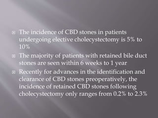
 Prepared by :- DR/Medhat Raafat Mehani
 Graduated from faculty of medicine
 Sohag university
 Secondary common duct stones, which, by
definition, originate in the gallbladder and pass
into the common duct, are usually cholesterol
stones and frequently become symptomatic
within weeks after cholecystectomy.
 The incidence of CBD stones in patients
undergoing elective cholecystectomy is 5% to
10%
 The majority of patients with retained bile duct
stones are seen within 6 weeks to 1 year
 Recently for advances in the identification and
clearance of CBD stones preoperatively, the
incidence of retained CBD stones following
cholecystectomy only ranges from 0.2% to 2.3%
Biliary disorders Pancreatic disorders Other
Gastrointestinal
Disorders
Extra-
intestinal
Disorders
Choledocholithiasis Pancreatitis Gastroesophage
al reflux disease
Psychiatric
disorders
Biliary stricture Pancreatic pseudocyst Esophageal
motor disorders
Coronary
artery disease
Bile duct injury + biliary
leak
Pancreatic malignancy Peptic ulcer
disease
Intercostal
neuritis
Sphincter of Oddi
dysfunction
Mesenteric
ischemia
Wound
neuroma
Stenosing papillitis Intra-abdominal
adhesions
Neurologic
disorders
Biliary tract malignancy Intestinal
malignancy
Unexplained
pain
syndromes
Choledochocele Irritable bowel
The patient will complain of :
 Attacks of sever dull aching pain in the rt
hypochondrium & epigastrium may be
associated with nausea & vomiting
 Obstructive jaundice that may wax and wane
because of the ball valve action of stone
 Fever& hypotention denoting further
complications like acute bacterial cholangitis
&acute pancreatitis
 Elevated serum bilirubin mainly direct and
elevated alkaline phosphatase level are most
often present even at a patient not clinically
jaundiced
 Dilatation of bile duct is most sensively shown
by ultrasonography but the stone itself will be
demostrated in 15% of cases at best
US view shows dilated CBD with a stone obstructing it
 Direct cholangiography either by ERCP or
percutaneous (PTC) is the definitive test
 Transtubal cholangiography may be tried if the
pt has T –tube inserted at the CBD
N.B:- Chongiography should be delayed & done
under antibiotic coverage when obtructed biliary
tree is infected as the pressure of injection may
cause septic shock
 MRCP :- is accurate but it is only dignostic not
therapeutic

ERCP shows missed stones at
CBD
Transtubal cholagiography shows large
CBD Stone completely obstruct it
Preoperative preparations:-
 I.V vit k & the pt should be
checked before operation
 fresh blood transfusion
 Broad spectrum
antibiotics(cepholosporins ) if there
is evidence of cholangitis
1-ERCP:
 Sphincterotomy by diathermy (The
location of the sphincterotomy should be
between the 11 and 1 o’clock position. to
avoid injury to the blood supply).
Removal of stone(s) by dormia basket.
2- percutaneous lithotomy :-
 If residaul stones discovered early and a T-tube
drains the bile duct, the tube tract can be
allowed to mature( devolop firm fibrous wall )
for about 4 to 6 wks and then can be used to
access the duct & stones.
 during the waiting period,monooctanoin,
(a solvent of cholesterol stones) can be infused
through the tube hoping to decrease the stone size
& facilitate its passage to the duodenum
Once the tract is mature enough to withstand
manipulation, tools such as dormia basket,
catheters, ballon dilators & even flexible
choledocoscope can be used to extract stones or
bush it into the duodenum .
 Lasers are the last tools to be used in this
fashion to break up stones .
3- extracorporial lithotripsy :-
 Electrohydraulic shock waves &US waves are
being used at a smaller but generally successful
experience of lithotripsy of CBD stones
 this techniqhe should be used for pts with large
stones cant be removed by endoscopic means
or for pts with contraindications to surgical or
endoscopic manipulations.
 Orally administered chenodeoxycholic A &
ursodeoxycholic A to reduce lithogenicity of
bile & facilitate further diminution & passage
of stone fragments
4- sugical lithotomy :-
Surgical removal of residual stones is indicated
only when other non operative alternatives are
not available , have failed or contraindicated
 Technique of CBD exploration :-
- Approuch to porta hepatis from the right is
likely to be easier and safer than direct anterior
approach used for exploration
- Routine preexploration cholangiogram is
advised to ascertain site & number of stones
- The bile duct is opened just above
the duodenum , the majority of
stones can be milked out manually
- 8 french catheter is passed upward
into the intrahepatic radicals &
downward into the duodenum with
vigorous infusion of saline will
wash further stones
- Progressively larger rubber
catheters or bakes dilatos are used
gently to dilate the papillary orifice
- A large T- tube at least 12 – 14
french is left in the CBD for P.O
cholagiography , if necessary, to
allow later removal of missed stones
& for drainage of bile
 About 5-10 % of pts have stones in the CBD at
time of cholecystectomy
 Clinical criteria(jaundice either present or past
& elevetedALP )+dilated ducts + palpable
stones + small stones are helpful to decide
wether to explore CBD or not
 Routine opeative cholangiography is adviced
by some added to operative time
 Intraoperative
choledocoscopy with
either rigid or flixible
can be used easly
especially with
experianced hands and
balloon catheter or
busket may be passed
through the scope
under direct vision to
remove the stone , this
procedure is costy and
this is its main
disadvantage

Flexible Choledochoscope
Rigid Choledochoscope
 When there are many stones in the biliary tree
and all of them cannot be removed , it is
generally safer to create side to side
choledocoduodenostomy so that possible
retained stones can be passed easly or at least
prevented from obstructing bile flow , it is
generally simpler than roux en y loop of
jejunum and transduodenal sphicteroplasty but
it needs dilated CBD at least 2 cm or more
A.Choledochotomy
and
duodenotomy. B. Sta
y sutures
placed. C. Tension
placed upon stay
sutures to prepare
longitudinal incision
for anastomosis to
duodenotomy.
1. Maingot's abdominal operations. (9th
edition),section 7 , page 1431
2. Blumgart's Surgery of the liver , biliary tract
&pancreas chapter 38, 633-641.e4
3. http://emedicine.medscape.com.
4. https://basicmedicalkey.com/choledochoenteric-
anastomosis-by-choledochoduodenostomy.
5. Bailey & love ,part eleven, chapter 67 ,page 1097

Missed biliary stones

  • 1.
     Prepared by:- DR/Medhat Raafat Mehani  Graduated from faculty of medicine  Sohag university
  • 2.
     Secondary commonduct stones, which, by definition, originate in the gallbladder and pass into the common duct, are usually cholesterol stones and frequently become symptomatic within weeks after cholecystectomy.
  • 3.
     The incidenceof CBD stones in patients undergoing elective cholecystectomy is 5% to 10%  The majority of patients with retained bile duct stones are seen within 6 weeks to 1 year  Recently for advances in the identification and clearance of CBD stones preoperatively, the incidence of retained CBD stones following cholecystectomy only ranges from 0.2% to 2.3%
  • 4.
    Biliary disorders Pancreaticdisorders Other Gastrointestinal Disorders Extra- intestinal Disorders Choledocholithiasis Pancreatitis Gastroesophage al reflux disease Psychiatric disorders Biliary stricture Pancreatic pseudocyst Esophageal motor disorders Coronary artery disease Bile duct injury + biliary leak Pancreatic malignancy Peptic ulcer disease Intercostal neuritis Sphincter of Oddi dysfunction Mesenteric ischemia Wound neuroma Stenosing papillitis Intra-abdominal adhesions Neurologic disorders Biliary tract malignancy Intestinal malignancy Unexplained pain syndromes Choledochocele Irritable bowel
  • 5.
    The patient willcomplain of :  Attacks of sever dull aching pain in the rt hypochondrium & epigastrium may be associated with nausea & vomiting  Obstructive jaundice that may wax and wane because of the ball valve action of stone  Fever& hypotention denoting further complications like acute bacterial cholangitis &acute pancreatitis
  • 6.
     Elevated serumbilirubin mainly direct and elevated alkaline phosphatase level are most often present even at a patient not clinically jaundiced  Dilatation of bile duct is most sensively shown by ultrasonography but the stone itself will be demostrated in 15% of cases at best
  • 7.
    US view showsdilated CBD with a stone obstructing it
  • 8.
     Direct cholangiographyeither by ERCP or percutaneous (PTC) is the definitive test  Transtubal cholangiography may be tried if the pt has T –tube inserted at the CBD N.B:- Chongiography should be delayed & done under antibiotic coverage when obtructed biliary tree is infected as the pressure of injection may cause septic shock  MRCP :- is accurate but it is only dignostic not therapeutic
  • 9.
     ERCP shows missedstones at CBD Transtubal cholagiography shows large CBD Stone completely obstruct it
  • 10.
    Preoperative preparations:-  I.Vvit k & the pt should be checked before operation  fresh blood transfusion  Broad spectrum antibiotics(cepholosporins ) if there is evidence of cholangitis 1-ERCP:  Sphincterotomy by diathermy (The location of the sphincterotomy should be between the 11 and 1 o’clock position. to avoid injury to the blood supply). Removal of stone(s) by dormia basket.
  • 12.
    2- percutaneous lithotomy:-  If residaul stones discovered early and a T-tube drains the bile duct, the tube tract can be allowed to mature( devolop firm fibrous wall ) for about 4 to 6 wks and then can be used to access the duct & stones.  during the waiting period,monooctanoin, (a solvent of cholesterol stones) can be infused
  • 13.
    through the tubehoping to decrease the stone size & facilitate its passage to the duodenum Once the tract is mature enough to withstand manipulation, tools such as dormia basket, catheters, ballon dilators & even flexible choledocoscope can be used to extract stones or bush it into the duodenum .  Lasers are the last tools to be used in this fashion to break up stones .
  • 14.
    3- extracorporial lithotripsy:-  Electrohydraulic shock waves &US waves are being used at a smaller but generally successful experience of lithotripsy of CBD stones  this techniqhe should be used for pts with large stones cant be removed by endoscopic means or for pts with contraindications to surgical or endoscopic manipulations.  Orally administered chenodeoxycholic A & ursodeoxycholic A to reduce lithogenicity of bile & facilitate further diminution & passage of stone fragments
  • 15.
    4- sugical lithotomy:- Surgical removal of residual stones is indicated only when other non operative alternatives are not available , have failed or contraindicated  Technique of CBD exploration :- - Approuch to porta hepatis from the right is likely to be easier and safer than direct anterior approach used for exploration - Routine preexploration cholangiogram is advised to ascertain site & number of stones
  • 16.
    - The bileduct is opened just above the duodenum , the majority of stones can be milked out manually - 8 french catheter is passed upward into the intrahepatic radicals & downward into the duodenum with vigorous infusion of saline will wash further stones - Progressively larger rubber catheters or bakes dilatos are used gently to dilate the papillary orifice - A large T- tube at least 12 – 14 french is left in the CBD for P.O cholagiography , if necessary, to allow later removal of missed stones & for drainage of bile
  • 17.
     About 5-10% of pts have stones in the CBD at time of cholecystectomy  Clinical criteria(jaundice either present or past & elevetedALP )+dilated ducts + palpable stones + small stones are helpful to decide wether to explore CBD or not  Routine opeative cholangiography is adviced by some added to operative time
  • 18.
     Intraoperative choledocoscopy with eitherrigid or flixible can be used easly especially with experianced hands and balloon catheter or busket may be passed through the scope under direct vision to remove the stone , this procedure is costy and this is its main disadvantage  Flexible Choledochoscope Rigid Choledochoscope
  • 19.
     When thereare many stones in the biliary tree and all of them cannot be removed , it is generally safer to create side to side choledocoduodenostomy so that possible retained stones can be passed easly or at least prevented from obstructing bile flow , it is generally simpler than roux en y loop of jejunum and transduodenal sphicteroplasty but it needs dilated CBD at least 2 cm or more
  • 20.
    A.Choledochotomy and duodenotomy. B. Sta ysutures placed. C. Tension placed upon stay sutures to prepare longitudinal incision for anastomosis to duodenotomy.
  • 21.
    1. Maingot's abdominaloperations. (9th edition),section 7 , page 1431 2. Blumgart's Surgery of the liver , biliary tract &pancreas chapter 38, 633-641.e4 3. http://emedicine.medscape.com. 4. https://basicmedicalkey.com/choledochoenteric- anastomosis-by-choledochoduodenostomy. 5. Bailey & love ,part eleven, chapter 67 ,page 1097