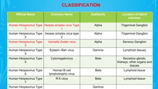
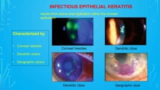
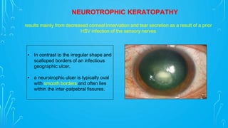
DR. AMEY TAMHANE
HERPES KERATITIS
Herpetic corneal disease
▪ HSV v VZV
▪ Clinical features
▪ Diagnosis
▪ Treatment
▪ Prophylaxis
OUTLINE
CHARACTERISTI
CS OF VIRUSES
Small, obligate, intracellular parasites.
Contain only one type of nucleic acid, either DNA or RNA but never
both.
Cannot replicate on their own.
Must infect and take over a host cell in order to replicate.
Multiply by a complex process and not by binary fission.
Unaffected by antibiotics
SIZE
MORPHOLOGY
MEDICALLY RELEVANT DNA
VIRUSES
MEDICALLY RELEVANT RNA VIRUSES
HERPES SIMPLEX
KERATITIS
Hippocrates is known to have described the cutaneous spread of herpes
simplex lesions
Greek word "herpes" to mean "to creep or crawl" in reference the
spreading nature of the herpetic skin lesions
1919- Lowenstein confirmed experimentally the infectious nature of
HSV that Shakespeare had only suspected.
(It is believed that Shakespeare mentioned oral herpes in Romeo and
Juliet)
1920-1930: Was found that HSV not only infects the skin, but also the
central nervous system.
1930: the property of HSV - latency
HISTORY
Official Name Common Name Subfamily Location of latent
infection
Human Herpesvirus Type
1
Herpes simplex virus Type
1
Alpha Trigeminal Ganglion
Human Herpesvirus Type
2
Herpes simplex virus type
2
Alpha Trigeminal Ganglion
Human Herpesvirus Type
3
Varicella Zoster virus Alpha Sensory Ganglion
Human Herpesvirus Type
4
Epstein- Barr virus Gamma Lymphoid tissues
Human Herpesvirus Type
5
Cytomegalovirus Beta Secretory glands,
Kidneys, other organs and
tissues
Human Herpesvirus Type
6
Human B cell
lymphotrophic virus
Beta Lymphoid tissue
Human Herpesvirus Type
7
R K virus Beta Lymphoid tissue
Human Herpesvirus Type Gamma
CLASSIFICATION
Establishes primary infection within the host
Enters sensory nerve terminals at peripheral sites
Enters the trigeminal nerve ganglion
Establishes latency
Reactivation of virus
Retrograde axonal transport
Triggering Factors
Most common 40-60
%
acquired in early
childhood : 2-5 years
of age via saliva,
skin,
respiratory
TRIGGERS FOR RECURRENT HSV
INFECTION
Ophthalmic
▪ Contact lens wear
▪ Eye injury
▪ Corneal grafting
▪ Laser eye surgery
▪ Cataract surgery
▪ Intravitreal injections
▪ Topical prostaglandin analogue
Systemic
▪ Stress
▪ Systemic infection/fever
▪ Sunlight exposure
▪ Menstruation
▪ Genetic factors - CSSG-1
FREQUENCY
How common is ocular herpes simplex ?
▪ Ocular herpes simplex infections have an incidence of 5–15 new cases per 100,000
people per year.
▪ Long-term studies of ocular HSV-1 infections have found the mean age for the first
episode to be 37 years.
▪ A single recurrence of ophthalmic infection is observed in 20–30% of patients within
2 years of the first infection.
▪ The rate of recurrence is believed to be slightly higher in men than in women.
CLINICAL FEATURES
Patients with HSV keratitis may complain of
▪ Pain
▪ Photophobia
▪ Blurred vision
▪ Tearing
▪ Redness
A history of prior episodes in patients with
recurrent disease may exist.
PHYSICAL
HSV keratitis may be divided into 4 categories
Infectious epithelial keratitis
Neurotrophic keratopathy
Stromal keratitis
Endothelitis
Characterized by
▪ Corneal vesicles
▪ Dendritic ulcers
▪ Geographic ulcers
INFECTIOUS EPITHELIAL KERATITIS
Geographic ulcer
Corneal Vesicles Dendritic Ulcer
Dendritic Ulcer
results from active viral replication within the corneal
epithelium
▪ In contrast to the irregular shape and
scalloped borders of an infectious
geographic ulcer,
▪ a neurotrophic ulcer is typically oval
with smooth borders and often lies
within the inter-palpebral fissures.
NEUROTROPHIC KERATOPATHY
results mainly from decreased corneal innervation and tear secretion as a result of a prior
HSV infection of the sensory nerves
2. Immune stromal keratitis
may present clinically with focal, multifocal, or
diffuse stroll opacities; immune rings;
neovascularization; or
ghost vessels at any level of the cornea.
1. Necrotizing stromal keratitis
characterized by dense stromal infiltrate, ulceration,
and necrosis causing corneal melting and
perforation.
CORNEAL STROMAL INFLAMMATION
arises from direct infection of the corneal stroma and the
resultant severe host inflammatory response
is an antibody-complement cascade triggered by retained
viral antigen within the stroma
• keratic precipitates (KP)
• stromal and epithelial edema
• absence of stroma infiltrate or neovascularization
▪ Disciform
▪ Diffuse
▪ Linear
ENDOTHELITIS
is believed to be primarily an immunologic reaction to an antigen in endothelial cells;
however,
the role of live virus has been speculated
• HSV keratitis is primarily a clinical diagnosis based on characteristic features of the corneal
lesion.
• Epithelial scrapings with Giemsa stain may show multi-nucleated giant cells . Viral cultures
obtained within several days of onset of disease and prior to antiviral therapy have a
sensitivity of up to 70%.
• Commercially available HSV antigen detection tests are very specific for detecting herpes
infection, but they are limited by their lower sensitivity.
• Polymerase chain reaction from a tear sample or from an anterior chamber tap may detect
viral DNA in cases of herpetic keratitis or kerato-uveitis.
WORKUP
Antiviral therapy, oral or topical, is an effective treatment of epithelial herpes infection
▪ Trifluridine 1% solution 9 times daily
▪ Acyclovir 3% ointment 5 times a day for 14-15 days.
▪ Ganciclovir (0.15%) ointment 5 times a day untill ulcer heals and then 3 times a day for 5
days.
▪ Vidarabine 3% ointment 5 times daily has equal efficacy in treating a dendritic ulcer;
however, trifluridine is more effective than vidarabine for treatment of a geographic ulcer.
MEDICAL CARE
HSV EPITHELIAL KERATITIS
No added benefit of oral antiviral with topical antiviral, but may be a better alternative to
topical for patients with pre existing ocular surface disease and who are at high risk for
toxicity from topical medications.
▪ acyclovir 400 mg 3-5 times daily or
▪ famcyclovir 250 mg 3 times daily or
▪ valacyclovir 500 mg 3 times daily
for a period of 10-21 days.
Normal dendrites heal in 1-3 weeks
If not, think toxicity, resistance or wrong diagnosis!
With epithelial ulceration
Limited dose of topical corticosteroid
PLUS therapeutic dose of oral antiviral
agent
Prednisolone 1% : twice daily PLUS
Acyclovir (Zovirax®) : 400 mg 3–5 times daily
or
Valacyclovir (Valtrex®) : 500 mg 3 times daily
or
Famciclovir (Famvir®) : 250 mg 3 times daily
for 7–10 days
The oral antiviral agent is reduced to
Without epithelial ulceration
Therapeutic dose of topical corticosteroid
PLUS prophylactic dose of oral antiviral
agent
Prednisolone 1% : 6–8 times daily tapered
over greater than 10 weeks PLUS
Acyclovir (Zovirax®) : 400 mg twice daily or
Valacyclovir (Valtrex®) : 500 mg twice daily
or
Famciclovir (Famvir®) : 250 mg twice daily
Oral antiviral agents in prophylactic doses
(above) should be maintained during
corticosteroid treatment.
STROMAL KERATITIS
Therapeutic dose of topical corticosteroid PLUS therapeutic dose of oral antiviral
Prednisolone 1% : 6–8 times daily PLUS
Acyclovir (Zovirax®) : 400 mg 3–5 times daily or
Valacyclovir (Valtrex®) : 500 mg 3 times daily or
Famciclovir (Famvir®) : 250 mg 3 times daily
The oral antiviral agent is reduced to prophylactic dose after 7–10 days and maintained as
long as topical corticosteroids are in use.
As disease comes under control, the topical corticosteroid can be tapered slowly.
ENDOTHELIAL KERATITIS
Potential indications:
• Multiple recurrences of any type of HSV keratitis, especially HSV stromal keratitis.
• Recurrent inflammation with scar / vascularization approaching visual axis.
PROPHILAXIS FOR RECURRENT HSV KERARTITIS
Type Prophylaxis regimen
HSV epithelial keratitis Initial episode = no prophylactic therapy
One or more recurrent episodes = oral therapy x 1 year
HSV stromal keratitis Initial episode = oral therapy x 1 year
Chronic disease = oral therapy (2 years to indefinite)
HSV endothelial
keratitis
Initial episode = oral therapy x 1 year
Multiple episodes = oral therapy (2 years to indefinite)
HSV keratouveitis Initial episode = oral therapy x 1 year
Multiple episodes = oral therapy (2 years to indefinite)
Post-phototherapeutic
keratectomy
oral therapy (1 year)
Post-keratoplasty
(any type)
oral therapy (2 years to indefinite)
PROPHILAXIS FOR RECURRENT HSV KERARTITIS
CURRENT ANTIVIRALS
Drug (Trade
Name)
Route Dose Frequency Adverse Effects
Trifluridine
(Viroptic)
Topical Drops 1% every 2 hours Follicular conjunctivitis,
epitheliopathy
Vidarabine (Ara-A) Topical ointment 3% 5 times a day As Above
Acyclovir
(Zovirax)
Topical ointment
Oral treatment
Oral prophylaxis
3%
400 mg
400 mg
5 times a day
5 times a day
twice a day
Headache, nausea,
nephrotoxicity,
neurotoxicity
Valacyclovir
(Valterex)
Oral treatment
Oral prophylaxis
500 mg
500 mg
3 times a day
twice a day
As above,
TTP & hemolytic ureic
syndrome in
immunosuppressed
Famciclovir
(Famvir)
Oral treatment
Oral prophylaxis
250 mg
250 mg
3 times a day
twice a day
As Acyclovir
SURGICAL THERAPY
• Surgical treatment is indicated when severe stromal
keratitis leads to stromal perforation or visually significant
stromal scarring.
• Surgical treatment of perforation may include application of
cyanoacrylate glue or penetrating keratoplasty (PK).
• Penetrating keratoplasty is the treatment when vision is
reduced due to stromal scar after HSV keratitis.
• Success of a PK is 60%–80% if there have been no signs
of active disease at least 6 months prior to surgery.
• Graft failure may develop in actively inflamed eyes
undergoing PK or in recipient corneas with vascularization
in >2 quadrants.
HERPES ZOSTER OPHTHALMICUS
( SHINGLES )
BACKGROUND
• Varicella-zoster virus (VZV) is a member of
the Herpesviridae family.
• It is the etiologic agent of varicella
(chickenpox), the primary infection, and
herpes zoster, the reactivation.
• Herpes zoster ophthalmicus involves the
tissues innervated by the ophthalmic division
of the trigeminal nerve .
• The sequelae of herpes zoster ophthalmicus
can be devastating and include chronic
ocular inflammation, visual loss, and
debilitating pain.
Establishes the primary infection within the
host
Enters the trigeminal nerve
ganglion
Establishes latency
Reactivation of
virus
Triggering Factors
The hallmark of herpes zoster ophthalmicus is a vesicular rash that
involves the first (ophthalmic) division of the fifth cranial nerve that presents
in a dermatomal distribution and respects the midline.
• Regarding primary infection, more than 90% of the
population is infected by adolescence, and approximately
100% are infected by 60 years of age.
• Affects about 10-20% of the population. 131 per 100,000
person/year in white individuals.
• According to Pavan-Langston's review, 1 million
consultations for herpes zoster occur per year; and
approximately 250,000 of the patients thus examined
develop herpes zoster ophthalmicus.
• A subset of 50% of these patients develops complications of
herpes zoster ophthalmicus. 100 deaths occur per year as a
result of complications from VZV infection.
FREQUENCY
CLINICAL
FEATURES
Symptoms of herpes zoster ophthalmicus may include
• skin/eyelid rash with eye pain,
• red eye (usually unilateral),
• decreased vision,
• fever, malaise, and tearing
• Recurrence is a characteristic feature of herpes zoster
ophthalmicus. Relapse may occur as late as 10 years
after onset.
PHYSICAL
• Vesicular rashes involving the
ophthalmic division of the
trigeminal nerve.
• Crusting begins on the fifth to sixth
day.
• In 1864, Hutchinson proposed that
the cutaneous involvement in the
distribution of the nasociliary nerve
heralds ocular involvement.
DENDRITIC KERATITIS
▪ In contrast to HSV dendrites, VZV
pseudodendrites are smaller in size, elevated
without central ulceration, they do not have
terminal bulbs, and have relative lack of central
staining.
▪ Elevated mucous plaques may occur weeks to
months after resolution of the original
pseudodendrites.
▪ Residual corneal hypoesthesia after the
infection can be severe.
▪ nummular – fine granular deposits under Bowman’s layer
▪ disciform – 3 weeks after the rash (occurs in 5% of cases)
▪ endothelial changes and KP
▪ anterior uveitis
▪ secondary glaucoma (16-56 %)
▪ rarely, posterior segment involvement: retinitis, acute retinal necrosis,
choroiditis, optic neuritis, optic atrophy
OTHER FEATURES
• Acute retinal necrosis caused
by VZV or less commonly by
HSV-1 & HSV-2, is a severe
ocular inflammatory
syndrome associated with a
poor (visual) prognosis.
Classically, posterior segment involvement
includes vitreous inflammation, retinal vascular
arteriolitis, and peripheral retinitis.
• The diagnosis of ARN is
usually made based upon
clinical characteristics.
• However, it may be
appropriate to analyze an
aqueous or vitreous sample
to test for presence of
members of the herpesvirus
family.
ACUTE RETINAL NECROSIS (ARN)
▪ Herpes zoster is diagnosed mostly on the basis of the characteristic pain and appearance
of the dermatomal rashes.
▪ More often than not, laboratory tests are unnecessary.
▪ On rare occasion, when a differential diagnosis of several skin lesions must be made,
confirmatory laboratory examinations may be undertaken.
▪ These tests include morphologic immuno-morphologic viral isolation and serologic cellular
immunity tests.
WORKUP
Include antiviral agents, systemic corticosteroids, antidepressants, and adequate pain
control.
Treatment of acute herpes zoster ophthalmicus is optimal if started within 72 hours of rash onset.
Oral antiviral drugs
Famciclovir 750 mg once a day for 7 days or
Valacyclovir 1 g tid for 7 days or
Acyclovir 800 mg 5 times / day for 7 days
If immunosuppressed, then acyclovir IV 10mg/kg 3 times per day
Tricyclic antidepressants nortriptyline, amitriptyline, or desipramine 25 mg (to inhibit acute and
prolonged postherpetic neuralgia PHN ).
Additional topical corticosteroids, lubricants, mucolytics, cycloplegics and glaucoma medications as
necessary
MEDICAL CARE
• Systemic antiviral agents include
Acyclovir (13 mg/kg divided every 8 h IV for 7 days, followed by 800 mg five times daily orally for
3–4 months)
• Antiviral agents administered intra-vitreally can provide beneficial adjunctive therapy,
especially if retinitis is threatening the macula or optic disc :
Ganciclovir (200-2000 ug per 0.1 mL)
Foscarnet (1.2-2.4 mg per 0.1 mL)
• Steroids may have a beneficial therapeutic effects if initiated 24-48 hours after the start of
antiviral therapy or once regression of retinal necrosis has been demonstrated :
TREATMENT OF ARN
▪ Post Herpetic Neuralgia 20-60% (age <40-60 y) occurs when damaged nerve fibres
send confused and exaggerated messages of pain.
▪ Skin Infections: If shingles blisters aren't properly treated, bacterial skin infections
may develop.
▪ Neuro-ophthalmic (8%) inflammation of the brain (encephalitis), spinal cord
(transverse myelitis), facial paralysis, or hearing or balance problems.
COMPLICATIONS
▪ HSV and VZV cause distinctive clinical pictures
▪ Each layer of the cornea may be affected with different manifestations
▪ Never start topical steroid in suspected herpes simplex keratitis without
antiviral cover
▪ Reduced corneal sensation can be a useful sign of previous disease
▪ Protect the neurotrophic cornea
TAKE HOME MESSAGE
THANK YOU

Herpes Simplex Keratitis .pptx

  • 1.
  • 2.
    Herpetic corneal disease ▪HSV v VZV ▪ Clinical features ▪ Diagnosis ▪ Treatment ▪ Prophylaxis OUTLINE
  • 3.
    CHARACTERISTI CS OF VIRUSES Small,obligate, intracellular parasites. Contain only one type of nucleic acid, either DNA or RNA but never both. Cannot replicate on their own. Must infect and take over a host cell in order to replicate. Multiply by a complex process and not by binary fission. Unaffected by antibiotics
  • 4.
  • 5.
  • 6.
  • 7.
  • 8.
  • 9.
    Hippocrates is knownto have described the cutaneous spread of herpes simplex lesions Greek word "herpes" to mean "to creep or crawl" in reference the spreading nature of the herpetic skin lesions 1919- Lowenstein confirmed experimentally the infectious nature of HSV that Shakespeare had only suspected. (It is believed that Shakespeare mentioned oral herpes in Romeo and Juliet) 1920-1930: Was found that HSV not only infects the skin, but also the central nervous system. 1930: the property of HSV - latency HISTORY
  • 10.
    Official Name CommonName Subfamily Location of latent infection Human Herpesvirus Type 1 Herpes simplex virus Type 1 Alpha Trigeminal Ganglion Human Herpesvirus Type 2 Herpes simplex virus type 2 Alpha Trigeminal Ganglion Human Herpesvirus Type 3 Varicella Zoster virus Alpha Sensory Ganglion Human Herpesvirus Type 4 Epstein- Barr virus Gamma Lymphoid tissues Human Herpesvirus Type 5 Cytomegalovirus Beta Secretory glands, Kidneys, other organs and tissues Human Herpesvirus Type 6 Human B cell lymphotrophic virus Beta Lymphoid tissue Human Herpesvirus Type 7 R K virus Beta Lymphoid tissue Human Herpesvirus Type Gamma CLASSIFICATION
  • 11.
    Establishes primary infectionwithin the host Enters sensory nerve terminals at peripheral sites Enters the trigeminal nerve ganglion Establishes latency Reactivation of virus Retrograde axonal transport Triggering Factors Most common 40-60 % acquired in early childhood : 2-5 years of age via saliva, skin, respiratory
  • 12.
    TRIGGERS FOR RECURRENTHSV INFECTION Ophthalmic ▪ Contact lens wear ▪ Eye injury ▪ Corneal grafting ▪ Laser eye surgery ▪ Cataract surgery ▪ Intravitreal injections ▪ Topical prostaglandin analogue Systemic ▪ Stress ▪ Systemic infection/fever ▪ Sunlight exposure ▪ Menstruation ▪ Genetic factors - CSSG-1
  • 13.
    FREQUENCY How common isocular herpes simplex ? ▪ Ocular herpes simplex infections have an incidence of 5–15 new cases per 100,000 people per year. ▪ Long-term studies of ocular HSV-1 infections have found the mean age for the first episode to be 37 years. ▪ A single recurrence of ophthalmic infection is observed in 20–30% of patients within 2 years of the first infection. ▪ The rate of recurrence is believed to be slightly higher in men than in women.
  • 14.
    CLINICAL FEATURES Patients withHSV keratitis may complain of ▪ Pain ▪ Photophobia ▪ Blurred vision ▪ Tearing ▪ Redness A history of prior episodes in patients with recurrent disease may exist.
  • 15.
    PHYSICAL HSV keratitis maybe divided into 4 categories Infectious epithelial keratitis Neurotrophic keratopathy Stromal keratitis Endothelitis
  • 16.
    Characterized by ▪ Cornealvesicles ▪ Dendritic ulcers ▪ Geographic ulcers INFECTIOUS EPITHELIAL KERATITIS Geographic ulcer Corneal Vesicles Dendritic Ulcer Dendritic Ulcer results from active viral replication within the corneal epithelium
  • 17.
    ▪ In contrastto the irregular shape and scalloped borders of an infectious geographic ulcer, ▪ a neurotrophic ulcer is typically oval with smooth borders and often lies within the inter-palpebral fissures. NEUROTROPHIC KERATOPATHY results mainly from decreased corneal innervation and tear secretion as a result of a prior HSV infection of the sensory nerves
  • 18.
    2. Immune stromalkeratitis may present clinically with focal, multifocal, or diffuse stroll opacities; immune rings; neovascularization; or ghost vessels at any level of the cornea. 1. Necrotizing stromal keratitis characterized by dense stromal infiltrate, ulceration, and necrosis causing corneal melting and perforation. CORNEAL STROMAL INFLAMMATION arises from direct infection of the corneal stroma and the resultant severe host inflammatory response is an antibody-complement cascade triggered by retained viral antigen within the stroma
  • 19.
    • keratic precipitates(KP) • stromal and epithelial edema • absence of stroma infiltrate or neovascularization ▪ Disciform ▪ Diffuse ▪ Linear ENDOTHELITIS is believed to be primarily an immunologic reaction to an antigen in endothelial cells; however, the role of live virus has been speculated
  • 20.
    • HSV keratitisis primarily a clinical diagnosis based on characteristic features of the corneal lesion. • Epithelial scrapings with Giemsa stain may show multi-nucleated giant cells . Viral cultures obtained within several days of onset of disease and prior to antiviral therapy have a sensitivity of up to 70%. • Commercially available HSV antigen detection tests are very specific for detecting herpes infection, but they are limited by their lower sensitivity. • Polymerase chain reaction from a tear sample or from an anterior chamber tap may detect viral DNA in cases of herpetic keratitis or kerato-uveitis. WORKUP
  • 21.
    Antiviral therapy, oralor topical, is an effective treatment of epithelial herpes infection ▪ Trifluridine 1% solution 9 times daily ▪ Acyclovir 3% ointment 5 times a day for 14-15 days. ▪ Ganciclovir (0.15%) ointment 5 times a day untill ulcer heals and then 3 times a day for 5 days. ▪ Vidarabine 3% ointment 5 times daily has equal efficacy in treating a dendritic ulcer; however, trifluridine is more effective than vidarabine for treatment of a geographic ulcer. MEDICAL CARE HSV EPITHELIAL KERATITIS
  • 22.
    No added benefitof oral antiviral with topical antiviral, but may be a better alternative to topical for patients with pre existing ocular surface disease and who are at high risk for toxicity from topical medications. ▪ acyclovir 400 mg 3-5 times daily or ▪ famcyclovir 250 mg 3 times daily or ▪ valacyclovir 500 mg 3 times daily for a period of 10-21 days. Normal dendrites heal in 1-3 weeks If not, think toxicity, resistance or wrong diagnosis!
  • 23.
    With epithelial ulceration Limiteddose of topical corticosteroid PLUS therapeutic dose of oral antiviral agent Prednisolone 1% : twice daily PLUS Acyclovir (Zovirax®) : 400 mg 3–5 times daily or Valacyclovir (Valtrex®) : 500 mg 3 times daily or Famciclovir (Famvir®) : 250 mg 3 times daily for 7–10 days The oral antiviral agent is reduced to Without epithelial ulceration Therapeutic dose of topical corticosteroid PLUS prophylactic dose of oral antiviral agent Prednisolone 1% : 6–8 times daily tapered over greater than 10 weeks PLUS Acyclovir (Zovirax®) : 400 mg twice daily or Valacyclovir (Valtrex®) : 500 mg twice daily or Famciclovir (Famvir®) : 250 mg twice daily Oral antiviral agents in prophylactic doses (above) should be maintained during corticosteroid treatment. STROMAL KERATITIS
  • 24.
    Therapeutic dose oftopical corticosteroid PLUS therapeutic dose of oral antiviral Prednisolone 1% : 6–8 times daily PLUS Acyclovir (Zovirax®) : 400 mg 3–5 times daily or Valacyclovir (Valtrex®) : 500 mg 3 times daily or Famciclovir (Famvir®) : 250 mg 3 times daily The oral antiviral agent is reduced to prophylactic dose after 7–10 days and maintained as long as topical corticosteroids are in use. As disease comes under control, the topical corticosteroid can be tapered slowly. ENDOTHELIAL KERATITIS
  • 25.
    Potential indications: • Multiplerecurrences of any type of HSV keratitis, especially HSV stromal keratitis. • Recurrent inflammation with scar / vascularization approaching visual axis. PROPHILAXIS FOR RECURRENT HSV KERARTITIS
  • 26.
    Type Prophylaxis regimen HSVepithelial keratitis Initial episode = no prophylactic therapy One or more recurrent episodes = oral therapy x 1 year HSV stromal keratitis Initial episode = oral therapy x 1 year Chronic disease = oral therapy (2 years to indefinite) HSV endothelial keratitis Initial episode = oral therapy x 1 year Multiple episodes = oral therapy (2 years to indefinite) HSV keratouveitis Initial episode = oral therapy x 1 year Multiple episodes = oral therapy (2 years to indefinite) Post-phototherapeutic keratectomy oral therapy (1 year) Post-keratoplasty (any type) oral therapy (2 years to indefinite) PROPHILAXIS FOR RECURRENT HSV KERARTITIS
  • 27.
    CURRENT ANTIVIRALS Drug (Trade Name) RouteDose Frequency Adverse Effects Trifluridine (Viroptic) Topical Drops 1% every 2 hours Follicular conjunctivitis, epitheliopathy Vidarabine (Ara-A) Topical ointment 3% 5 times a day As Above Acyclovir (Zovirax) Topical ointment Oral treatment Oral prophylaxis 3% 400 mg 400 mg 5 times a day 5 times a day twice a day Headache, nausea, nephrotoxicity, neurotoxicity Valacyclovir (Valterex) Oral treatment Oral prophylaxis 500 mg 500 mg 3 times a day twice a day As above, TTP & hemolytic ureic syndrome in immunosuppressed Famciclovir (Famvir) Oral treatment Oral prophylaxis 250 mg 250 mg 3 times a day twice a day As Acyclovir
  • 28.
    SURGICAL THERAPY • Surgicaltreatment is indicated when severe stromal keratitis leads to stromal perforation or visually significant stromal scarring. • Surgical treatment of perforation may include application of cyanoacrylate glue or penetrating keratoplasty (PK). • Penetrating keratoplasty is the treatment when vision is reduced due to stromal scar after HSV keratitis. • Success of a PK is 60%–80% if there have been no signs of active disease at least 6 months prior to surgery. • Graft failure may develop in actively inflamed eyes undergoing PK or in recipient corneas with vascularization in >2 quadrants.
  • 29.
  • 30.
    BACKGROUND • Varicella-zoster virus(VZV) is a member of the Herpesviridae family. • It is the etiologic agent of varicella (chickenpox), the primary infection, and herpes zoster, the reactivation. • Herpes zoster ophthalmicus involves the tissues innervated by the ophthalmic division of the trigeminal nerve . • The sequelae of herpes zoster ophthalmicus can be devastating and include chronic ocular inflammation, visual loss, and debilitating pain.
  • 31.
    Establishes the primaryinfection within the host Enters the trigeminal nerve ganglion Establishes latency Reactivation of virus Triggering Factors The hallmark of herpes zoster ophthalmicus is a vesicular rash that involves the first (ophthalmic) division of the fifth cranial nerve that presents in a dermatomal distribution and respects the midline.
  • 32.
    • Regarding primaryinfection, more than 90% of the population is infected by adolescence, and approximately 100% are infected by 60 years of age. • Affects about 10-20% of the population. 131 per 100,000 person/year in white individuals. • According to Pavan-Langston's review, 1 million consultations for herpes zoster occur per year; and approximately 250,000 of the patients thus examined develop herpes zoster ophthalmicus. • A subset of 50% of these patients develops complications of herpes zoster ophthalmicus. 100 deaths occur per year as a result of complications from VZV infection. FREQUENCY
  • 33.
    CLINICAL FEATURES Symptoms of herpeszoster ophthalmicus may include • skin/eyelid rash with eye pain, • red eye (usually unilateral), • decreased vision, • fever, malaise, and tearing • Recurrence is a characteristic feature of herpes zoster ophthalmicus. Relapse may occur as late as 10 years after onset.
  • 34.
    PHYSICAL • Vesicular rashesinvolving the ophthalmic division of the trigeminal nerve. • Crusting begins on the fifth to sixth day. • In 1864, Hutchinson proposed that the cutaneous involvement in the distribution of the nasociliary nerve heralds ocular involvement.
  • 35.
    DENDRITIC KERATITIS ▪ Incontrast to HSV dendrites, VZV pseudodendrites are smaller in size, elevated without central ulceration, they do not have terminal bulbs, and have relative lack of central staining. ▪ Elevated mucous plaques may occur weeks to months after resolution of the original pseudodendrites. ▪ Residual corneal hypoesthesia after the infection can be severe.
  • 36.
    ▪ nummular –fine granular deposits under Bowman’s layer ▪ disciform – 3 weeks after the rash (occurs in 5% of cases) ▪ endothelial changes and KP ▪ anterior uveitis ▪ secondary glaucoma (16-56 %) ▪ rarely, posterior segment involvement: retinitis, acute retinal necrosis, choroiditis, optic neuritis, optic atrophy OTHER FEATURES
  • 37.
    • Acute retinalnecrosis caused by VZV or less commonly by HSV-1 & HSV-2, is a severe ocular inflammatory syndrome associated with a poor (visual) prognosis. Classically, posterior segment involvement includes vitreous inflammation, retinal vascular arteriolitis, and peripheral retinitis. • The diagnosis of ARN is usually made based upon clinical characteristics. • However, it may be appropriate to analyze an aqueous or vitreous sample to test for presence of members of the herpesvirus family. ACUTE RETINAL NECROSIS (ARN)
  • 38.
    ▪ Herpes zosteris diagnosed mostly on the basis of the characteristic pain and appearance of the dermatomal rashes. ▪ More often than not, laboratory tests are unnecessary. ▪ On rare occasion, when a differential diagnosis of several skin lesions must be made, confirmatory laboratory examinations may be undertaken. ▪ These tests include morphologic immuno-morphologic viral isolation and serologic cellular immunity tests. WORKUP
  • 39.
    Include antiviral agents,systemic corticosteroids, antidepressants, and adequate pain control. Treatment of acute herpes zoster ophthalmicus is optimal if started within 72 hours of rash onset. Oral antiviral drugs Famciclovir 750 mg once a day for 7 days or Valacyclovir 1 g tid for 7 days or Acyclovir 800 mg 5 times / day for 7 days If immunosuppressed, then acyclovir IV 10mg/kg 3 times per day Tricyclic antidepressants nortriptyline, amitriptyline, or desipramine 25 mg (to inhibit acute and prolonged postherpetic neuralgia PHN ). Additional topical corticosteroids, lubricants, mucolytics, cycloplegics and glaucoma medications as necessary MEDICAL CARE
  • 40.
    • Systemic antiviralagents include Acyclovir (13 mg/kg divided every 8 h IV for 7 days, followed by 800 mg five times daily orally for 3–4 months) • Antiviral agents administered intra-vitreally can provide beneficial adjunctive therapy, especially if retinitis is threatening the macula or optic disc : Ganciclovir (200-2000 ug per 0.1 mL) Foscarnet (1.2-2.4 mg per 0.1 mL) • Steroids may have a beneficial therapeutic effects if initiated 24-48 hours after the start of antiviral therapy or once regression of retinal necrosis has been demonstrated : TREATMENT OF ARN
  • 41.
    ▪ Post HerpeticNeuralgia 20-60% (age <40-60 y) occurs when damaged nerve fibres send confused and exaggerated messages of pain. ▪ Skin Infections: If shingles blisters aren't properly treated, bacterial skin infections may develop. ▪ Neuro-ophthalmic (8%) inflammation of the brain (encephalitis), spinal cord (transverse myelitis), facial paralysis, or hearing or balance problems. COMPLICATIONS
  • 42.
    ▪ HSV andVZV cause distinctive clinical pictures ▪ Each layer of the cornea may be affected with different manifestations ▪ Never start topical steroid in suspected herpes simplex keratitis without antiviral cover ▪ Reduced corneal sensation can be a useful sign of previous disease ▪ Protect the neurotrophic cornea TAKE HOME MESSAGE
  • 43.