SlideShare a Scribd company logo
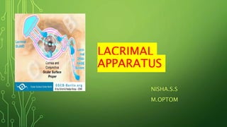
LACRIMAL
APPARATUS
NISHA.S.S
M.OPTOM
LACRIMAL APPARATUS
• The lacrimal apparatus comprises the structures concerned with the
formation of tears( the main lacrimal gland and accessory lacrimal glands)
and its transport.
• The lacrimal passage includes:
• Puncta
• Canaliculi
• Lacrimal sac
• Nasolacrimal duct.
LACRIMAL GLANDS
Main lacrimal gland
• The main lacrimal gland is situated in the fossa for lacrimal gland,
formed by the orbital plate of frontal bone, in the anterolateral part of the
roof of orbit.
• The gland is divided into two parts-the superior orbital and the inferior
palpebral- which are continuous with each other posteriorly.
The orbital part of lacrimal gland
• It is large, about the size and shape of a small almond. It has two surfaces (superior and
inferior), two borders (anterior and posterior) and two extremities (medial and lateral).
• Superior surface of the orbital part is convex and lies in contact with the periorbita lining the
part of the frontal bone forming the fossa for the lacrimal gland.
• Inferior surface of the orbital part is concave and lies on the levator palpebrae superioris
muscle.
• Anterior border is sharp and seems within and parallel to the orbital margin. It lies in contact
with the septum orbitale.
• Posterior border is round and becomes continuous with the palpebral part of the gland.
• Lateral extremity rests on the lateral rectus muscle.
• Medial extremity is related to the levator palpebrae superioris muscle.
Palpebral part of lacrimal gland
• It is small (about one-third the size of orbital part) and consists of only
two or three lobules.
• It is situated upon the course of the ducts of orbital part from which it
is separated by the levator palpebrae superioris muscle, which is
related to it superiorly.
• Inferiorly, the gland lies in relation to the superior fornix.
• The gland is compressed from above downwards and can be seen
through the conjunctiva when the upper lid is everted.
• Posteriorly, it is continuous with the orbital part.
Ducts of lacrimal gland
• About 10-12 ducts pass downwards from the main lacrimal gland
to open in the lateral part of the superior fornix.
• One or two ducts also open in the lateral part of the inferior fornix.
• Since all the ducts pass through the palpebral part of the gland,
therefore, excision of the palpebral part alone amounts to excision
of the entire gland as far as the secretory function of the gland is
concerned.
HISTOLOGICAL STRUCTURE OF THE LACRIMAL GLAND
• Lacrimal gland is a branched tubuloalveolar (serous acinous) gland. Microscopically, it
consists of glandular tissue, stroma and septa .
• Glandular tissue consists of ducts arranged in lobes and lobules.
• Acini are lined by a single layer of pyramidal cells mounted on a basement membrane. These
cells are surrounded by a layer of flattened myoepithelial cells. The pyramidal cells are of the
serous type with eosinophilic secretory granules and a round nucleus situated towards the
base. These cells secrete the tears, expelled by the contraction of myofibrils.
• Ductules and ducts : The secretion of the acinar units is drained by connecting channels which
to begin with are intralobular, then these become extralobular and lastly open in the ducts. The
ducts are lined by two layers of epithelial cells the inner lining is formed by thick cylindrical
cells and the outer layer is of flattened cells.
• Stroma of the lacrimal gland is formed by mesodermal tissue which contains connective
tissue, elastic tissue which contains connective tissue, elastic tissue, lymphoid tissue, plasma
cells, rich nerve terminals and blood vessels.
BLOOD SUPPLY
• Arterial supply: Main lacrimal gland is supplied by lacrimal artery, a
branch of ophthalmic artery. Sometimes a branch of the transverse
facial artery may also supply the gland.
• Lacrimal veins draining the gland join the ophthalmic vein.
NERVE SUPPLY
• Main lacrimal gland has three modes of innervations:
1. Sensory nerve supply comes from the lacrimal nerve, a branch of
ophthalmic division of the fifth cranial nerve.
2. Sympathetic nerve supply arises from the superior cervical sympathetic
ganglion as postganglionic fibres which from the carotid plexus of the cervical
sympathetics. From the sympathetic plexus around the internal carotid artery,
some fibres join the deep petrosal nerve, then the nerve of pterygoid gland,
and then ultimately reach the lacrimal gland through the lacrimal nerve .
ACCESSORY LACRIMAL GLANDS
These include:
• Glands of Krause
• Glands of Wolfring
• Intraorbital glands
• Glands in the caruncle and plica semilunaris
• Glands of Krause: These are microscopic glands lying in the
subconjunctival tissue of the foraices .These are about 40-42 in the upper
fornix and about 6-8 in the lower fornix. Their ducts unite to form a long duct
which opens in the fornix.
• Glands of Wolfring: These are microscopic glands present along the upper
border of superior tarsus (2-5 in number), and lower border of inferior
tarsus (2-3 in number).
• Rudimentary accessory lacrimal glands: These are present in the caruncle,
plica semilunaris and infraorbital region.
LACRIMAL PASSAGES
 LACRIMAL PASSAGES
1. Lacrimal puncta
• These are two small rounded or oval openings one each on the upper and lower eyelids,
at the junction of ciliary and lacrimal portion of the lid margin .
• The upper upon a slight elevation called lacrimal papilla, which becomes prominent in
old age." and the lower puncta lie about 6 mm and 6.5 mm lateral to the inner canthus,
respectively. Thus when the eyelids are closed, the puncta do not overlap each other,
but the upper punctum lies medical to the lower punctum.
• The upper punctum is directed downwards and backwards, while the lower punctum is
directed upwards and backward..
• The puncta are surrounded by a ring of dense fibrous tissue .With each blink, the puncta
slide in the groove between the plica semilunaris and the eyeball.
2. Lacrimal canaliculi
• The superior and inferior canaliculi join the puncta to the lacrimal sac.
• Each canaliculus is 0.5 mm in diameter and has two parts, vertical (2 mm) and
horizontal (8 mm), which lie at right angles to each other . At the junction of the two
portions, there is slight dilatation called the ampulla.
• The horizontal part of each canaliculus converges towards the medial canthus. The
point of opening into the sac lies at the middle of the lateral surface of the sac about
2.5 mm from its apex.
3. Lacrimal sac
• Location: It lies in the lacrimal fossa located in the anterior part of medial orbital
wall. The lacrimal fossa is formed by lacrimal bone and frontal process of the
maxilla.
• Dimension: The lacrimal sac when distended is about 15 mm in length and 5-6
mm in breadth with a capacity of about 20cm.
4. Nasolacrimal duct (NLD)
It continues downward from the neck of the lacrimal sac to its opening in the
inferior meatus of the nose .
• It is about 18 mm (may vary from 12-24 mm) in length and about 3 mm in
diameter. The upper end of the NLD is its narrowest part. Direction of the NLD
is downwards, backwards and laterally. Externally, its location is represented
by a line joining inner canthus with the ala of nose.
• The opening of the NLD in the inferior meatus is situated at a depth of about
30-40 mm from the anterior nares.
Structure of the lacrimal sac and NLD
• Epithelium: The lacrimal sac and NLD are lined by 2 layers of cells. The superficial layer is
of non-ciliated columnar cells and contains goblet cells. The deep layer is of flattened
cells.
• Subepithelial tissue contains lymphocytes.
• Fibroelastic tissue of the lacrimal sac becomes continuous with that of the canaliculi.
• Plexus of vessels is well developed around the NLD, forming an erectile tissue resembling
in structure with that on the inferior concha. Engorgement of these vessels is said to be
sufficient to cause obstruction of the NLD and produce epiphora.
VESSELS AND NERVES
Blood supply of the lacrimal passages
• Arterial supply to the lacrimal passage is derived from superior and inferior palpebral arteries
(branches of ophthalmic artery), angular artery, infraorbital artery and nasal branches of
sphenopalatine artery.
• Venous drainage occurs into the angular vein and infraorbital vein from above and into the nasal
vein from below.
Nerve supply
• Sensory nerve supply to the lacrimal sac and NLD comes from the infratrochlear nerve and the
anterior superior alveolar nerves.
THANKYOU

More Related Content

Similar to LACRIMAL APPARATUS.pptx

Lacrimal system ppt.
Lacrimal system ppt.Lacrimal system ppt.
Lacrimal system ppt.
University of Gondar
 
lacrimal sys -1.pdf
lacrimal sys -1.pdflacrimal sys -1.pdf
lacrimal sys -1.pdf
MohammadShoaib60236
 
lacrimal system ppt
lacrimal system pptlacrimal system ppt
lacrimal system ppt
Maaz ul haq
 
maazulhaq-231119060417-ed8ae7bd.pdf
maazulhaq-231119060417-ed8ae7bd.pdfmaazulhaq-231119060417-ed8ae7bd.pdf
maazulhaq-231119060417-ed8ae7bd.pdf
MohammadShoaib60236
 
Uveal tract anatomy
Uveal tract anatomyUveal tract anatomy
Uveal tract anatomy
Othman Al-Abbadi
 
lacrimal app.pptx
lacrimal app.pptxlacrimal app.pptx
lacrimal app.pptx
drsaavi87
 
LACRIMAL APPARATUS- ANATOMY & PHYSIOLOGY
LACRIMAL APPARATUS- ANATOMY & PHYSIOLOGYLACRIMAL APPARATUS- ANATOMY & PHYSIOLOGY
LACRIMAL APPARATUS- ANATOMY & PHYSIOLOGY
SAPHAL PANTHI
 
anatomy of conjunctiva. Ophthalmology slides
anatomy of conjunctiva. Ophthalmology slidesanatomy of conjunctiva. Ophthalmology slides
anatomy of conjunctiva. Ophthalmology slides
RahulGupta39947
 
LACRIMAL APPARATUS.pptx
LACRIMAL APPARATUS.pptxLACRIMAL APPARATUS.pptx
LACRIMAL APPARATUS.pptx
Nasrinsultanarmc
 
Anatomy and Physiology of Lacrimal system
Anatomy and Physiology of Lacrimal system Anatomy and Physiology of Lacrimal system
Anatomy and Physiology of Lacrimal system
SristiThakur
 
Anatomy of the orbit (nerves and vessels)
Anatomy of the orbit (nerves and vessels)Anatomy of the orbit (nerves and vessels)
Anatomy of the orbit (nerves and vessels)
Dr. Mohammad Mahmoud
 
Lacrimal apparatus anatomy by Dr sravani
Lacrimal apparatus anatomy by Dr sravaniLacrimal apparatus anatomy by Dr sravani
Lacrimal apparatus anatomy by Dr sravani
Akkala Sravani
 
Anatomy of Conjunctiva.pptx
Anatomy of Conjunctiva.pptxAnatomy of Conjunctiva.pptx
Anatomy of Conjunctiva.pptx
ShraddhaKadam71
 
lacrimal secretion.pptx
lacrimal secretion.pptxlacrimal secretion.pptx
lacrimal secretion.pptx
BARNABASMUGABI
 
Anatomy of the eyelids
Anatomy of the eyelids Anatomy of the eyelids
Anatomy of the eyelids
SAMEEKSHA AGRAWAL
 
ANAT PPT.pptx
ANAT PPT.pptxANAT PPT.pptx
ANAT PPT.pptx
Lydiahkawira1
 
Surgical Anatomy of Orbit
Surgical Anatomy of OrbitSurgical Anatomy of Orbit
Surgical Anatomy of Orbit
Godwin Alex Kiruba
 
Limbus and Sclera.pptx
Limbus and Sclera.pptxLimbus and Sclera.pptx
Limbus and Sclera.pptx
ShraddhaKadam71
 
Inner ear embryology and anatomy
Inner ear embryology and anatomyInner ear embryology and anatomy
Inner ear embryology and anatomy
chandu Chandu
 
Anatomy of the eyelids.
Anatomy of the eyelids.Anatomy of the eyelids.
Anatomy of the eyelids.
samirlal
 

Similar to LACRIMAL APPARATUS.pptx (20)

Lacrimal system ppt.
Lacrimal system ppt.Lacrimal system ppt.
Lacrimal system ppt.
 
lacrimal sys -1.pdf
lacrimal sys -1.pdflacrimal sys -1.pdf
lacrimal sys -1.pdf
 
lacrimal system ppt
lacrimal system pptlacrimal system ppt
lacrimal system ppt
 
maazulhaq-231119060417-ed8ae7bd.pdf
maazulhaq-231119060417-ed8ae7bd.pdfmaazulhaq-231119060417-ed8ae7bd.pdf
maazulhaq-231119060417-ed8ae7bd.pdf
 
Uveal tract anatomy
Uveal tract anatomyUveal tract anatomy
Uveal tract anatomy
 
lacrimal app.pptx
lacrimal app.pptxlacrimal app.pptx
lacrimal app.pptx
 
LACRIMAL APPARATUS- ANATOMY & PHYSIOLOGY
LACRIMAL APPARATUS- ANATOMY & PHYSIOLOGYLACRIMAL APPARATUS- ANATOMY & PHYSIOLOGY
LACRIMAL APPARATUS- ANATOMY & PHYSIOLOGY
 
anatomy of conjunctiva. Ophthalmology slides
anatomy of conjunctiva. Ophthalmology slidesanatomy of conjunctiva. Ophthalmology slides
anatomy of conjunctiva. Ophthalmology slides
 
LACRIMAL APPARATUS.pptx
LACRIMAL APPARATUS.pptxLACRIMAL APPARATUS.pptx
LACRIMAL APPARATUS.pptx
 
Anatomy and Physiology of Lacrimal system
Anatomy and Physiology of Lacrimal system Anatomy and Physiology of Lacrimal system
Anatomy and Physiology of Lacrimal system
 
Anatomy of the orbit (nerves and vessels)
Anatomy of the orbit (nerves and vessels)Anatomy of the orbit (nerves and vessels)
Anatomy of the orbit (nerves and vessels)
 
Lacrimal apparatus anatomy by Dr sravani
Lacrimal apparatus anatomy by Dr sravaniLacrimal apparatus anatomy by Dr sravani
Lacrimal apparatus anatomy by Dr sravani
 
Anatomy of Conjunctiva.pptx
Anatomy of Conjunctiva.pptxAnatomy of Conjunctiva.pptx
Anatomy of Conjunctiva.pptx
 
lacrimal secretion.pptx
lacrimal secretion.pptxlacrimal secretion.pptx
lacrimal secretion.pptx
 
Anatomy of the eyelids
Anatomy of the eyelids Anatomy of the eyelids
Anatomy of the eyelids
 
ANAT PPT.pptx
ANAT PPT.pptxANAT PPT.pptx
ANAT PPT.pptx
 
Surgical Anatomy of Orbit
Surgical Anatomy of OrbitSurgical Anatomy of Orbit
Surgical Anatomy of Orbit
 
Limbus and Sclera.pptx
Limbus and Sclera.pptxLimbus and Sclera.pptx
Limbus and Sclera.pptx
 
Inner ear embryology and anatomy
Inner ear embryology and anatomyInner ear embryology and anatomy
Inner ear embryology and anatomy
 
Anatomy of the eyelids.
Anatomy of the eyelids.Anatomy of the eyelids.
Anatomy of the eyelids.
 

Recently uploaded

Pneumothorax and role of Physiotherapy in it.
Pneumothorax and role of Physiotherapy in it.Pneumothorax and role of Physiotherapy in it.
Pneumothorax and role of Physiotherapy in it.
Vishal kr Thakur
 
FACIAL NERVE
FACIAL NERVEFACIAL NERVE
FACIAL NERVE
aditigupta1117
 
Professional Secrecy: Forensic Medicine Lecture
Professional Secrecy: Forensic Medicine LectureProfessional Secrecy: Forensic Medicine Lecture
Professional Secrecy: Forensic Medicine Lecture
DIVYANSHU740006
 
Surface guided deep inspiration breath hold (SG-DIBH) in ultra-hypofractionat...
Surface guided deep inspiration breath hold (SG-DIBH) in ultra-hypofractionat...Surface guided deep inspiration breath hold (SG-DIBH) in ultra-hypofractionat...
Surface guided deep inspiration breath hold (SG-DIBH) in ultra-hypofractionat...
SGRT Community
 
1比1制作(uofm毕业证书)美国密歇根大学毕业证学位证书原版一模一样
1比1制作(uofm毕业证书)美国密歇根大学毕业证学位证书原版一模一样1比1制作(uofm毕业证书)美国密歇根大学毕业证学位证书原版一模一样
1比1制作(uofm毕业证书)美国密歇根大学毕业证学位证书原版一模一样
5sj7jxf7
 
TEST BANK FOR Health Assessment in Nursing 7th Edition by Weber Chapters 1 - ...
TEST BANK FOR Health Assessment in Nursing 7th Edition by Weber Chapters 1 - ...TEST BANK FOR Health Assessment in Nursing 7th Edition by Weber Chapters 1 - ...
TEST BANK FOR Health Assessment in Nursing 7th Edition by Weber Chapters 1 - ...
rightmanforbloodline
 
Exploring the Benefits of Binaural Hearing: Why Two Hearing Aids Are Better T...
Exploring the Benefits of Binaural Hearing: Why Two Hearing Aids Are Better T...Exploring the Benefits of Binaural Hearing: Why Two Hearing Aids Are Better T...
Exploring the Benefits of Binaural Hearing: Why Two Hearing Aids Are Better T...
Ear Solutions (ESPL)
 
Innovative Minds France's Most Impactful Healthcare Leaders.pdf
Innovative Minds France's Most Impactful Healthcare Leaders.pdfInnovative Minds France's Most Impactful Healthcare Leaders.pdf
Innovative Minds France's Most Impactful Healthcare Leaders.pdf
eurohealthleaders
 
CHAPTER 1 SEMESTER V COMMUNICATION TECHNIQUES FOR CHILDREN.pdf
CHAPTER 1 SEMESTER V  COMMUNICATION TECHNIQUES FOR CHILDREN.pdfCHAPTER 1 SEMESTER V  COMMUNICATION TECHNIQUES FOR CHILDREN.pdf
CHAPTER 1 SEMESTER V COMMUNICATION TECHNIQUES FOR CHILDREN.pdf
Sachin Sharma
 
一比一原版(USF毕业证)旧金山大学毕业证如何办理
一比一原版(USF毕业证)旧金山大学毕业证如何办理一比一原版(USF毕业证)旧金山大学毕业证如何办理
一比一原版(USF毕业证)旧金山大学毕业证如何办理
40fortunate
 
Pediatric Emergency Care for Children | Apollo Hospital
Pediatric Emergency Care for Children | Apollo HospitalPediatric Emergency Care for Children | Apollo Hospital
Pediatric Emergency Care for Children | Apollo Hospital
Apollo 24/7 Adult & Paediatric Emergency Services
 
Luxurious Spa In Ajman Chandrima Massage Center
Luxurious Spa In Ajman Chandrima Massage CenterLuxurious Spa In Ajman Chandrima Massage Center
Luxurious Spa In Ajman Chandrima Massage Center
Chandrima Spa Ajman
 
Mental Health and Physical Wellbeing.pdf
Mental Health and Physical Wellbeing.pdfMental Health and Physical Wellbeing.pdf
Mental Health and Physical Wellbeing.pdf
shindesupriya013
 
National Rural Health Mission(NRHM).pptx
National Rural Health Mission(NRHM).pptxNational Rural Health Mission(NRHM).pptx
National Rural Health Mission(NRHM).pptx
Jyoti Chand
 
Friendly Massage in Ajman - Malayali Kerala Spa Ajman
Friendly Massage in Ajman - Malayali Kerala Spa AjmanFriendly Massage in Ajman - Malayali Kerala Spa Ajman
Friendly Massage in Ajman - Malayali Kerala Spa Ajman
Malayali Kerala Spa Ajman
 
PrudentRx: A Resource for Patient Education and Engagement
PrudentRx: A Resource for Patient Education and EngagementPrudentRx: A Resource for Patient Education and Engagement
PrudentRx: A Resource for Patient Education and Engagement
PrudentRx Program
 
Hypertension and it's role of physiotherapy in it.
Hypertension and it's role of physiotherapy in it.Hypertension and it's role of physiotherapy in it.
Hypertension and it's role of physiotherapy in it.
Vishal kr Thakur
 
CAPNOGRAPHY and CAPNOMETRY/ ETCO2 .pptx
CAPNOGRAPHY and CAPNOMETRY/ ETCO2  .pptxCAPNOGRAPHY and CAPNOMETRY/ ETCO2  .pptx
CAPNOGRAPHY and CAPNOMETRY/ ETCO2 .pptx
Nursing Station
 
geriatric changes in endocrine system.pdf
geriatric changes in endocrine system.pdfgeriatric changes in endocrine system.pdf
geriatric changes in endocrine system.pdf
Yes No
 
DELIRIUM BY DR JAGMOHAN PRAJAPATI.......
DELIRIUM BY DR JAGMOHAN PRAJAPATI.......DELIRIUM BY DR JAGMOHAN PRAJAPATI.......
DELIRIUM BY DR JAGMOHAN PRAJAPATI.......
DR Jag Mohan Prajapati
 

Recently uploaded (20)

Pneumothorax and role of Physiotherapy in it.
Pneumothorax and role of Physiotherapy in it.Pneumothorax and role of Physiotherapy in it.
Pneumothorax and role of Physiotherapy in it.
 
FACIAL NERVE
FACIAL NERVEFACIAL NERVE
FACIAL NERVE
 
Professional Secrecy: Forensic Medicine Lecture
Professional Secrecy: Forensic Medicine LectureProfessional Secrecy: Forensic Medicine Lecture
Professional Secrecy: Forensic Medicine Lecture
 
Surface guided deep inspiration breath hold (SG-DIBH) in ultra-hypofractionat...
Surface guided deep inspiration breath hold (SG-DIBH) in ultra-hypofractionat...Surface guided deep inspiration breath hold (SG-DIBH) in ultra-hypofractionat...
Surface guided deep inspiration breath hold (SG-DIBH) in ultra-hypofractionat...
 
1比1制作(uofm毕业证书)美国密歇根大学毕业证学位证书原版一模一样
1比1制作(uofm毕业证书)美国密歇根大学毕业证学位证书原版一模一样1比1制作(uofm毕业证书)美国密歇根大学毕业证学位证书原版一模一样
1比1制作(uofm毕业证书)美国密歇根大学毕业证学位证书原版一模一样
 
TEST BANK FOR Health Assessment in Nursing 7th Edition by Weber Chapters 1 - ...
TEST BANK FOR Health Assessment in Nursing 7th Edition by Weber Chapters 1 - ...TEST BANK FOR Health Assessment in Nursing 7th Edition by Weber Chapters 1 - ...
TEST BANK FOR Health Assessment in Nursing 7th Edition by Weber Chapters 1 - ...
 
Exploring the Benefits of Binaural Hearing: Why Two Hearing Aids Are Better T...
Exploring the Benefits of Binaural Hearing: Why Two Hearing Aids Are Better T...Exploring the Benefits of Binaural Hearing: Why Two Hearing Aids Are Better T...
Exploring the Benefits of Binaural Hearing: Why Two Hearing Aids Are Better T...
 
Innovative Minds France's Most Impactful Healthcare Leaders.pdf
Innovative Minds France's Most Impactful Healthcare Leaders.pdfInnovative Minds France's Most Impactful Healthcare Leaders.pdf
Innovative Minds France's Most Impactful Healthcare Leaders.pdf
 
CHAPTER 1 SEMESTER V COMMUNICATION TECHNIQUES FOR CHILDREN.pdf
CHAPTER 1 SEMESTER V  COMMUNICATION TECHNIQUES FOR CHILDREN.pdfCHAPTER 1 SEMESTER V  COMMUNICATION TECHNIQUES FOR CHILDREN.pdf
CHAPTER 1 SEMESTER V COMMUNICATION TECHNIQUES FOR CHILDREN.pdf
 
一比一原版(USF毕业证)旧金山大学毕业证如何办理
一比一原版(USF毕业证)旧金山大学毕业证如何办理一比一原版(USF毕业证)旧金山大学毕业证如何办理
一比一原版(USF毕业证)旧金山大学毕业证如何办理
 
Pediatric Emergency Care for Children | Apollo Hospital
Pediatric Emergency Care for Children | Apollo HospitalPediatric Emergency Care for Children | Apollo Hospital
Pediatric Emergency Care for Children | Apollo Hospital
 
Luxurious Spa In Ajman Chandrima Massage Center
Luxurious Spa In Ajman Chandrima Massage CenterLuxurious Spa In Ajman Chandrima Massage Center
Luxurious Spa In Ajman Chandrima Massage Center
 
Mental Health and Physical Wellbeing.pdf
Mental Health and Physical Wellbeing.pdfMental Health and Physical Wellbeing.pdf
Mental Health and Physical Wellbeing.pdf
 
National Rural Health Mission(NRHM).pptx
National Rural Health Mission(NRHM).pptxNational Rural Health Mission(NRHM).pptx
National Rural Health Mission(NRHM).pptx
 
Friendly Massage in Ajman - Malayali Kerala Spa Ajman
Friendly Massage in Ajman - Malayali Kerala Spa AjmanFriendly Massage in Ajman - Malayali Kerala Spa Ajman
Friendly Massage in Ajman - Malayali Kerala Spa Ajman
 
PrudentRx: A Resource for Patient Education and Engagement
PrudentRx: A Resource for Patient Education and EngagementPrudentRx: A Resource for Patient Education and Engagement
PrudentRx: A Resource for Patient Education and Engagement
 
Hypertension and it's role of physiotherapy in it.
Hypertension and it's role of physiotherapy in it.Hypertension and it's role of physiotherapy in it.
Hypertension and it's role of physiotherapy in it.
 
CAPNOGRAPHY and CAPNOMETRY/ ETCO2 .pptx
CAPNOGRAPHY and CAPNOMETRY/ ETCO2  .pptxCAPNOGRAPHY and CAPNOMETRY/ ETCO2  .pptx
CAPNOGRAPHY and CAPNOMETRY/ ETCO2 .pptx
 
geriatric changes in endocrine system.pdf
geriatric changes in endocrine system.pdfgeriatric changes in endocrine system.pdf
geriatric changes in endocrine system.pdf
 
DELIRIUM BY DR JAGMOHAN PRAJAPATI.......
DELIRIUM BY DR JAGMOHAN PRAJAPATI.......DELIRIUM BY DR JAGMOHAN PRAJAPATI.......
DELIRIUM BY DR JAGMOHAN PRAJAPATI.......
 

LACRIMAL APPARATUS.pptx

  • 2. LACRIMAL APPARATUS • The lacrimal apparatus comprises the structures concerned with the formation of tears( the main lacrimal gland and accessory lacrimal glands) and its transport. • The lacrimal passage includes: • Puncta • Canaliculi • Lacrimal sac • Nasolacrimal duct.
  • 3. LACRIMAL GLANDS Main lacrimal gland • The main lacrimal gland is situated in the fossa for lacrimal gland, formed by the orbital plate of frontal bone, in the anterolateral part of the roof of orbit. • The gland is divided into two parts-the superior orbital and the inferior palpebral- which are continuous with each other posteriorly.
  • 4. The orbital part of lacrimal gland • It is large, about the size and shape of a small almond. It has two surfaces (superior and inferior), two borders (anterior and posterior) and two extremities (medial and lateral). • Superior surface of the orbital part is convex and lies in contact with the periorbita lining the part of the frontal bone forming the fossa for the lacrimal gland. • Inferior surface of the orbital part is concave and lies on the levator palpebrae superioris muscle. • Anterior border is sharp and seems within and parallel to the orbital margin. It lies in contact with the septum orbitale. • Posterior border is round and becomes continuous with the palpebral part of the gland. • Lateral extremity rests on the lateral rectus muscle. • Medial extremity is related to the levator palpebrae superioris muscle.
  • 5. Palpebral part of lacrimal gland • It is small (about one-third the size of orbital part) and consists of only two or three lobules. • It is situated upon the course of the ducts of orbital part from which it is separated by the levator palpebrae superioris muscle, which is related to it superiorly. • Inferiorly, the gland lies in relation to the superior fornix. • The gland is compressed from above downwards and can be seen through the conjunctiva when the upper lid is everted. • Posteriorly, it is continuous with the orbital part.
  • 6.
  • 7.
  • 8. Ducts of lacrimal gland • About 10-12 ducts pass downwards from the main lacrimal gland to open in the lateral part of the superior fornix. • One or two ducts also open in the lateral part of the inferior fornix. • Since all the ducts pass through the palpebral part of the gland, therefore, excision of the palpebral part alone amounts to excision of the entire gland as far as the secretory function of the gland is concerned.
  • 9.
  • 10. HISTOLOGICAL STRUCTURE OF THE LACRIMAL GLAND • Lacrimal gland is a branched tubuloalveolar (serous acinous) gland. Microscopically, it consists of glandular tissue, stroma and septa . • Glandular tissue consists of ducts arranged in lobes and lobules. • Acini are lined by a single layer of pyramidal cells mounted on a basement membrane. These cells are surrounded by a layer of flattened myoepithelial cells. The pyramidal cells are of the serous type with eosinophilic secretory granules and a round nucleus situated towards the base. These cells secrete the tears, expelled by the contraction of myofibrils. • Ductules and ducts : The secretion of the acinar units is drained by connecting channels which to begin with are intralobular, then these become extralobular and lastly open in the ducts. The ducts are lined by two layers of epithelial cells the inner lining is formed by thick cylindrical cells and the outer layer is of flattened cells. • Stroma of the lacrimal gland is formed by mesodermal tissue which contains connective tissue, elastic tissue which contains connective tissue, elastic tissue, lymphoid tissue, plasma cells, rich nerve terminals and blood vessels.
  • 11.
  • 12. BLOOD SUPPLY • Arterial supply: Main lacrimal gland is supplied by lacrimal artery, a branch of ophthalmic artery. Sometimes a branch of the transverse facial artery may also supply the gland. • Lacrimal veins draining the gland join the ophthalmic vein.
  • 13. NERVE SUPPLY • Main lacrimal gland has three modes of innervations: 1. Sensory nerve supply comes from the lacrimal nerve, a branch of ophthalmic division of the fifth cranial nerve. 2. Sympathetic nerve supply arises from the superior cervical sympathetic ganglion as postganglionic fibres which from the carotid plexus of the cervical sympathetics. From the sympathetic plexus around the internal carotid artery, some fibres join the deep petrosal nerve, then the nerve of pterygoid gland, and then ultimately reach the lacrimal gland through the lacrimal nerve .
  • 14. ACCESSORY LACRIMAL GLANDS These include: • Glands of Krause • Glands of Wolfring • Intraorbital glands • Glands in the caruncle and plica semilunaris
  • 15.
  • 16.
  • 17. • Glands of Krause: These are microscopic glands lying in the subconjunctival tissue of the foraices .These are about 40-42 in the upper fornix and about 6-8 in the lower fornix. Their ducts unite to form a long duct which opens in the fornix. • Glands of Wolfring: These are microscopic glands present along the upper border of superior tarsus (2-5 in number), and lower border of inferior tarsus (2-3 in number). • Rudimentary accessory lacrimal glands: These are present in the caruncle, plica semilunaris and infraorbital region.
  • 19.  LACRIMAL PASSAGES 1. Lacrimal puncta • These are two small rounded or oval openings one each on the upper and lower eyelids, at the junction of ciliary and lacrimal portion of the lid margin . • The upper upon a slight elevation called lacrimal papilla, which becomes prominent in old age." and the lower puncta lie about 6 mm and 6.5 mm lateral to the inner canthus, respectively. Thus when the eyelids are closed, the puncta do not overlap each other, but the upper punctum lies medical to the lower punctum. • The upper punctum is directed downwards and backwards, while the lower punctum is directed upwards and backward.. • The puncta are surrounded by a ring of dense fibrous tissue .With each blink, the puncta slide in the groove between the plica semilunaris and the eyeball.
  • 20.
  • 21. 2. Lacrimal canaliculi • The superior and inferior canaliculi join the puncta to the lacrimal sac. • Each canaliculus is 0.5 mm in diameter and has two parts, vertical (2 mm) and horizontal (8 mm), which lie at right angles to each other . At the junction of the two portions, there is slight dilatation called the ampulla. • The horizontal part of each canaliculus converges towards the medial canthus. The point of opening into the sac lies at the middle of the lateral surface of the sac about 2.5 mm from its apex.
  • 22.
  • 23. 3. Lacrimal sac • Location: It lies in the lacrimal fossa located in the anterior part of medial orbital wall. The lacrimal fossa is formed by lacrimal bone and frontal process of the maxilla. • Dimension: The lacrimal sac when distended is about 15 mm in length and 5-6 mm in breadth with a capacity of about 20cm.
  • 24. 4. Nasolacrimal duct (NLD) It continues downward from the neck of the lacrimal sac to its opening in the inferior meatus of the nose . • It is about 18 mm (may vary from 12-24 mm) in length and about 3 mm in diameter. The upper end of the NLD is its narrowest part. Direction of the NLD is downwards, backwards and laterally. Externally, its location is represented by a line joining inner canthus with the ala of nose. • The opening of the NLD in the inferior meatus is situated at a depth of about 30-40 mm from the anterior nares.
  • 25.
  • 26. Structure of the lacrimal sac and NLD • Epithelium: The lacrimal sac and NLD are lined by 2 layers of cells. The superficial layer is of non-ciliated columnar cells and contains goblet cells. The deep layer is of flattened cells. • Subepithelial tissue contains lymphocytes. • Fibroelastic tissue of the lacrimal sac becomes continuous with that of the canaliculi. • Plexus of vessels is well developed around the NLD, forming an erectile tissue resembling in structure with that on the inferior concha. Engorgement of these vessels is said to be sufficient to cause obstruction of the NLD and produce epiphora.
  • 27. VESSELS AND NERVES Blood supply of the lacrimal passages • Arterial supply to the lacrimal passage is derived from superior and inferior palpebral arteries (branches of ophthalmic artery), angular artery, infraorbital artery and nasal branches of sphenopalatine artery. • Venous drainage occurs into the angular vein and infraorbital vein from above and into the nasal vein from below. Nerve supply • Sensory nerve supply to the lacrimal sac and NLD comes from the infratrochlear nerve and the anterior superior alveolar nerves.