SlideShare a Scribd company logo
1
RAJKUMARI AMRIT KAUR COLLEGE OF NURSING
LAJPAT NAGAR, NEW DELHI
MATERIAL ON
DISCUSSION OF FETAL SKULL
SUBJECT: OBSTETRICS AND GYANECOLOGICAL NURSING
TOPIC: FETAL SKULL
DATE OF SUBMISSION : 22nd
AUGUST 2023
SUBMITTED TO: SUBMITTED BY:
Mrs. ANUGRAH MILTON Ms. KOMAL UPRETI
HEAD OF DEPARTMENT M.Sc. NURSING
OBSTETRICS & GYNAECOLOGICAL NURSING FIRST YEAR
RAK COLLEGE OF NURSING
2
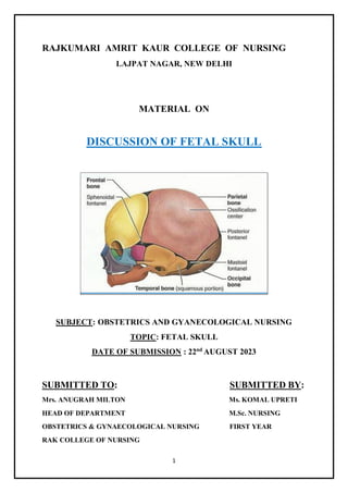
FETAL SKULL
INTRODUCTION
Understanding the anatomy of the fetal skull and its diameter will help us
recognise how a labour is progressing, and whether the baby's head is 'presenting'
correctly as it comes down the birth canal. This will give you a better
understanding of whether a normal vaginal delivery is likely, or if the mother
needs referral because the descent of the baby's head is not & making sufficient
progress.
 The fetal head is large in relation to the fetal body compared with the adult.
 The fetal skull is the most difficult part of the baby to pass through the
mother's pelvic canal, due to the hard bony nature of the skull. Adaptation
between the fetal skull and the pelvis is necessary to allow the head to pass
through the pelvis during labour without complications.
OBJECTIVES
 Define the fetal skull.
 Enumerate the importance of fetal skull.
 Describe the divisions and regions of the fetal skull.
 Enlist the bones which constitute the fetal head.
 Elaborate the sutures and fontanelles in fetal skull and their importance.
 Describe the various diameters and circumferences of the fetal skull and
their significance.
 Discuss the moulding and its relevance.
 Identify types of injury that can occur to the fetal skull during labour and
birth.
DEFINITION OF FETAL SKULL
Figure 1 FETAL SKULL
Fetal skull is the part of fetal body which encase and
protect the brain.
Skull is compressible to some extent because it made up
of thin flat bones which forms the vault. This is anchored
to the rigid and incompressible bones at the base of the
skull.
FIG 1: FETAL SKULL
3
IMPORTANCE OF FETAL SKULL
1. Largest part of the fetal body.
2. Most frequent presenting part of the fetus during delivery.
3. Least compressible among all the fetal parts.
DIVISONS OF FETAL SKULL
The fetal skull has divided into 3 major parts:
o Vault
o Base
o Face
Fig 2 : Fetal skull showing different regions
VAULT: The vault is the large, dome-shaped part above an imaginary line
drawn between the orbital ridges and the nape of the neck.
BASE: The base comprises bones that are firmly united to protect the vital
centres in the medulla oblongata.
FACE: The face is composed of 14 small bones that are also firmly united and
non-compressible.
4
REGIONS OF FETAL SKULL
The fetal skull has divided into different regions which are of obstetrical
importance:
o Vertex
o Brow
o Face
o Sinciput
o Occiput
VERTEX
It is a quadrangular area bounded anteriorly by the bregma and coronal sutures
behind the lambda and lambdoid sutures and laterally by lines passing through
the parietal eminences.
BROW
It is an area bounded on one side by the anterior fontanelle and coronal sutures
and on the other side by the root of the nose and the supra orbital ridges of the
either side. The base comprises bones that are firmly united to protect the vital
centres in the medulla oblongata.
FACE
It is the area bounded on one side by root of the nose and supra orbital ridges and
on the other, by the junction of the floor of the mouth and neck. The point between
the eyebrows is known as glabella.
SINCIPUT
It is the area lying in front of the anterior fontanelle and corresponds to the area
of brow.
OCCIPUT
It is the area limited to the occipital lobe.
5
Fig 3 : Fetal skull showing different regions and landmarks of obstetrical significance
BONES OF FETAL HEAD
Fetal head is consists of 7 bones:
 2 frontal bone
 2 parietal bone
 2 temporal bone
 1 occipital bone
Fig 4 : Bones of fetal head
6
SUTURES AND FONTANELLES
Flat bones of the vault are united together by non-ossified membranes attached
to the margins of the bones , these are called sutures and fontanelles.
These sutures and fontanelles are of obstetric significance because they allow
moulding of the fetal head during labour.
SUTURES:
Four types of sutures are found in the fetal skull:-
1. Sagittal or longitudinal suture: Lies between two parietal bones.
2. Coronal suture: Between parietal and frontal bones on either side.
3. Frontal suture: Lies between two frontal bones.
4. Lambdoid suture: Separate the occipital bone and the two parietal bones.
Fig 5 : Image showing the sutures and fontanelles
IMPORTANCE OF SUTURES:
(a) It permits gliding movement of one bone over the other during moulding
of the head, a phenomenon of significance while the head passes through
the pelvis during labour.
(b) Digital palpation of sagittal suture during internal examination in labour
gives an idea of the manner of engagement of the head, degree of internal
rotation of the head and degree of moulding of the head.
7
FONTANELLES:
Wide gap in the suture line is called fontanelle. There are six fontanelles in
number but out of these six only two are of obstetrics significance.
1. Posterior fontanelle
2. Anterior fontanelle
POSTERIOR FONTANELLE
 The posterior fontanelle or lambda is situated at the junction of the
lambdoidal and sagittal sutures.
 It is small, triangular in shape and can be recognized vaginally
because a suture leaves from each of the three angles.
 It normally closes by 6 weeks of age.
Fig 6: Image showing anterior and posterior fontanelle
ANTERIOR FONTANELLE
 The anterior fontanelle or bregma is found at the junction of the
sagittal, coronal and frontal sutures.
8
 It is broad, kite or diamond shaped and recognizable vaginally because a
suture leaves from each of the four corners.
 It measures 3–4 cm long and 1.5–2 cm wide and normally closes
by 18 months of age.
 Pulsations of cerebral vessels can be felt through this fontanelle.
IMPORTANCE OF FONTANELLES:
(a) Its palpation through internal examination denotes the degree of flexion of
the head.
(b) It facilitates moulding of the head.
(c) As it remains membranous long after birth, it helps in accommodating the
marked brain growth; the brain becoming almost double its size during the
first year of life.
(d) Palpation of the floor reflects intracranial status: depressed in dehydration,
elevated in raised intracranial tension.
(e) Collection of blood and exchange transfusion, on rare occasion, can be
performed through it via the superior longitudinal sinus.
(f) Cerebrospinal fluid can be drawn, although rarely, through the angle of the
anterior fontanel from the lateral ventricle.
(g) Denotes the position of head in relation to maternal pelvis.
DIAMETERS OF SKULL
The engaging diameter of the fetal skull depends on the degree of flexion present.
According to measurement whole skull is divided into two i.e.
 Anterior posterior diameter
 Transverse diameter.
9
1. ANTERIOR POSTERIOR DIAMETER
Fig 7 : Anterior posterior diameters of the fetal head
(a) Suboccipito bregmatic (SOB)
 Extends from the nape of the neck to the centre of the bregma.
 9.5cm in diameter.
 Attitude of the head is Complete flexion.
 Presentation is Vertex.
(b) Sub-occipitofrontal (SOF)
 Extends from the nape of the neck to the anterior end of the anterior
fontanelle or the centre of the sinciput.
 10cm in diameter.
 Attitude of the head is Incomplete flexion.
 Presentation is Vertex.
(c) Occipitofrontal (OF)
 Measured from the occipital eminence to the glabella.
10
 11.5cm in diameter.
 Attitude of the head is Marked deflexion.
 Presentation is Vertex.
(d) Mento vertical (MV)
 Measured from the point of the chin to the highest point on
the sagittal suture.
 14cm in diameter.
 Attitude of the head is Partial extension.
 Presentation is Brow.
(e) Sub-mento vertical (SMV)
 Measured from the point where the chin joins the neck to the
highest point on the vertex.
 11.5cm in diameter.
 Attitude of the head is Incomplete extension.
 Presentation is Face.
(f) Sub-mento bregmatic (SMB)
 Measured from the point where the chin joins the neck to the
centre of the bregma.
 9.5cm in diameter.
 Attitude of the head is Complete extension. Presentation is Face.
Fig 8: Image showing measurement of anterior posterior diameter
11
2. TRANSVERSE DIAMETER
Fig 9 :Transverse diameter of fetal head
(a)Biparietal diameter
 Extends between two parietal eminences. Whatever may be the
position of the head, the diameter nearly always engages.
 9.5cm in diameter.
(b)Bitemporal diameter
 It is the distance between the antero-inferior ends of the coronal
suture.
 8.2cm in diameter.

(c) Super- subparietal diameter
 It extends from a point placed below one parietal eminence to a point
placed above the other parietal eminence of the opposite side.
 8.5cm in diameter.

(d)Bimastoid diameter
 It is the distance between the tips of mastoid process.
 7.5cm in diameter.
12
CIRCUMFERENCES:
Circumference of the plane of the diameter of engagement differs according to
the attitude of the head.
ATTITUDE
OF THE
HEAD
PLANE OF
ENGAGEMENT
SHAPE CIRCUMFERENCE
Complete
Flexion
Biparietal sub
occpito bregmatic
Almost round 27.5cm
Deflexed Biparietal sub
occpito frontal
Oval 34cm
Incomplete
extension
Biparietal mento
vertical
Bigger oval 37.5cm
Complete
extension
Biparietal sub
mento bregmatic
Almost round 27.5cm
MOULDING
 The overlapping of the fetal skull bones at the suture line.
(Wylie, 2005, p.201)
 Bones of vault allow slight bending and override one another at the sutures.
 Skull changes shape from ovoid to cylindrical.
 Causes presenting diameter to decrease while increasing the diameter at
right angles.
 It is the alteration of the shape of the fore-coming head while passing
through the resistant birth passage during labour.
(D C Dutta,2013,p.86)
MECHANISM OF MOULDING:
There is compression of the engaging diameter of the head with corresponding
elongation of the diameter at right angle to it.
13
Thus, in well flexed head of the anterior vertex presentation, the engaging
suboccipito bregmatic diameter is compressed with elongation of the head in
mento-vertical diameter which is at right angle to suboccipito bregmatic.
Moulding disappear within few hours after birth.
GRADING:
There are three gradings:-
Grade-1—the bones touching but not overlapping.
Grade-2— overlapping but easily separated.
Grade-3—fixed overlapping.
IMPORTANCE OF MOULDING:
 Slight moulding is good and beneficial because it enables the head to pass
more easily, through the birth canal.
 Shape of the moulding can be a useful information about the position of
the head occupied in the pelvis.
INJURIES DURING THE DELIVERY OF HEAD
Fig 10: image showing caput succedaneum
CAPUT SUCCEDANEUM
 Oedematous swelling that
occurs on the presenting
part.
 Present at birth.
 Can cross over the
sutures.
 Soft swelling and will
indent with pressure.
 Decreases following
delivery.
 No treatment is required.
 Will disappear
approximately 24-48hrs
after birth.
 Moulding will also be
apparent.
14
Fig 11: Image showing cephalohematoma
CEPHALOHEMATOMA
 Bleeding that occurs
between the bones of the
fetal skull and
periosteum.
 Occurs 12-72 hours after
birth.
 The swelling is restricted
to one area and will not
indent with pressure.
 Does not cross over the
sutures.
 Can be bilateral.
 Persist for a few weeks
and occasionally months.
15
BIBLIOGRAPHY
 Burden, B. and Sapsed, M.S., The Fetal Skull. In: Macdonald, S.,Magill-
Cuerden (eds.) Mayes' Midwifery 14th ed. London: Ballière Tindall, 2011,
page no: 96-104.
 Wylie, L., Essential anatomy and physiology in maternity care. 2nd ed.
Edinburgh: Elsevier,2005,page no: 201-206.
 Shirish S Sheth, Essential of Obstetrics, 1st Edition, Jaypee Brothers
Medical Publishers, New Delhi,2004, page no.: 102 - 104.
 Diane M, Myles Textbook for Midwives, 14th edition, Elsevier Publisher,
London, 2008, page no: 118-123.
 Dutta D C, Textbook of obstetrics,7th
edition, New central book agency (P)
ltd. ,London ,2013, page no: 83-87.
 Shirish N Daftary, Manual of obstetrics,2nd
edition, Elsevier publication,
2007, Page no: 68-72.
REFERENCES FOR THE IMAGES
IMAGES REFERENCE
Figure 1 & 2 https://www.google.com>fetalskull//
Figure 3 & 9  Dutta D C, Textbook of
obstetrics,7th
edition, New central
book agency (P) ltd. ,London ,2013,
page no: 83-87.
Figure 4  Diane M, Myles Textbook for
Midwives, 14th edition, Elsevier
Publisher, London, 2008, page no:
118-123.
Figure 5 & 6  Wylie, L., Essential anatomy and
physiology in maternity care. 2nd
ed. Edinburgh: Elsevier,2005,page
no: 201-206.
Figure 7 & 8  Shirish N Daftary, Manual of
obstetrics,2nd
edition, Elsevier
publication, 2007, Page no: 68-72.
16
Figure 10 & 11  Burden, B. and Sapsed, M.S.,The
FetalSkull.In:Macdonald,S.,Magill-
Cuerden (eds.) Mayes' Midwifery
14th ed. London: Ballière Tindall,
2011, page no:

More Related Content

What's hot

Destructive operation
Destructive operationDestructive operation
Destructive operation
PRANATI PATRA
 
Fetal skull
Fetal skullFetal skull
Fetal skull
Kshyanaprava Behera
 
Destructive operations
Destructive operationsDestructive operations
Destructive operations
Neethu Satheesan
 
Fetal skull
Fetal skullFetal skull
Fetal skull
Arya Anish
 
Placenta examination
Placenta examinationPlacenta examination
Placenta examination
1302011987
 
Induction of labour
Induction of labourInduction of labour
Induction of labour
SREEVIDYA UMMADISETTI
 
Medical induction of labour
Medical induction of labourMedical induction of labour
Medical induction of labour
Kasturi Ramasamy
 
Female pelvis
Female pelvisFemale pelvis
Female pelvis
Kshyanaprava Behera
 
Antenatal care deepti ppt
Antenatal care deepti pptAntenatal care deepti ppt
Antenatal care deepti ppt
nidhi maurya
 
Current trends in midwifery &; obstetrical nursing
Current trends in midwifery &; obstetrical nursingCurrent trends in midwifery &; obstetrical nursing
Current trends in midwifery &; obstetrical nursing
Abhilasha verma
 
Introduction of midwifery and obstetrical nursing
Introduction of midwifery and obstetrical nursingIntroduction of midwifery and obstetrical nursing
Introduction of midwifery and obstetrical nursing
HARSH786249
 
Female Pelvis
Female PelvisFemale Pelvis
Female Pelvis
Kanchan Mehra
 
Female pelvis
Female pelvisFemale pelvis
Female pelvis
SrujaniDash1
 
Newborn care
Newborn careNewborn care
Newborn care
ABHIJIT BHOYAR
 
First stage management of labour
First stage management of labourFirst stage management of labour
First stage management of labour
Birat Medical College, Kathmandu University, Nepal
 
Labour 1st stage
Labour 1st stageLabour 1st stage
Labour 1st stage
Amandeep Jhinjar
 
Placenta & cord abnormality
Placenta & cord abnormalityPlacenta & cord abnormality
Placenta & cord abnormality
Abhilasha verma
 

What's hot (20)

Antenatal care
Antenatal care Antenatal care
Antenatal care
 
Destructive operation
Destructive operationDestructive operation
Destructive operation
 
Fetal skull
Fetal skullFetal skull
Fetal skull
 
Destructive operations
Destructive operationsDestructive operations
Destructive operations
 
Fetal skull
Fetal skullFetal skull
Fetal skull
 
Placenta examination
Placenta examinationPlacenta examination
Placenta examination
 
Induction of labour
Induction of labourInduction of labour
Induction of labour
 
Placenta examination
Placenta examinationPlacenta examination
Placenta examination
 
Medical induction of labour
Medical induction of labourMedical induction of labour
Medical induction of labour
 
Female pelvis
Female pelvisFemale pelvis
Female pelvis
 
Antenatal care deepti ppt
Antenatal care deepti pptAntenatal care deepti ppt
Antenatal care deepti ppt
 
Fetal skull
Fetal skull Fetal skull
Fetal skull
 
Current trends in midwifery &; obstetrical nursing
Current trends in midwifery &; obstetrical nursingCurrent trends in midwifery &; obstetrical nursing
Current trends in midwifery &; obstetrical nursing
 
Introduction of midwifery and obstetrical nursing
Introduction of midwifery and obstetrical nursingIntroduction of midwifery and obstetrical nursing
Introduction of midwifery and obstetrical nursing
 
Female Pelvis
Female PelvisFemale Pelvis
Female Pelvis
 
Female pelvis
Female pelvisFemale pelvis
Female pelvis
 
Newborn care
Newborn careNewborn care
Newborn care
 
First stage management of labour
First stage management of labourFirst stage management of labour
First stage management of labour
 
Labour 1st stage
Labour 1st stageLabour 1st stage
Labour 1st stage
 
Placenta & cord abnormality
Placenta & cord abnormalityPlacenta & cord abnormality
Placenta & cord abnormality
 

Similar to FETAL SKULL DISCUSSION (1).pdf

Fetal skull
Fetal skull Fetal skull
Fetal skull
mariem ahmed
 
FETAL SKULL PPT (1).pptx
FETAL SKULL PPT (1).pptxFETAL SKULL PPT (1).pptx
FETAL SKULL PPT (1).pptx
PRIYANKASINGH102278
 
FETAL SKULL PPT .pptx
FETAL SKULL PPT .pptxFETAL SKULL PPT .pptx
FETAL SKULL PPT .pptx
Komal Upreti
 
Fetal skull
Fetal skullFetal skull
Fetal skull
Jasleen Kaur
 
FETAL SKULL BY FELI.pdf
FETAL SKULL BY FELI.pdfFETAL SKULL BY FELI.pdf
FETAL SKULL BY FELI.pdf
Bright89
 
FOETAL SKULL.pptx
FOETAL SKULL.pptxFOETAL SKULL.pptx
fetal skull ppt workshop obg.pptx
fetal skull ppt workshop obg.pptxfetal skull ppt workshop obg.pptx
fetal skull ppt workshop obg.pptx
DivyaPandey734483
 
Feotal skull presentation
Feotal skull presentationFeotal skull presentation
Feotal skull presentation
Rekha Dobal
 
Fetal skull
Fetal skullFetal skull
Fetal skull
Kavitha Reddy
 
Foetal skull and maternal pelvis
Foetal skull and maternal pelvisFoetal skull and maternal pelvis
Foetal skull and maternal pelvis
Sneha Tiwari
 
Fetal Skull obstetrics and gynecological
Fetal Skull obstetrics and gynecologicalFetal Skull obstetrics and gynecological
Fetal Skull obstetrics and gynecological
Thangamjayarani
 
FETAL SKULL AND MATERNAL PELVIS (Dr. Utpala Mazumder).pptx
FETAL SKULL AND MATERNAL PELVIS (Dr. Utpala Mazumder).pptxFETAL SKULL AND MATERNAL PELVIS (Dr. Utpala Mazumder).pptx
FETAL SKULL AND MATERNAL PELVIS (Dr. Utpala Mazumder).pptx
UtpalaMazumder
 
fetal skull
 fetal skull fetal skull
fetal skull
yashasvikashiv
 
Fetal head diameters
Fetal head diametersFetal head diameters
Fetal head diameters
mohammad tailakh
 
Fetal skull
Fetal skullFetal skull
Fetal skull
parekhkishan1
 
The fetal skull
The fetal skullThe fetal skull
The fetal skull
Kishan Parekh
 
fetus and gynaecoid pelvis
 fetus and gynaecoid pelvis  fetus and gynaecoid pelvis
fetus and gynaecoid pelvis
tariggally
 
fetalskullppt-160707124535-converted.pptx
fetalskullppt-160707124535-converted.pptxfetalskullppt-160707124535-converted.pptx
fetalskullppt-160707124535-converted.pptx
AkhilaLukose
 
Female bony pelvis and fetal skull for undergraduate
Female   bony pelvis and fetal skull for undergraduateFemale   bony pelvis and fetal skull for undergraduate
Female bony pelvis and fetal skull for undergraduate
Faculty of Medicine,Zagazig University,EGYPT
 
Fetal skull
Fetal skullFetal skull
Fetal skull
anishreshma
 

Similar to FETAL SKULL DISCUSSION (1).pdf (20)

Fetal skull
Fetal skull Fetal skull
Fetal skull
 
FETAL SKULL PPT (1).pptx
FETAL SKULL PPT (1).pptxFETAL SKULL PPT (1).pptx
FETAL SKULL PPT (1).pptx
 
FETAL SKULL PPT .pptx
FETAL SKULL PPT .pptxFETAL SKULL PPT .pptx
FETAL SKULL PPT .pptx
 
Fetal skull
Fetal skullFetal skull
Fetal skull
 
FETAL SKULL BY FELI.pdf
FETAL SKULL BY FELI.pdfFETAL SKULL BY FELI.pdf
FETAL SKULL BY FELI.pdf
 
FOETAL SKULL.pptx
FOETAL SKULL.pptxFOETAL SKULL.pptx
FOETAL SKULL.pptx
 
fetal skull ppt workshop obg.pptx
fetal skull ppt workshop obg.pptxfetal skull ppt workshop obg.pptx
fetal skull ppt workshop obg.pptx
 
Feotal skull presentation
Feotal skull presentationFeotal skull presentation
Feotal skull presentation
 
Fetal skull
Fetal skullFetal skull
Fetal skull
 
Foetal skull and maternal pelvis
Foetal skull and maternal pelvisFoetal skull and maternal pelvis
Foetal skull and maternal pelvis
 
Fetal Skull obstetrics and gynecological
Fetal Skull obstetrics and gynecologicalFetal Skull obstetrics and gynecological
Fetal Skull obstetrics and gynecological
 
FETAL SKULL AND MATERNAL PELVIS (Dr. Utpala Mazumder).pptx
FETAL SKULL AND MATERNAL PELVIS (Dr. Utpala Mazumder).pptxFETAL SKULL AND MATERNAL PELVIS (Dr. Utpala Mazumder).pptx
FETAL SKULL AND MATERNAL PELVIS (Dr. Utpala Mazumder).pptx
 
fetal skull
 fetal skull fetal skull
fetal skull
 
Fetal head diameters
Fetal head diametersFetal head diameters
Fetal head diameters
 
Fetal skull
Fetal skullFetal skull
Fetal skull
 
The fetal skull
The fetal skullThe fetal skull
The fetal skull
 
fetus and gynaecoid pelvis
 fetus and gynaecoid pelvis  fetus and gynaecoid pelvis
fetus and gynaecoid pelvis
 
fetalskullppt-160707124535-converted.pptx
fetalskullppt-160707124535-converted.pptxfetalskullppt-160707124535-converted.pptx
fetalskullppt-160707124535-converted.pptx
 
Female bony pelvis and fetal skull for undergraduate
Female   bony pelvis and fetal skull for undergraduateFemale   bony pelvis and fetal skull for undergraduate
Female bony pelvis and fetal skull for undergraduate
 
Fetal skull
Fetal skullFetal skull
Fetal skull
 

More from Komal Upreti

CHECKLIST ON CPR.pdf
CHECKLIST ON CPR.pdfCHECKLIST ON CPR.pdf
CHECKLIST ON CPR.pdf
Komal Upreti
 
CPR_PRESENTATION.pptx
CPR_PRESENTATION.pptxCPR_PRESENTATION.pptx
CPR_PRESENTATION.pptx
Komal Upreti
 
DEMONSTRATION CPR.pdf
DEMONSTRATION CPR.pdfDEMONSTRATION CPR.pdf
DEMONSTRATION CPR.pdf
Komal Upreti
 
stroke 5th semestr final.pptx
stroke 5th semestr final.pptxstroke 5th semestr final.pptx
stroke 5th semestr final.pptx
Komal Upreti
 
NEUROREHABILITATION.pptx
NEUROREHABILITATION.pptxNEUROREHABILITATION.pptx
NEUROREHABILITATION.pptx
Komal Upreti
 
myastheniagravis-130317015902-phpapp02.pptx
myastheniagravis-130317015902-phpapp02.pptxmyastheniagravis-130317015902-phpapp02.pptx
myastheniagravis-130317015902-phpapp02.pptx
Komal Upreti
 
alzheimersdisease final.pptx
alzheimersdisease final.pptxalzheimersdisease final.pptx
alzheimersdisease final.pptx
Komal Upreti
 
HEADACHE (CEPHAGIA).pptx
HEADACHE (CEPHAGIA).pptxHEADACHE (CEPHAGIA).pptx
HEADACHE (CEPHAGIA).pptx
Komal Upreti
 
psychiatric emergencies.pptx
psychiatric emergencies.pptxpsychiatric emergencies.pptx
psychiatric emergencies.pptx
Komal Upreti
 
ACUTE RESPIRATORY INFECTIONS.pptx
ACUTE RESPIRATORY INFECTIONS.pptxACUTE RESPIRATORY INFECTIONS.pptx
ACUTE RESPIRATORY INFECTIONS.pptx
Komal Upreti
 

More from Komal Upreti (10)

CHECKLIST ON CPR.pdf
CHECKLIST ON CPR.pdfCHECKLIST ON CPR.pdf
CHECKLIST ON CPR.pdf
 
CPR_PRESENTATION.pptx
CPR_PRESENTATION.pptxCPR_PRESENTATION.pptx
CPR_PRESENTATION.pptx
 
DEMONSTRATION CPR.pdf
DEMONSTRATION CPR.pdfDEMONSTRATION CPR.pdf
DEMONSTRATION CPR.pdf
 
stroke 5th semestr final.pptx
stroke 5th semestr final.pptxstroke 5th semestr final.pptx
stroke 5th semestr final.pptx
 
NEUROREHABILITATION.pptx
NEUROREHABILITATION.pptxNEUROREHABILITATION.pptx
NEUROREHABILITATION.pptx
 
myastheniagravis-130317015902-phpapp02.pptx
myastheniagravis-130317015902-phpapp02.pptxmyastheniagravis-130317015902-phpapp02.pptx
myastheniagravis-130317015902-phpapp02.pptx
 
alzheimersdisease final.pptx
alzheimersdisease final.pptxalzheimersdisease final.pptx
alzheimersdisease final.pptx
 
HEADACHE (CEPHAGIA).pptx
HEADACHE (CEPHAGIA).pptxHEADACHE (CEPHAGIA).pptx
HEADACHE (CEPHAGIA).pptx
 
psychiatric emergencies.pptx
psychiatric emergencies.pptxpsychiatric emergencies.pptx
psychiatric emergencies.pptx
 
ACUTE RESPIRATORY INFECTIONS.pptx
ACUTE RESPIRATORY INFECTIONS.pptxACUTE RESPIRATORY INFECTIONS.pptx
ACUTE RESPIRATORY INFECTIONS.pptx
 

Recently uploaded

Model Attribute Check Company Auto Property
Model Attribute  Check Company Auto PropertyModel Attribute  Check Company Auto Property
Model Attribute Check Company Auto Property
Celine George
 
How libraries can support authors with open access requirements for UKRI fund...
How libraries can support authors with open access requirements for UKRI fund...How libraries can support authors with open access requirements for UKRI fund...
How libraries can support authors with open access requirements for UKRI fund...
Jisc
 
aaaaaaaaaaaaaaaaaaaaaaaaaaaaaaaaaaaaaaaaa
aaaaaaaaaaaaaaaaaaaaaaaaaaaaaaaaaaaaaaaaaaaaaaaaaaaaaaaaaaaaaaaaaaaaaaaaaaaaaaaaaa
aaaaaaaaaaaaaaaaaaaaaaaaaaaaaaaaaaaaaaaaa
siemaillard
 
Operation Blue Star - Saka Neela Tara
Operation Blue Star   -  Saka Neela TaraOperation Blue Star   -  Saka Neela Tara
Operation Blue Star - Saka Neela Tara
Balvir Singh
 
Overview on Edible Vaccine: Pros & Cons with Mechanism
Overview on Edible Vaccine: Pros & Cons with MechanismOverview on Edible Vaccine: Pros & Cons with Mechanism
Overview on Edible Vaccine: Pros & Cons with Mechanism
DeeptiGupta154
 
GIÁO ÁN DẠY THÊM (KẾ HOẠCH BÀI BUỔI 2) - TIẾNG ANH 8 GLOBAL SUCCESS (2 CỘT) N...
GIÁO ÁN DẠY THÊM (KẾ HOẠCH BÀI BUỔI 2) - TIẾNG ANH 8 GLOBAL SUCCESS (2 CỘT) N...GIÁO ÁN DẠY THÊM (KẾ HOẠCH BÀI BUỔI 2) - TIẾNG ANH 8 GLOBAL SUCCESS (2 CỘT) N...
GIÁO ÁN DẠY THÊM (KẾ HOẠCH BÀI BUỔI 2) - TIẾNG ANH 8 GLOBAL SUCCESS (2 CỘT) N...
Nguyen Thanh Tu Collection
 
Students, digital devices and success - Andreas Schleicher - 27 May 2024..pptx
Students, digital devices and success - Andreas Schleicher - 27 May 2024..pptxStudents, digital devices and success - Andreas Schleicher - 27 May 2024..pptx
Students, digital devices and success - Andreas Schleicher - 27 May 2024..pptx
EduSkills OECD
 
Fish and Chips - have they had their chips
Fish and Chips - have they had their chipsFish and Chips - have they had their chips
Fish and Chips - have they had their chips
GeoBlogs
 
The Roman Empire A Historical Colossus.pdf
The Roman Empire A Historical Colossus.pdfThe Roman Empire A Historical Colossus.pdf
The Roman Empire A Historical Colossus.pdf
kaushalkr1407
 
special B.ed 2nd year old paper_20240531.pdf
special B.ed 2nd year old paper_20240531.pdfspecial B.ed 2nd year old paper_20240531.pdf
special B.ed 2nd year old paper_20240531.pdf
Special education needs
 
Thesis Statement for students diagnonsed withADHD.ppt
Thesis Statement for students diagnonsed withADHD.pptThesis Statement for students diagnonsed withADHD.ppt
Thesis Statement for students diagnonsed withADHD.ppt
EverAndrsGuerraGuerr
 
Chapter 3 - Islamic Banking Products and Services.pptx
Chapter 3 - Islamic Banking Products and Services.pptxChapter 3 - Islamic Banking Products and Services.pptx
Chapter 3 - Islamic Banking Products and Services.pptx
Mohd Adib Abd Muin, Senior Lecturer at Universiti Utara Malaysia
 
How to Split Bills in the Odoo 17 POS Module
How to Split Bills in the Odoo 17 POS ModuleHow to Split Bills in the Odoo 17 POS Module
How to Split Bills in the Odoo 17 POS Module
Celine George
 
Unit 2- Research Aptitude (UGC NET Paper I).pdf
Unit 2- Research Aptitude (UGC NET Paper I).pdfUnit 2- Research Aptitude (UGC NET Paper I).pdf
Unit 2- Research Aptitude (UGC NET Paper I).pdf
Thiyagu K
 
The Art Pastor's Guide to Sabbath | Steve Thomason
The Art Pastor's Guide to Sabbath | Steve ThomasonThe Art Pastor's Guide to Sabbath | Steve Thomason
The Art Pastor's Guide to Sabbath | Steve Thomason
Steve Thomason
 
TESDA TM1 REVIEWER FOR NATIONAL ASSESSMENT WRITTEN AND ORAL QUESTIONS WITH A...
TESDA TM1 REVIEWER  FOR NATIONAL ASSESSMENT WRITTEN AND ORAL QUESTIONS WITH A...TESDA TM1 REVIEWER  FOR NATIONAL ASSESSMENT WRITTEN AND ORAL QUESTIONS WITH A...
TESDA TM1 REVIEWER FOR NATIONAL ASSESSMENT WRITTEN AND ORAL QUESTIONS WITH A...
EugeneSaldivar
 
How to Create Map Views in the Odoo 17 ERP
How to Create Map Views in the Odoo 17 ERPHow to Create Map Views in the Odoo 17 ERP
How to Create Map Views in the Odoo 17 ERP
Celine George
 
Language Across the Curriculm LAC B.Ed.
Language Across the  Curriculm LAC B.Ed.Language Across the  Curriculm LAC B.Ed.
Language Across the Curriculm LAC B.Ed.
Atul Kumar Singh
 
Polish students' mobility in the Czech Republic
Polish students' mobility in the Czech RepublicPolish students' mobility in the Czech Republic
Polish students' mobility in the Czech Republic
Anna Sz.
 
Cambridge International AS A Level Biology Coursebook - EBook (MaryFosbery J...
Cambridge International AS  A Level Biology Coursebook - EBook (MaryFosbery J...Cambridge International AS  A Level Biology Coursebook - EBook (MaryFosbery J...
Cambridge International AS A Level Biology Coursebook - EBook (MaryFosbery J...
AzmatAli747758
 

Recently uploaded (20)

Model Attribute Check Company Auto Property
Model Attribute  Check Company Auto PropertyModel Attribute  Check Company Auto Property
Model Attribute Check Company Auto Property
 
How libraries can support authors with open access requirements for UKRI fund...
How libraries can support authors with open access requirements for UKRI fund...How libraries can support authors with open access requirements for UKRI fund...
How libraries can support authors with open access requirements for UKRI fund...
 
aaaaaaaaaaaaaaaaaaaaaaaaaaaaaaaaaaaaaaaaa
aaaaaaaaaaaaaaaaaaaaaaaaaaaaaaaaaaaaaaaaaaaaaaaaaaaaaaaaaaaaaaaaaaaaaaaaaaaaaaaaaa
aaaaaaaaaaaaaaaaaaaaaaaaaaaaaaaaaaaaaaaaa
 
Operation Blue Star - Saka Neela Tara
Operation Blue Star   -  Saka Neela TaraOperation Blue Star   -  Saka Neela Tara
Operation Blue Star - Saka Neela Tara
 
Overview on Edible Vaccine: Pros & Cons with Mechanism
Overview on Edible Vaccine: Pros & Cons with MechanismOverview on Edible Vaccine: Pros & Cons with Mechanism
Overview on Edible Vaccine: Pros & Cons with Mechanism
 
GIÁO ÁN DẠY THÊM (KẾ HOẠCH BÀI BUỔI 2) - TIẾNG ANH 8 GLOBAL SUCCESS (2 CỘT) N...
GIÁO ÁN DẠY THÊM (KẾ HOẠCH BÀI BUỔI 2) - TIẾNG ANH 8 GLOBAL SUCCESS (2 CỘT) N...GIÁO ÁN DẠY THÊM (KẾ HOẠCH BÀI BUỔI 2) - TIẾNG ANH 8 GLOBAL SUCCESS (2 CỘT) N...
GIÁO ÁN DẠY THÊM (KẾ HOẠCH BÀI BUỔI 2) - TIẾNG ANH 8 GLOBAL SUCCESS (2 CỘT) N...
 
Students, digital devices and success - Andreas Schleicher - 27 May 2024..pptx
Students, digital devices and success - Andreas Schleicher - 27 May 2024..pptxStudents, digital devices and success - Andreas Schleicher - 27 May 2024..pptx
Students, digital devices and success - Andreas Schleicher - 27 May 2024..pptx
 
Fish and Chips - have they had their chips
Fish and Chips - have they had their chipsFish and Chips - have they had their chips
Fish and Chips - have they had their chips
 
The Roman Empire A Historical Colossus.pdf
The Roman Empire A Historical Colossus.pdfThe Roman Empire A Historical Colossus.pdf
The Roman Empire A Historical Colossus.pdf
 
special B.ed 2nd year old paper_20240531.pdf
special B.ed 2nd year old paper_20240531.pdfspecial B.ed 2nd year old paper_20240531.pdf
special B.ed 2nd year old paper_20240531.pdf
 
Thesis Statement for students diagnonsed withADHD.ppt
Thesis Statement for students diagnonsed withADHD.pptThesis Statement for students diagnonsed withADHD.ppt
Thesis Statement for students diagnonsed withADHD.ppt
 
Chapter 3 - Islamic Banking Products and Services.pptx
Chapter 3 - Islamic Banking Products and Services.pptxChapter 3 - Islamic Banking Products and Services.pptx
Chapter 3 - Islamic Banking Products and Services.pptx
 
How to Split Bills in the Odoo 17 POS Module
How to Split Bills in the Odoo 17 POS ModuleHow to Split Bills in the Odoo 17 POS Module
How to Split Bills in the Odoo 17 POS Module
 
Unit 2- Research Aptitude (UGC NET Paper I).pdf
Unit 2- Research Aptitude (UGC NET Paper I).pdfUnit 2- Research Aptitude (UGC NET Paper I).pdf
Unit 2- Research Aptitude (UGC NET Paper I).pdf
 
The Art Pastor's Guide to Sabbath | Steve Thomason
The Art Pastor's Guide to Sabbath | Steve ThomasonThe Art Pastor's Guide to Sabbath | Steve Thomason
The Art Pastor's Guide to Sabbath | Steve Thomason
 
TESDA TM1 REVIEWER FOR NATIONAL ASSESSMENT WRITTEN AND ORAL QUESTIONS WITH A...
TESDA TM1 REVIEWER  FOR NATIONAL ASSESSMENT WRITTEN AND ORAL QUESTIONS WITH A...TESDA TM1 REVIEWER  FOR NATIONAL ASSESSMENT WRITTEN AND ORAL QUESTIONS WITH A...
TESDA TM1 REVIEWER FOR NATIONAL ASSESSMENT WRITTEN AND ORAL QUESTIONS WITH A...
 
How to Create Map Views in the Odoo 17 ERP
How to Create Map Views in the Odoo 17 ERPHow to Create Map Views in the Odoo 17 ERP
How to Create Map Views in the Odoo 17 ERP
 
Language Across the Curriculm LAC B.Ed.
Language Across the  Curriculm LAC B.Ed.Language Across the  Curriculm LAC B.Ed.
Language Across the Curriculm LAC B.Ed.
 
Polish students' mobility in the Czech Republic
Polish students' mobility in the Czech RepublicPolish students' mobility in the Czech Republic
Polish students' mobility in the Czech Republic
 
Cambridge International AS A Level Biology Coursebook - EBook (MaryFosbery J...
Cambridge International AS  A Level Biology Coursebook - EBook (MaryFosbery J...Cambridge International AS  A Level Biology Coursebook - EBook (MaryFosbery J...
Cambridge International AS A Level Biology Coursebook - EBook (MaryFosbery J...
 

FETAL SKULL DISCUSSION (1).pdf

  • 1. 1 RAJKUMARI AMRIT KAUR COLLEGE OF NURSING LAJPAT NAGAR, NEW DELHI MATERIAL ON DISCUSSION OF FETAL SKULL SUBJECT: OBSTETRICS AND GYANECOLOGICAL NURSING TOPIC: FETAL SKULL DATE OF SUBMISSION : 22nd AUGUST 2023 SUBMITTED TO: SUBMITTED BY: Mrs. ANUGRAH MILTON Ms. KOMAL UPRETI HEAD OF DEPARTMENT M.Sc. NURSING OBSTETRICS & GYNAECOLOGICAL NURSING FIRST YEAR RAK COLLEGE OF NURSING
  • 2. 2 FETAL SKULL INTRODUCTION Understanding the anatomy of the fetal skull and its diameter will help us recognise how a labour is progressing, and whether the baby's head is 'presenting' correctly as it comes down the birth canal. This will give you a better understanding of whether a normal vaginal delivery is likely, or if the mother needs referral because the descent of the baby's head is not & making sufficient progress.  The fetal head is large in relation to the fetal body compared with the adult.  The fetal skull is the most difficult part of the baby to pass through the mother's pelvic canal, due to the hard bony nature of the skull. Adaptation between the fetal skull and the pelvis is necessary to allow the head to pass through the pelvis during labour without complications. OBJECTIVES  Define the fetal skull.  Enumerate the importance of fetal skull.  Describe the divisions and regions of the fetal skull.  Enlist the bones which constitute the fetal head.  Elaborate the sutures and fontanelles in fetal skull and their importance.  Describe the various diameters and circumferences of the fetal skull and their significance.  Discuss the moulding and its relevance.  Identify types of injury that can occur to the fetal skull during labour and birth. DEFINITION OF FETAL SKULL Figure 1 FETAL SKULL Fetal skull is the part of fetal body which encase and protect the brain. Skull is compressible to some extent because it made up of thin flat bones which forms the vault. This is anchored to the rigid and incompressible bones at the base of the skull. FIG 1: FETAL SKULL
  • 3. 3 IMPORTANCE OF FETAL SKULL 1. Largest part of the fetal body. 2. Most frequent presenting part of the fetus during delivery. 3. Least compressible among all the fetal parts. DIVISONS OF FETAL SKULL The fetal skull has divided into 3 major parts: o Vault o Base o Face Fig 2 : Fetal skull showing different regions VAULT: The vault is the large, dome-shaped part above an imaginary line drawn between the orbital ridges and the nape of the neck. BASE: The base comprises bones that are firmly united to protect the vital centres in the medulla oblongata. FACE: The face is composed of 14 small bones that are also firmly united and non-compressible.
  • 4. 4 REGIONS OF FETAL SKULL The fetal skull has divided into different regions which are of obstetrical importance: o Vertex o Brow o Face o Sinciput o Occiput VERTEX It is a quadrangular area bounded anteriorly by the bregma and coronal sutures behind the lambda and lambdoid sutures and laterally by lines passing through the parietal eminences. BROW It is an area bounded on one side by the anterior fontanelle and coronal sutures and on the other side by the root of the nose and the supra orbital ridges of the either side. The base comprises bones that are firmly united to protect the vital centres in the medulla oblongata. FACE It is the area bounded on one side by root of the nose and supra orbital ridges and on the other, by the junction of the floor of the mouth and neck. The point between the eyebrows is known as glabella. SINCIPUT It is the area lying in front of the anterior fontanelle and corresponds to the area of brow. OCCIPUT It is the area limited to the occipital lobe.
  • 5. 5 Fig 3 : Fetal skull showing different regions and landmarks of obstetrical significance BONES OF FETAL HEAD Fetal head is consists of 7 bones:  2 frontal bone  2 parietal bone  2 temporal bone  1 occipital bone Fig 4 : Bones of fetal head
  • 6. 6 SUTURES AND FONTANELLES Flat bones of the vault are united together by non-ossified membranes attached to the margins of the bones , these are called sutures and fontanelles. These sutures and fontanelles are of obstetric significance because they allow moulding of the fetal head during labour. SUTURES: Four types of sutures are found in the fetal skull:- 1. Sagittal or longitudinal suture: Lies between two parietal bones. 2. Coronal suture: Between parietal and frontal bones on either side. 3. Frontal suture: Lies between two frontal bones. 4. Lambdoid suture: Separate the occipital bone and the two parietal bones. Fig 5 : Image showing the sutures and fontanelles IMPORTANCE OF SUTURES: (a) It permits gliding movement of one bone over the other during moulding of the head, a phenomenon of significance while the head passes through the pelvis during labour. (b) Digital palpation of sagittal suture during internal examination in labour gives an idea of the manner of engagement of the head, degree of internal rotation of the head and degree of moulding of the head.
  • 7. 7 FONTANELLES: Wide gap in the suture line is called fontanelle. There are six fontanelles in number but out of these six only two are of obstetrics significance. 1. Posterior fontanelle 2. Anterior fontanelle POSTERIOR FONTANELLE  The posterior fontanelle or lambda is situated at the junction of the lambdoidal and sagittal sutures.  It is small, triangular in shape and can be recognized vaginally because a suture leaves from each of the three angles.  It normally closes by 6 weeks of age. Fig 6: Image showing anterior and posterior fontanelle ANTERIOR FONTANELLE  The anterior fontanelle or bregma is found at the junction of the sagittal, coronal and frontal sutures.
  • 8. 8  It is broad, kite or diamond shaped and recognizable vaginally because a suture leaves from each of the four corners.  It measures 3–4 cm long and 1.5–2 cm wide and normally closes by 18 months of age.  Pulsations of cerebral vessels can be felt through this fontanelle. IMPORTANCE OF FONTANELLES: (a) Its palpation through internal examination denotes the degree of flexion of the head. (b) It facilitates moulding of the head. (c) As it remains membranous long after birth, it helps in accommodating the marked brain growth; the brain becoming almost double its size during the first year of life. (d) Palpation of the floor reflects intracranial status: depressed in dehydration, elevated in raised intracranial tension. (e) Collection of blood and exchange transfusion, on rare occasion, can be performed through it via the superior longitudinal sinus. (f) Cerebrospinal fluid can be drawn, although rarely, through the angle of the anterior fontanel from the lateral ventricle. (g) Denotes the position of head in relation to maternal pelvis. DIAMETERS OF SKULL The engaging diameter of the fetal skull depends on the degree of flexion present. According to measurement whole skull is divided into two i.e.  Anterior posterior diameter  Transverse diameter.
  • 9. 9 1. ANTERIOR POSTERIOR DIAMETER Fig 7 : Anterior posterior diameters of the fetal head (a) Suboccipito bregmatic (SOB)  Extends from the nape of the neck to the centre of the bregma.  9.5cm in diameter.  Attitude of the head is Complete flexion.  Presentation is Vertex. (b) Sub-occipitofrontal (SOF)  Extends from the nape of the neck to the anterior end of the anterior fontanelle or the centre of the sinciput.  10cm in diameter.  Attitude of the head is Incomplete flexion.  Presentation is Vertex. (c) Occipitofrontal (OF)  Measured from the occipital eminence to the glabella.
  • 10. 10  11.5cm in diameter.  Attitude of the head is Marked deflexion.  Presentation is Vertex. (d) Mento vertical (MV)  Measured from the point of the chin to the highest point on the sagittal suture.  14cm in diameter.  Attitude of the head is Partial extension.  Presentation is Brow. (e) Sub-mento vertical (SMV)  Measured from the point where the chin joins the neck to the highest point on the vertex.  11.5cm in diameter.  Attitude of the head is Incomplete extension.  Presentation is Face. (f) Sub-mento bregmatic (SMB)  Measured from the point where the chin joins the neck to the centre of the bregma.  9.5cm in diameter.  Attitude of the head is Complete extension. Presentation is Face. Fig 8: Image showing measurement of anterior posterior diameter
  • 11. 11 2. TRANSVERSE DIAMETER Fig 9 :Transverse diameter of fetal head (a)Biparietal diameter  Extends between two parietal eminences. Whatever may be the position of the head, the diameter nearly always engages.  9.5cm in diameter. (b)Bitemporal diameter  It is the distance between the antero-inferior ends of the coronal suture.  8.2cm in diameter.  (c) Super- subparietal diameter  It extends from a point placed below one parietal eminence to a point placed above the other parietal eminence of the opposite side.  8.5cm in diameter.  (d)Bimastoid diameter  It is the distance between the tips of mastoid process.  7.5cm in diameter.
  • 12. 12 CIRCUMFERENCES: Circumference of the plane of the diameter of engagement differs according to the attitude of the head. ATTITUDE OF THE HEAD PLANE OF ENGAGEMENT SHAPE CIRCUMFERENCE Complete Flexion Biparietal sub occpito bregmatic Almost round 27.5cm Deflexed Biparietal sub occpito frontal Oval 34cm Incomplete extension Biparietal mento vertical Bigger oval 37.5cm Complete extension Biparietal sub mento bregmatic Almost round 27.5cm MOULDING  The overlapping of the fetal skull bones at the suture line. (Wylie, 2005, p.201)  Bones of vault allow slight bending and override one another at the sutures.  Skull changes shape from ovoid to cylindrical.  Causes presenting diameter to decrease while increasing the diameter at right angles.  It is the alteration of the shape of the fore-coming head while passing through the resistant birth passage during labour. (D C Dutta,2013,p.86) MECHANISM OF MOULDING: There is compression of the engaging diameter of the head with corresponding elongation of the diameter at right angle to it.
  • 13. 13 Thus, in well flexed head of the anterior vertex presentation, the engaging suboccipito bregmatic diameter is compressed with elongation of the head in mento-vertical diameter which is at right angle to suboccipito bregmatic. Moulding disappear within few hours after birth. GRADING: There are three gradings:- Grade-1—the bones touching but not overlapping. Grade-2— overlapping but easily separated. Grade-3—fixed overlapping. IMPORTANCE OF MOULDING:  Slight moulding is good and beneficial because it enables the head to pass more easily, through the birth canal.  Shape of the moulding can be a useful information about the position of the head occupied in the pelvis. INJURIES DURING THE DELIVERY OF HEAD Fig 10: image showing caput succedaneum CAPUT SUCCEDANEUM  Oedematous swelling that occurs on the presenting part.  Present at birth.  Can cross over the sutures.  Soft swelling and will indent with pressure.  Decreases following delivery.  No treatment is required.  Will disappear approximately 24-48hrs after birth.  Moulding will also be apparent.
  • 14. 14 Fig 11: Image showing cephalohematoma CEPHALOHEMATOMA  Bleeding that occurs between the bones of the fetal skull and periosteum.  Occurs 12-72 hours after birth.  The swelling is restricted to one area and will not indent with pressure.  Does not cross over the sutures.  Can be bilateral.  Persist for a few weeks and occasionally months.
  • 15. 15 BIBLIOGRAPHY  Burden, B. and Sapsed, M.S., The Fetal Skull. In: Macdonald, S.,Magill- Cuerden (eds.) Mayes' Midwifery 14th ed. London: Ballière Tindall, 2011, page no: 96-104.  Wylie, L., Essential anatomy and physiology in maternity care. 2nd ed. Edinburgh: Elsevier,2005,page no: 201-206.  Shirish S Sheth, Essential of Obstetrics, 1st Edition, Jaypee Brothers Medical Publishers, New Delhi,2004, page no.: 102 - 104.  Diane M, Myles Textbook for Midwives, 14th edition, Elsevier Publisher, London, 2008, page no: 118-123.  Dutta D C, Textbook of obstetrics,7th edition, New central book agency (P) ltd. ,London ,2013, page no: 83-87.  Shirish N Daftary, Manual of obstetrics,2nd edition, Elsevier publication, 2007, Page no: 68-72. REFERENCES FOR THE IMAGES IMAGES REFERENCE Figure 1 & 2 https://www.google.com>fetalskull// Figure 3 & 9  Dutta D C, Textbook of obstetrics,7th edition, New central book agency (P) ltd. ,London ,2013, page no: 83-87. Figure 4  Diane M, Myles Textbook for Midwives, 14th edition, Elsevier Publisher, London, 2008, page no: 118-123. Figure 5 & 6  Wylie, L., Essential anatomy and physiology in maternity care. 2nd ed. Edinburgh: Elsevier,2005,page no: 201-206. Figure 7 & 8  Shirish N Daftary, Manual of obstetrics,2nd edition, Elsevier publication, 2007, Page no: 68-72.
  • 16. 16 Figure 10 & 11  Burden, B. and Sapsed, M.S.,The FetalSkull.In:Macdonald,S.,Magill- Cuerden (eds.) Mayes' Midwifery 14th ed. London: Ballière Tindall, 2011, page no: