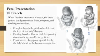
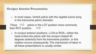
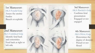
Normal labor usually begins within 2 weeks (before or after) the estimated delivery date. In a first pregnancy, labor usually lasts 12 to 18 hours on average; subsequent labors are often shorter, averaging 6 to 8 hours.
Russian Call Girls Hyderabad Saloni 9907093804 Independent Escort Service Hyd...
NORMAL LABOR AND DELIVERY
1. Normal Labor & Delivery
BY: ROMMEL LUIS C. ISRAEL III
1
BY: ROMMEL LUIS C. ISRAEL III
2. Normal labor
Labor is the process that leads to childbirth. It begins with
the onset of regular uterine contractions and ends with
delivery of the newborn and expulsion of the placenta.
Pregnancy and birth are physiological processes, and thus,
labor and delivery should be considered normal for most
women.
2
3. Mechanisms of labor
The process of adaptation or
accommodation of suitable portions
for the fetus’ head to the various
pelvic planes of the mother is
necessary to insure the completion
of childbirth.
3
4. Fetal lie
The relationship of the fetal long axis
to that of the mother
1. Longitudinal– present more than
99%
2. Transverse Predisposing –
multiparity, placenta previa,
hydramnios, uterine anomaly
3. Oblique – 45 degree angle, unstable
and converts to
transverse/longitudinal
4
5. Fetal Presentation
01 Cephalic
Presenting part – portion of fetal body that is foremost
within birth canal or close proximity to it.
1. Occiput fontanelle - vertex/occiput
2. Face
3. Anterior fontanelle - sinciput
4. Brow
5. Shoulder is presenting part when transverse lie.
Term fetus usually presents with vertex as uterus is pear
shaped Until 32w, amniotic cavity is large compared to fetus.
Later, walls are apposed to fetal parts as ratio of amniotic fluid
decreases compared to fetal mass.
5
6. Fetal Presentation
02 Breech
When the fetus presents as a breech, the three
general configurations are frank, complete, and
footling presentations.
1. Complete breech -Legs folded with feet at
the level of the baby's bottom
2. Footling breech - One or both feet pointing
down so the legs would emerge first.
3. Frank breech - Legs point up with feet by
the baby's head so the bottom emerges first.
6
1
2
3
7. Fetal Attitude (posture)
Normal - Back becomes markedly
convex, head sharply flexed over
abdomen, legs bent at knees
The image shows the differences in
attitude of the fetal body in vertex,
sinciput, brow, and face
presentations.
7
Note changes in fetal attitude in relation to fetal
vertex as the fetal head becomes less flexed.
8. Fetal Position
● Relationship of an arbitrarily chosen
portion of the fetal presenting part to the
right or left side of the birth canal.
○ Fetal occiput = vertex presentation
○ Chin = face presentation
○ Sacrum = breech
○ Acromion = shoulder presentation
● With each presentation, there may be two
positions—right or left
● Then, the relationship of a given portion of
the presenting part to the anterior (A),
transverse (T), or posterior (P) portion of
the maternal pelvis is considered.
8
9. Normal Labor Characteristics
● Labor: uterine contractions that bring about
demonstrable effacement and dilatation of the cervix
● Painful contractions become regular
● Admission to the labor unit
● Painful uterine contractions accompanied by any one
of the following:
○ (1) ruptured membranes,
○ (2) bloody "show,"
○ (3) complete cervical effacement.
9
10. Occiput Anterior Presentation
● In most cases, VerteX pelvis with the sagittal suture lying
in the transverse pelvic diameter.
Fetus pelvis in the LOT position more commonly
than ROT position.
● In occiput anterior positions—LOA or ROA—either the
head enters the pelvis with the occiput rotated 45
degrees anteriorly from the transverse position, or this
rotation occurs subsequently. The mechanism of labor in
all these presentations is usually similar.
10
11. Occiput Anterior Presentation (continued….)
The positional changes of the presenting part required to navigate the pelvic canal constitute the,
● Mechanisms of labor
● Cardinal movements of labor
Engagement
Descent
Flexion
Internal rotation
Extension
External rotation
Expulsion
During labor, these movements not only are sequential but also show great temporal overlap.
(ex: as part of engagement, there is both flexion and descent of the head. It is impossible for
the movements to be completed unless the presenting part descends simultaneously)
11
12. Diagnosis
Leopold’s Maneuver
● A method used to diagnose fetal position
and presentation.
● Inexpensive, non-invasive and easily
performed.
● May be difficult to perform in mothers who
are obese, have polyhydramnios and if the
placenta is anteriorly implanted.
● Usually done in the 3rd trimester.
12
13. Before the procedure:
- Explain the procedure to the
patient.
- Obtain consent.
- Let the patient empty her bladder.
- Assist in changing of clothes.
- Provide privacy.
- Position the mother in a dorsal
recumbent position (supine with
knees flexed)
13
14. 14
1st Maneuver:
AKA Fundal Grip
- Assess the uterine
fundus
- Breech or cephalic
3rd Maneuver:
AKA Pawlick’s Grip
- Confirms fetal
presentation
- Engaged or not
engaged
2nd Maneuver:
AKA Umbilical Grip
- Identify fetal back
and extremities
- Fetal back at right or
left side
4th Maneuver:
AKA Pelvic Grip
- Determines the
degree of descent
- Flexed or not
flexed
15. Cardinal Movements
Engagement, Descent, Flexion, Internal
rotation, Extension, External rotation, and
Expulsion
The cardinal movements, refer to the changes in
position of fetal head during its passage through
the birth canal.
Because of the asymmetry of the shape of both
the fetal head and the maternal bony pelvis, such
rotations are required for the fetus to successfully
negotiate the birth canal.
Although labor and birth comprise a continuous
process, seven discrete cardinal movements of
the fetus are described: engagement, descent,
flexion, internal rotation, extension, external
rotation or restitution, and expulsion.
15
16. Cardinal Movements
● Engagement: Biparietal diameter (greatest
transverse diameter) passes through pelvic
inlet.
○ Asynclitism: Anterior or posterior deflection of fetal
sagittal suture.
● Descent: FIRST REQUISITE for birth
Brought about by
○ pressure of amniotic fluid,
○ direct pressure of fundus upon breech with contractions
○ Bearing down efforts of maternal abdominal muscles
○ Extension and straightening of fetal body.
● Flexion: Longer occipitofrontal diameter 16
17. Cardinal Movements
● Internal rotation: Occiput gradually moves
towards symphysis pubis ESSENTIAL FOR
COMPLETION OF LABOR except if fetus
unusually small.
● Extension: Two forces Uterus pushes fetus
posteriorly Resistant pelvic floor and symphysis act
anteriorly.
● External rotation: Head undergoes restitution
Serves to bring bisacromial diameter into relation
with anteroposterior diameter of pelvic outlet.
● Expulsion: Anterior shoulder appears under
symphysis pubis then posterior shoulder
17
19. First stage of labor
Preparatory division
● Cervix dilates little, connective tissue components change considerably
● Sedation and conduction analgesia can ARREST dilatational division (active phase) – dilatation is most rapid
● Unaffected by sedation
● Pelvic division – deceleration phase of cervical dilatation Cardinal fetal movements occur during this stage.
19
Latent phase
● Starts when mother perceives regular contractions and ends once 3 to 5cm
● Prolonged latent phase
○ Nullipara = >20h
○ Multipara = >14
20. First stage of labor
Active phase
● Cervical dilatation of 3 to 6 cm or more in the presence of uterine contractions
● Mean duration in nulliparas was 4.9h. Statistical maximum of 11.7h
● Descent begins at 7 to 8 cm in nulliparas and most rapid after 8cm
● Active-phase abnormalities
○ Protraction - Slow rate of cervical dilatation
■ Nulliparas <1.2cm/h dilatation or <1cm descent/h
■ Multiparas <1.5cm/h dilatation or <2cm descent/h
○ Arrest of dilatation
■ 2 hours with no cervical change
○ Arrest of descent
■ 1 hour without fetal descent
● Epidural analgesia lengthens active phase by 1 hour.
20
21. Second stage of labor
● Begins with complete cervical dilatation and ends with fetal delivery
○ 50 minutes for nulliparas
○ 20 minutes for multiparas.
● Duration of labor
○ Parity and cervical dilatation at admission determined length of spontaneous labor.
● Identification of labor
○ In the absence of ruptured membranes or bleeding, uterine contractions 5 minutes apart for 1
hour = more than or equal to 12 contractions per hour
○ Unless there has been bleeding in excess of bloody show, a vaginal exam is performed
○ Number of vaginal exams correlates with infection-related morbidity especially in early
membrane rupture
21
23. Management of 1st stage of labor
1. Intrapartum fetal monitoring
2. Maternal monitoring
3. Oral intake
4. Intravenous fluids
5. Maternal position
6. Rupture of membranes
7. Urinary Bladder function
23
24. 1. Intrapartum fetal monitoring
● Fetal heart rate (FHR) - check after contraction at least every 30 mins.
○ If continuous monitoring is used, tracing is evaluated at least every 30 mins.
24
2. Maternal monitoring
● Temperature, pulse & blood pressure - evaluated at least every 4 hrs.
○ Check the temperature every hour
■ If membranes have been ruptured for many hours before the labor onset
■ Borderline temperature elevation
25. 3. Oral intake
● Food & liquids with particulate should be withheld during active labor &
delivery.
● Women with uncomplicated labor
○ Oral intake of moderate amount of clear liquids is reasonable
● Significant risk for cesarean delivery
○ Liquids: 2 hrs prior
○ Food: 6-8 hrs prior
25
26. 4. Intravenous fluids
● Venous access is advantageous during the immediate puerperium to administer oxytocin prophylactically at times
● Longer labors
○ Administration of glucose, sodium & water to the otherwise fasting woman at the rate of 60 to 120 ml/hr
■ Prevents dehydration & acidosis
26
5. Maternal position
● Laboring woman may assume the position she finds more comfortable.
● Lying supine is typically avoided
○ To avert aortocaval compression & its potential to lower uterine perfusion.
27. 6. Rupture of membranes
● Prolonged membrane rupture
○ >18 hrs
○ Antimicrobial administration for prevention of group B streptococcal infection - recommended
■ Lowers the rate of chorioamnionitis & endometriosis
27
7. Urinary bladder function
● During labor, suprapubic region is inspected & palpated to detect distention
● If bladder is readily seen or palpated above the symphysis pubis → encourage the px to void
● If the bladder is distended & voiding is not possible → catheterization is indicated
28. Management of 2nd stage of labor
● Full cervical dilation → signifies the onset of 2nd stage of labor
● Woman typically begins to bear down
● Uterine contractions & accompanying expulsive force
○ lasts for 1 min & recur at an interval no longer than 90s
● Monitoring intervals of fetal heart rate
○ FHR: check at least every 15 mins
○ If continuous monitoring is used, tracing is evaluated at least every 15 mins.
● Bearing down is reflexive & spontaneous (most cases).
● Next uterine contraction begins
○ Instruct the patient to exert downward pressure as though she were straining at stool
● As the head descend through the pelvis
○ Perineum begins to bulge & the overlying skin becomes stretched
○ Scalp of the fetus may be visible through the vulvar opening
○ At this time, the woman & her fetus are prepared for delivery.
28
29. Preparation for delivery
● Vulvar & perineal cleansing
● Sterile drapes may be placed in such a way that only immediate area around the
vulva is exposed.
● Scrubbing, gowning, gloving and donning protective mask and eye.
○ Protect both the laboring woman and the one assisting during birth from infectious
agents
29
30. Fundal Height Measurement
30
Procedure:
● Inform the patient on the procedure
● Ask the patient to empty the bladder prior to
the examination. A full bladder can change
fundal height measurements by several
centimeters.
● Let the patient lay down on her back with her
legs out in front of you. Using a tape measure
that measures centimeters, place the zero
marker at the top of the uterus.
● Move the tape measure vertically down your
stomach and place the other end at the top of
your pubic bone. This is your fundal height
measurement.
31. Fundal Height Measurement
31
● Starting at 24 weeks, your fundal height should be about the
same number of centimeters as the number of weeks you’ve
been pregnant.
● Your fundal height may be off by up to 2 centimeters in
either direction and still be considered normal.
○ Example: if you’re 30 weeks pregnant, a fundal
height of 28 to 32 centimeters is considered to be a
normal range.
● Factors that could affect the fundal height:
○ You have a BMI of 30 or more.
○ You have a history of uterine fibroids.
○ Your bladder is full.
○ You’re not lying on your back when you take the
measurement.
32. Internal Examination or Vaginal examination
● Indications:
○ To diagnose the pregnancy
○ To see any rupture of membranes
○ Cephalopelvic disproportion
○ Onset of labor by checking the cervix
■ Cervical dilation
■ Cervical effacement
■ State of membrane
■ Fetal presenting part
■ Fetal position
■ Fetal station
■ Cervical position and consistency
32
33. Internal Examination or Vaginal examination
● Procedure:
○ Inform the patient on the procedure
○ Ask the patient to empty the bladder prior to the
examination
○ Place the patient in a dorsal lithotomy position
○ Maintain asepsis throughout the procedure
○ Glove hands and apply lubricant to the fingers
○ Extend the index and middle fingers, flex the ring and little
finger. Thumb should be pointing upward.
○ Separate the labia with the other hand
○ Insert the index and middle finger of the examining hand
carefully into the vagina
○ Determine the cervical dilatation
33
41. Delivery of the Placenta – 3rd Stage of Labor
41
● Third-stage labor begins immediately after fetal birth and ends with placental delivery.
● GOALS:
○ Delivery of an Intact Placenta
○ Avoidance of Uterine Inversion (Postpartum Hemorrhage)
● Immediately after newborn birth, uterine fundal size and consistency are examined
● If the uterus remains firm, no unusual bleeding:
○ wait until the placenta separates
○ massage is not employed
○ frequently palpate the fundus
○ Umbilical cord traction must not be used
BY: ROMMEL LUIS C. ISRAEL III
42. Signs of Placental Separation
42
● Sudden gush of blood into the vagina
● globular and firmer fundus
● lengthening of the umbilical cord
● elevation of the uterus into the abdomen.
These signs show within minutes after newborn delivery,
median time of 4-12 minutes
BY: ROMMEL LUIS C. ISRAEL III
43. Delivery of the Placenta
43
● Once placenta is detached from the uterine wall, it should be determined that the uterus
is firmly contracted
● Mother may be asked to bear down, and the intraabdominal pressure often expels the
placenta into the vagina
● Once uterus is firmly contracted, pressure is exerted by a hand wrapped around the
fundus to propel detached placenta into the vagina
BY: ROMMEL LUIS C. ISRAEL III
44. ● Umbilical cord is kept slightly taught but is not pulled
● Heel of the hand exerts downward pressure between the
symphysis pubis and uterine fundus
● Once the placenta passes through the introitus, pressure on the
uterus is relieved
● Placenta is then gently lifted away
● If the membranes begin to tear, they are grasped w/ a clamp and
removed by gentle teasing
Delivery of the Placenta
44
BY: ROMMEL LUIS C. ISRAEL III
45. Management of the Third Stage of Labor
45
● Expectant Management
1. Involves waiting for placental separation signs and allowing the placenta to deliver either
spontaneously or aided by nipple stimulation or gravity (WHO, 2012)
● Active Management – goal is to limit postpartum hemorrhage
1. Early cord clamping
2. Controlled cord traction during placental delivery
3. Immediate administration of prophylactic oxytocin by IM within 1 min after delivery of baby
BY: ROMMEL LUIS C. ISRAEL III
46. Management of the Third Stage of Labor
46
Uterotonics plays an essential role to decrease postpartum blood loss.
Recommended Doses :
1. Oxytocin – 10 ”u”/500 ml NSS (20”u”/1000 ml NSS) - continous IV drip OR 5 “u” IV bolus
2. Ergometrine – 200-250 mcg IM OR 100-125 mcg IV bolus
BY: ROMMEL LUIS C. ISRAEL III
47. Manual Delivery of the Placenta
If there is risk bleeding and placenta can’t be delivered spontaneously, manual removal of the placenta
is indicated, commonly indicated with preterm delivery.
One hand grasps the fundus then the other hand is inserted into the uterine cavity and the fingers are
swept from side to side as they are advanced
47
BY: ROMMEL LUIS C. ISRAEL III
48. Manual Delivery of the Placenta
48
When the placenta has detached, it is grasped and removed
BY: ROMMEL LUIS C. ISRAEL III
49. Immediate Postpartum Care
49
-Hour immediately after the delivery of the placenta is critical, lacerations are
repaired during this time
-Postpartum hemorrhage may occur even if uterotonics administered due to uterine
atony and hematomas may expand
-Maternal Blood Pressure and pulse should be monitored after delivery and every
15 minutes for the first 2 hours
-Placenta, membranes and umbilical cord are examined for completeness and
anomalies
BY: ROMMEL LUIS C. ISRAEL III
50. Immediate Postpartum Care
50
Birth Canal Lacerations
-Lower genital tract lacerations may involve the cervix, vagina or perineum
-Perineal tears may follow any vaginal delivery and classified by their depth
-3RD & 4TH degree lacerations are considered higher order lacerations and are associated
with greater blood loss, puerperal pain and wound disruption of infection risk
BY: ROMMEL LUIS C. ISRAEL III
55. Episiotomy
55
Purpose - facilitate second stage of labor to improve maternal and neonatal outcome
• Maternal benefit - Reduced risk of perineal trauma, subsequent pelvic floor
dysfunction and prolapse, urinary incontinence, fecal incontinence and sexual
dysfunction
• Fetal benefit Shortened second stage of labor
BY: ROMMEL LUIS C. ISRAEL III
56. Episiotomy indications
56
● -Shoulder dystocia
● -Breech delivery
● -Macrosomic fetuses
● -operative vaginal deliveries,
● Persistent OP positions
● -markedly short perineal length
● other instances in which failure to perform an episiotomy will result in significant
perineal rupture.
BY: ROMMEL LUIS C. ISRAEL III
57. Episiotomy
57
-Incision may be made in the midline,
creating a median or midline
episiotomy or begin off the midline and
directed laterally and downward away
from the rectum known as mediolateral
epiosotomy
BY: ROMMEL LUIS C. ISRAEL III
58. Laceration and Episiotomy Repairs
58
-Typically, perineal repairs are deferred until the placenta has been delivered.
This permits undivided attention to the signs of placental separation and
delivery.
-Adequate analgesia is imperative, and women without regional analgesia can
experience high levels of pain during perineal suturing.
-Locally injected lidocaine can be used solely or as a supplement to bilateral
pudendal nerve blockade. In those with epidural analgesia, additional dosing
may be necessary.
BY: ROMMEL LUIS C. ISRAEL III
59. Laceration and Episiotomy Repairs
59
- First-degree lacerations do not always require
repair, and sutures are placed to control bleeding or
restore anatomy.
-Second-degree laceration correction as well as
midline and mediolateral episiotomy repairs include
similar steps. Namely, these close the vaginal
epithelium and reapproximate the bulbospongiosus
and superficial transverse perineal muscles during
restoration of the perineal body
BY: ROMMEL LUIS C. ISRAEL III
61. For third-degree laceration
repair, two methods are
available to repair the
external anal sphincter.
The first is preferably the
end-to-end technique and
the overlapping technique.
61
BY: ROMMEL LUIS C. ISRAEL III
62. With fourth-degree laceration
repairs, the torn edges of the rectal
mucosa are reapproximated.
At a point 1 cm proximal to the
wound apex, sutures are placed
approximately 0.5 cm apart in the
rectal muscularis and do not enter
the anorectal lumen.
Clinicians often use 4–0 polyglactin
910 or chromic gut for this running
suture line
62
BY: ROMMEL LUIS C. ISRAEL III
63. 63
Mediolateral Episiotomy Repair
A. The vaginal epithelium and deeper tissues are closed
with a single, continuous, locking suture. The angle is
approximately 45° since the perineum is no longer
distended.
B. After the vaginal component of the laceration is
repaired, deeper perineal tissues are reapproximated
by a single, continuous, nonlocking suture. Small
episiotomies may not require this deeper layer.
C. With a similar continuous, nonlocking technique, the
superficial transverse perineal and bulbospongiosus
muscles are reapproximated.
D. Last, the perineal skin is closed using a subcuticular
stitch.
BY: ROMMEL LUIS C. ISRAEL III
64. Midline Episiotomy Repair
64
A. An anchor stitch is placed above the wound apex to
begin a running, locking closure with 2–0 suture to
close the vaginal epithelium and deeper tissues and
reapproximate the hymeneal ring
B. A transition stitch redirects suturing from the vagina
to the perineum.
C. The superficial transverse perineal and
bulbospongiosus muscles are reapproximated using a
continuous, nonlocking technique with the same length
of suture. This aids restoration of the perineal body for
long-term support.
D. The continuous suture is then carried upward as a
subcuticular stitch. The final knot is tied proximal to the
hymeneal ring.
BY: ROMMEL LUIS C. ISRAEL III
65. Perineal Laceration Care
65
-Initially, locally applied ice packs help reduce swelling and allay
discomfort
-In subsequent days, warm sitz baths aid comfort and hygiene
-Oral analgesics containing codeine provide considerable relief. For
lesser degree of discomfort, NSAID tablets can be given
-For those with second-degree lacerations or anal sphincter tears,
intercourse is
usually advised until after the first puerperal visit at 6 weeks.
BY: ROMMEL LUIS C. ISRAEL III
Editor's Notes
Transverse lie. Right acromiodorsoposterior (RADP). The shoulder of the fetus is to the mother’s right, and the back is posterior.
A vaginal examination (speculum or digital examination) is not part of a routine obstetric examination but may be indicated to diagnose the pregnancy, to see any rupture of membranes, cephalopelvic disproportion, and to determine the onset of labour by checking cervix to assess:
Determine the cervical dilatation
Cervical dilatation is determined by estimating the average diameter of the cervical opening by sweeping the examining fingers from the margin of the cervical opening on one side to that on the opposite side. The diameter traversed is estimated in cm. The cervix is said to be fully dilated when diameter measures 10 cm.
Determine cervical effacement
The degree of cervical effacement is usually expressed in terms of the length of the cervical canal compared with that of an uneffaced cervix. When the length of the cervix is reduced by 1/2, it is 50% effaced. When the cervix becomes as thin as the adjacent uterine segment, it is completely or 100% effaced.
Determine cervical position
The position of the cervix is determined by the relation of the cervical os to the fetal head. It is categorized as posterior, midposition, or anterior.
Determine cervical consistency
As soft, firm, or intermediate between the two.
Determine state of the membrane
Whether intact or ruptured
Determine fetal presenting part, fetal position, fetal station.
The presenting part is palpated and differentiation whether it is vertex, face, or breech is accomplished. If the vertex is presenting the fingers are directed posteriorly and then swept forward over the fetal head towards the symphysis pubis. The linear course of the sagittal suture is delineated. The position of the two fontanels are ascertained. The fingers are passed to the most anterior extension of the sagittal suture and the fontanel examined is identified. Then with the sweeping motion the fingers are passed along the suture to the other end of the head until the other fontanel is felt and differentiated. Identify the triangular-shaped posterior fontanel and the diamond-shaped anterior fontanel.
Determine fetal station.
The station or extent to which the presenting part has descended into the pelvis can also be established. Determine the lowermost portion of the fetal head in relation to the ischial spines.
with end-to-end approximation of the external anal
sphincter (EAS), a suture is placed through the EAS muscle, and four to six simple
interrupted 2–0 or 3–0 sutures of polyglactin 910 are placed at the 3, 6, 9, and 12
o’clock positions through the perisphincter connective tissue. To begin, disrupted
ends of the striated EAS muscle and capsule are identified and grasped. The first
suture is placed posteriorly to maintain clear exposure. Another suture is then
placed inferiorly at the 6 o’clock position. The sphincter muscle fibers are next
reapposed by a figure-of-eight stitch. Last, the remainder of the fascia is closed
with a stitch placed anterior to the sphincter cylinder and again with once placed
superior to it.