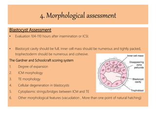
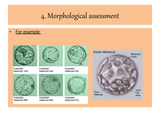
Embryo Life
Dr. Yasmin Magdi Abd-Elkreem
Embryo: The collection of cells
(2n) that has developed from
the fertilized oocyte (male n +
female n), before all the major
organs have developed.
In vitro
In vivo
.Steps oh human embryo
Pre-implantation
development:
1. Fertilization
2. cleavage
3. blastulation
Embryo Selection
• Most Optimal selection profile would be through taking into
consideration:
1. Chromosomal integrity.
2. Appropriate genomic activation.
3. Metabolic activity.
4. Morphological changes and dynamics appropriate for the culture
period Microscopic embryo assessment most used technique for
being non-invasive, non-traumatic, simple, cost effectiveness and
correlated with implantation and pregnancy rates techniques which
help assess embryos without causing damage .
1. Chromosomal integrity
• based on preimplantation genetic diagnosis (PGD), a procedure used to test
early human embryos for serious inherited genetic conditions and chromosomal
abnormalities.
• Normal embryos were those in which each cell had two chromosomes of each
kind.
Aneuploid :embryos could be monosomic or trisomic.
Monosomic : embryos were those in which the same chromosome was missing in
each cell.
Trisomic : embryos were those in which each cell had three chromosomes of the
same type instead of two.
Haploid : Embryos in which all the cells had only a single chromosome of each
kind.
Polyploid :embryos had three or more copies of each chromosome in each cell.
1. Chromosomal integrity
PGD is recommended when:
1. Familial Single Gene Disorders
2. Familial Sex-linked Disorders
3. Familial Chromosomal Disorders
4. Non-familial Chromosomal Disorders Associated with Advanced
Reproductive Age
5. Non-familial Chromosomal Disorders Associated with Infertility
(recurrent implantation failure )
6. Sex Selection
7. Select embryos with a genetic impairment seen in a parent
Two types of assessment techniques are common:
a. chromosome “painting” (or FISH)
b. genetic testing for specific disease loci (PCR or gene chips)
2. Appropriate genomic activation.
• Regulation of gene expression includes a wide range of mechanisms that are
used by cells to increase or decrease the production of specific gene products
(protein or RNA), and is informally termed gene regulation.
• With the emergence of new technologies such as Omics, the gene expression
profiling of human oocytes, embryos, and hESCs has been performed and
generated a flood of data related to the molecular signature of early embryo
development.
3. Metabolic activity
• Adenosine triphosphate (ATP) is the universal currency of energy in
biological systems (supplied by the Mitochondria).
• Too high and too low ATP production can be lethal to the cell, and any
mitochondrial dysfunction may be critical to the embryo.
• spectrophotometric tests are used to measure metabolomic changes , in
proteomic (proteins) profiling, in amino acid profiling, amino acid
depletion and production , respiration-rate measurement.
• Pyruvate/Glucose uptake
• Amino acids
• Oxygen consumption (Respirometry)
Production
Glucose
Pyruvate
Amino Acids
Other Sugars
Oxygen
Other Peptides & Factors
μl drop of defined
culture medium
Modified from: Gardner and Leese(1993) Assessment of embryo metabolism and viability. In: Handbook of In Vitro Fertilization.
EdsTrounson& Gardner CRC Press. pp195-211.
Lactate
Ammonium
Amino Acids
Enzymes, eg LDH
sHLA-G
HOXA10 regulator
PAFMetabolomics /
Proteomics
3. Metabolic activity
Uptake
4. Morphological assessment
• The best available method for embryo selection.
• Microscopic embryo assessment most used technique
• non-invasive, non-traumatic
• Simple
• cost effectiveness
• correlated with implantation and pregnancy rates.
4. Morphological assessment
Observation
Timing (hours post-
insemination)
Expected stage of
development
Fertilization check 17 ± 1 h Pronuclear stage
Syngamy check 23 ± 1 h
Up to 50% in syngamy
(up to 20% at the 2-cell stage)
Early cleavage check
26 ± 1 h post-ICSI
28 ± 1 h post-IVF
2-cell stage
Day 2 assessment 44 ± 1 h 4-cell stage
Day 3 assessment 68 ± 1 h 8-cell stage
Day 4 assessment 92 ± 2 h Morula
Day 5 assessment 116 ± 2 h Blastocyst
 Standardized timing relative to insemination time
Report cell number/stage and grade (separately) – with time post-insemination
4. Morphological assessment
Pronuclear Embryo Assessment
• Evaluation 16-18 hours following oocyte insemination (IVF) or ICSI.
• Pronucleus stadium/PN : the distribution pattern of the nucleoli (precursor
bodies) can be determined through an expensive digitalized, image-generating
method.
• Normal Fertilization: is assessed by two centrally positioned, juxtaposed PNs
with clearly defined membranes and two polar bodies.
• There are different systems for 2PN scoring:
A] Zygote-Score System (Revised zygote scoring)
B] Initial zygote scoring system
C] Ideal Features described by Tesarik and Greco
4. Morphological assessment
Cleavage Embryo Assessment
The embryo classification systems are based on the evaluation of
1. The number of blastomeres.
2. The degree of fragmentation.
3. The symmetry of the blastomeres.
4. the presence of multinucleation.
5. The compaction status.
4. Morphological assessment
24-28 Hours : first cleavage, 2-cells stage
40-44 Hours: number of blastomeres should be 4-6, symmetric, fragmentation of
less than 20%, and no multinucleated blastomeres.
64-68 Hours: number of blastomeres should be 8-12 , symmetric, fragmentation
less than 20%, and no multinucleated blastomeres.
4. Morphological assessment
Blastocyst Assessment
• Evaluation 104-110 hours after insemination or ICSI.
• Blastocyst cavity should be full, inner cell mass should be numerous and tightly packed,
trophectoderm should be numerous and cohesive.
The Gardner and Schoolcraft scoring system
1. Degree of expansion
2. ICM morphology
3. TE morphology
4. Cellular degeneration in blastocysts
5. Cytoplasmic strings/bridges between ICM and TE
6. Other morphological features (vacuolation , More than one point of natural hatching)
4. Morphological assessment
• For example:
Conclusions……….
• Embryo assessment is one of the most critical procedures that play a role in the
success of IVF/ART
• Traditional embryo assessment is challenged by different factors, ie, subjectivity, low
efficiency
• “Non-invasive” morphological assessment may provide valuable additional
information to optimize embryo assessment and maximize the chances of IVF
success
Thank You for your time…….
For contact:
E-mail: Yas.magdi@hotmail.com

In Vitro embryo life

  • 1.
    Embryo Life Dr. YasminMagdi Abd-Elkreem
  • 2.
    Embryo: The collectionof cells (2n) that has developed from the fertilized oocyte (male n + female n), before all the major organs have developed. In vitro In vivo .Steps oh human embryo Pre-implantation development: 1. Fertilization 2. cleavage 3. blastulation
  • 3.
    Embryo Selection • MostOptimal selection profile would be through taking into consideration: 1. Chromosomal integrity. 2. Appropriate genomic activation. 3. Metabolic activity. 4. Morphological changes and dynamics appropriate for the culture period Microscopic embryo assessment most used technique for being non-invasive, non-traumatic, simple, cost effectiveness and correlated with implantation and pregnancy rates techniques which help assess embryos without causing damage .
  • 4.
    1. Chromosomal integrity •based on preimplantation genetic diagnosis (PGD), a procedure used to test early human embryos for serious inherited genetic conditions and chromosomal abnormalities. • Normal embryos were those in which each cell had two chromosomes of each kind. Aneuploid :embryos could be monosomic or trisomic. Monosomic : embryos were those in which the same chromosome was missing in each cell. Trisomic : embryos were those in which each cell had three chromosomes of the same type instead of two. Haploid : Embryos in which all the cells had only a single chromosome of each kind. Polyploid :embryos had three or more copies of each chromosome in each cell.
  • 5.
    1. Chromosomal integrity PGDis recommended when: 1. Familial Single Gene Disorders 2. Familial Sex-linked Disorders 3. Familial Chromosomal Disorders 4. Non-familial Chromosomal Disorders Associated with Advanced Reproductive Age 5. Non-familial Chromosomal Disorders Associated with Infertility (recurrent implantation failure ) 6. Sex Selection 7. Select embryos with a genetic impairment seen in a parent
  • 6.
    Two types ofassessment techniques are common: a. chromosome “painting” (or FISH) b. genetic testing for specific disease loci (PCR or gene chips)
  • 7.
    2. Appropriate genomicactivation. • Regulation of gene expression includes a wide range of mechanisms that are used by cells to increase or decrease the production of specific gene products (protein or RNA), and is informally termed gene regulation. • With the emergence of new technologies such as Omics, the gene expression profiling of human oocytes, embryos, and hESCs has been performed and generated a flood of data related to the molecular signature of early embryo development.
  • 8.
    3. Metabolic activity •Adenosine triphosphate (ATP) is the universal currency of energy in biological systems (supplied by the Mitochondria). • Too high and too low ATP production can be lethal to the cell, and any mitochondrial dysfunction may be critical to the embryo. • spectrophotometric tests are used to measure metabolomic changes , in proteomic (proteins) profiling, in amino acid profiling, amino acid depletion and production , respiration-rate measurement. • Pyruvate/Glucose uptake • Amino acids • Oxygen consumption (Respirometry)
  • 9.
    Production Glucose Pyruvate Amino Acids Other Sugars Oxygen OtherPeptides & Factors μl drop of defined culture medium Modified from: Gardner and Leese(1993) Assessment of embryo metabolism and viability. In: Handbook of In Vitro Fertilization. EdsTrounson& Gardner CRC Press. pp195-211. Lactate Ammonium Amino Acids Enzymes, eg LDH sHLA-G HOXA10 regulator PAFMetabolomics / Proteomics 3. Metabolic activity Uptake
  • 10.
    4. Morphological assessment •The best available method for embryo selection. • Microscopic embryo assessment most used technique • non-invasive, non-traumatic • Simple • cost effectiveness • correlated with implantation and pregnancy rates.
  • 11.
    4. Morphological assessment Observation Timing(hours post- insemination) Expected stage of development Fertilization check 17 ± 1 h Pronuclear stage Syngamy check 23 ± 1 h Up to 50% in syngamy (up to 20% at the 2-cell stage) Early cleavage check 26 ± 1 h post-ICSI 28 ± 1 h post-IVF 2-cell stage Day 2 assessment 44 ± 1 h 4-cell stage Day 3 assessment 68 ± 1 h 8-cell stage Day 4 assessment 92 ± 2 h Morula Day 5 assessment 116 ± 2 h Blastocyst  Standardized timing relative to insemination time Report cell number/stage and grade (separately) – with time post-insemination
  • 12.
    4. Morphological assessment PronuclearEmbryo Assessment • Evaluation 16-18 hours following oocyte insemination (IVF) or ICSI. • Pronucleus stadium/PN : the distribution pattern of the nucleoli (precursor bodies) can be determined through an expensive digitalized, image-generating method. • Normal Fertilization: is assessed by two centrally positioned, juxtaposed PNs with clearly defined membranes and two polar bodies. • There are different systems for 2PN scoring: A] Zygote-Score System (Revised zygote scoring) B] Initial zygote scoring system C] Ideal Features described by Tesarik and Greco
  • 13.
    4. Morphological assessment CleavageEmbryo Assessment The embryo classification systems are based on the evaluation of 1. The number of blastomeres. 2. The degree of fragmentation. 3. The symmetry of the blastomeres. 4. the presence of multinucleation. 5. The compaction status.
  • 14.
    4. Morphological assessment 24-28Hours : first cleavage, 2-cells stage 40-44 Hours: number of blastomeres should be 4-6, symmetric, fragmentation of less than 20%, and no multinucleated blastomeres. 64-68 Hours: number of blastomeres should be 8-12 , symmetric, fragmentation less than 20%, and no multinucleated blastomeres.
  • 15.
    4. Morphological assessment BlastocystAssessment • Evaluation 104-110 hours after insemination or ICSI. • Blastocyst cavity should be full, inner cell mass should be numerous and tightly packed, trophectoderm should be numerous and cohesive. The Gardner and Schoolcraft scoring system 1. Degree of expansion 2. ICM morphology 3. TE morphology 4. Cellular degeneration in blastocysts 5. Cytoplasmic strings/bridges between ICM and TE 6. Other morphological features (vacuolation , More than one point of natural hatching)
  • 16.
  • 17.
    Conclusions………. • Embryo assessmentis one of the most critical procedures that play a role in the success of IVF/ART • Traditional embryo assessment is challenged by different factors, ie, subjectivity, low efficiency • “Non-invasive” morphological assessment may provide valuable additional information to optimize embryo assessment and maximize the chances of IVF success
  • 18.
    Thank You foryour time……. For contact: E-mail: Yas.magdi@hotmail.com