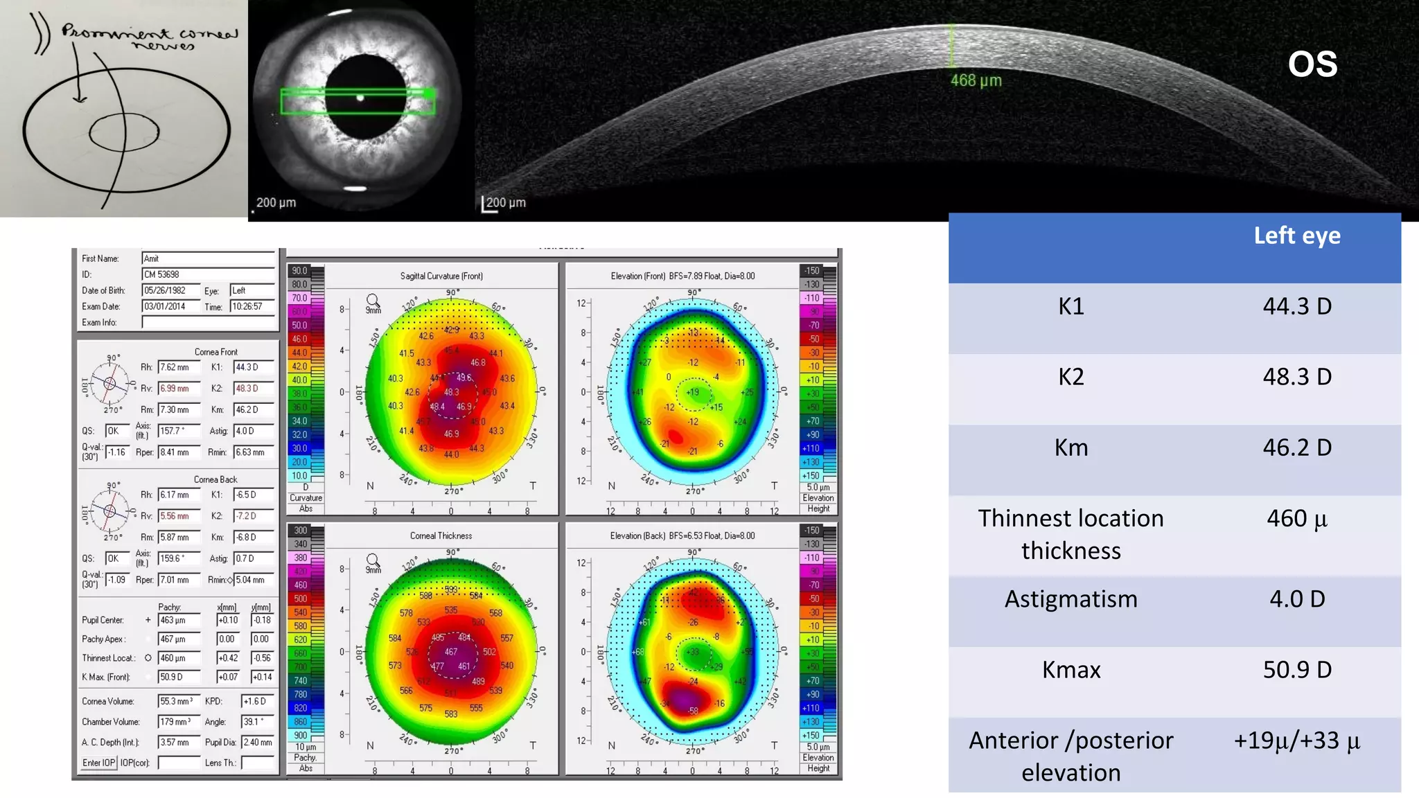
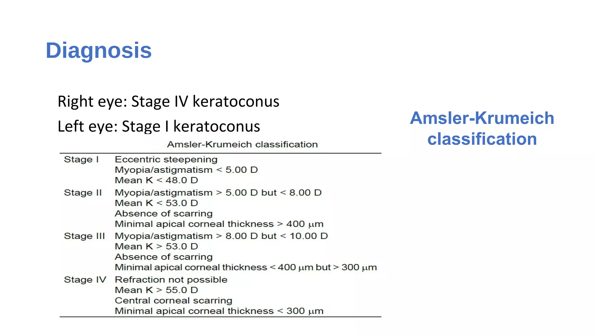
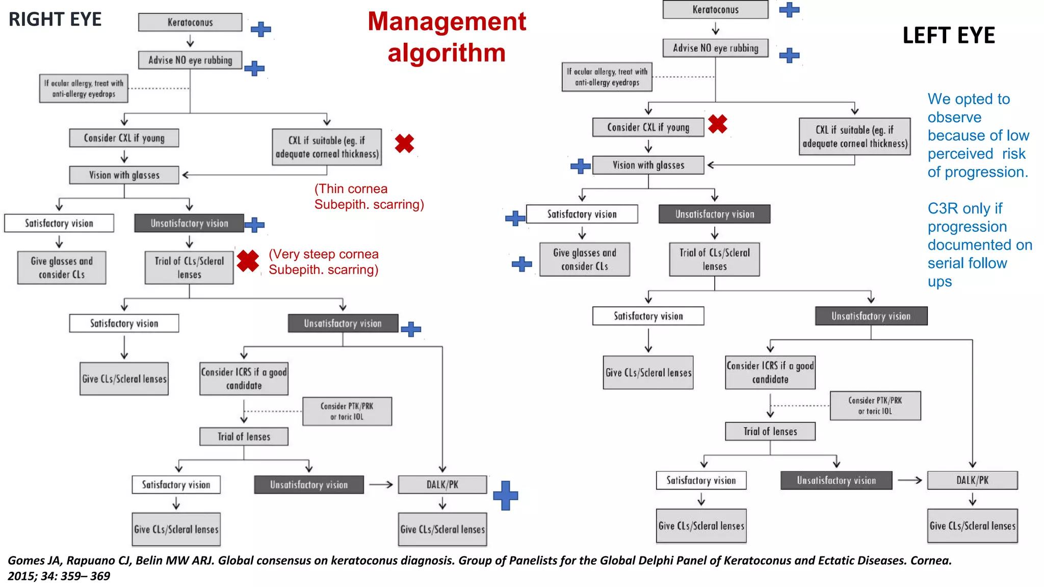
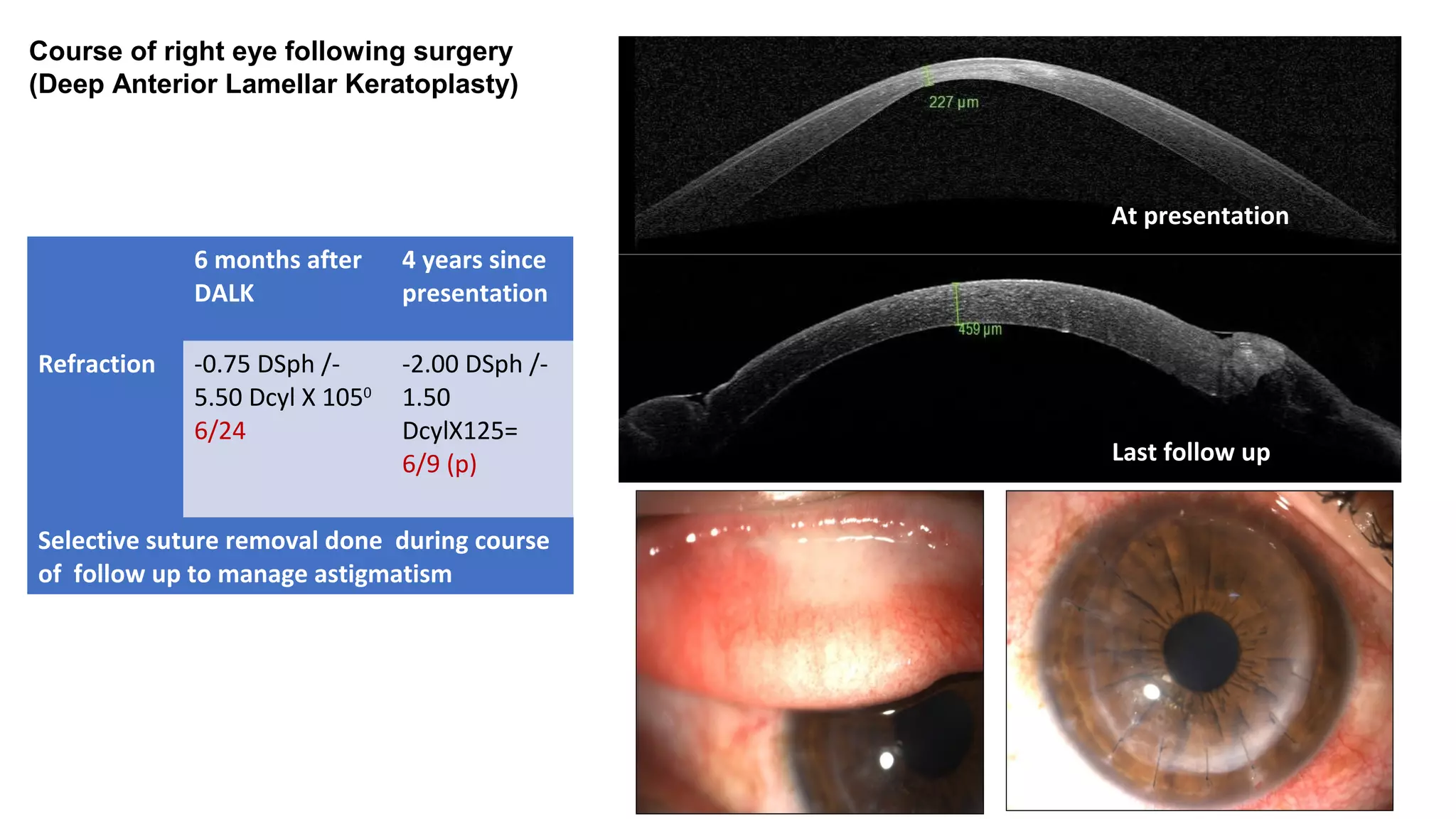
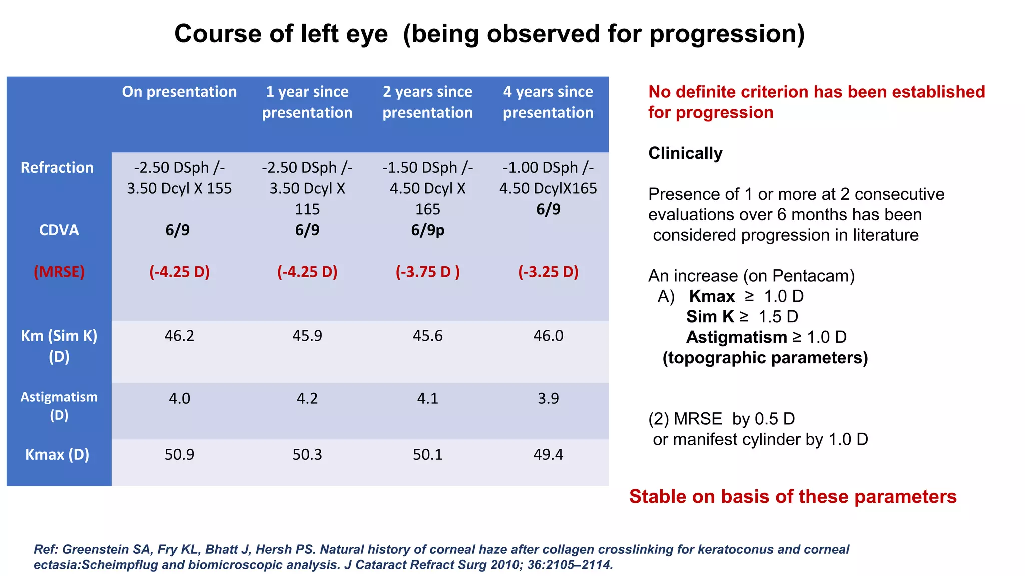
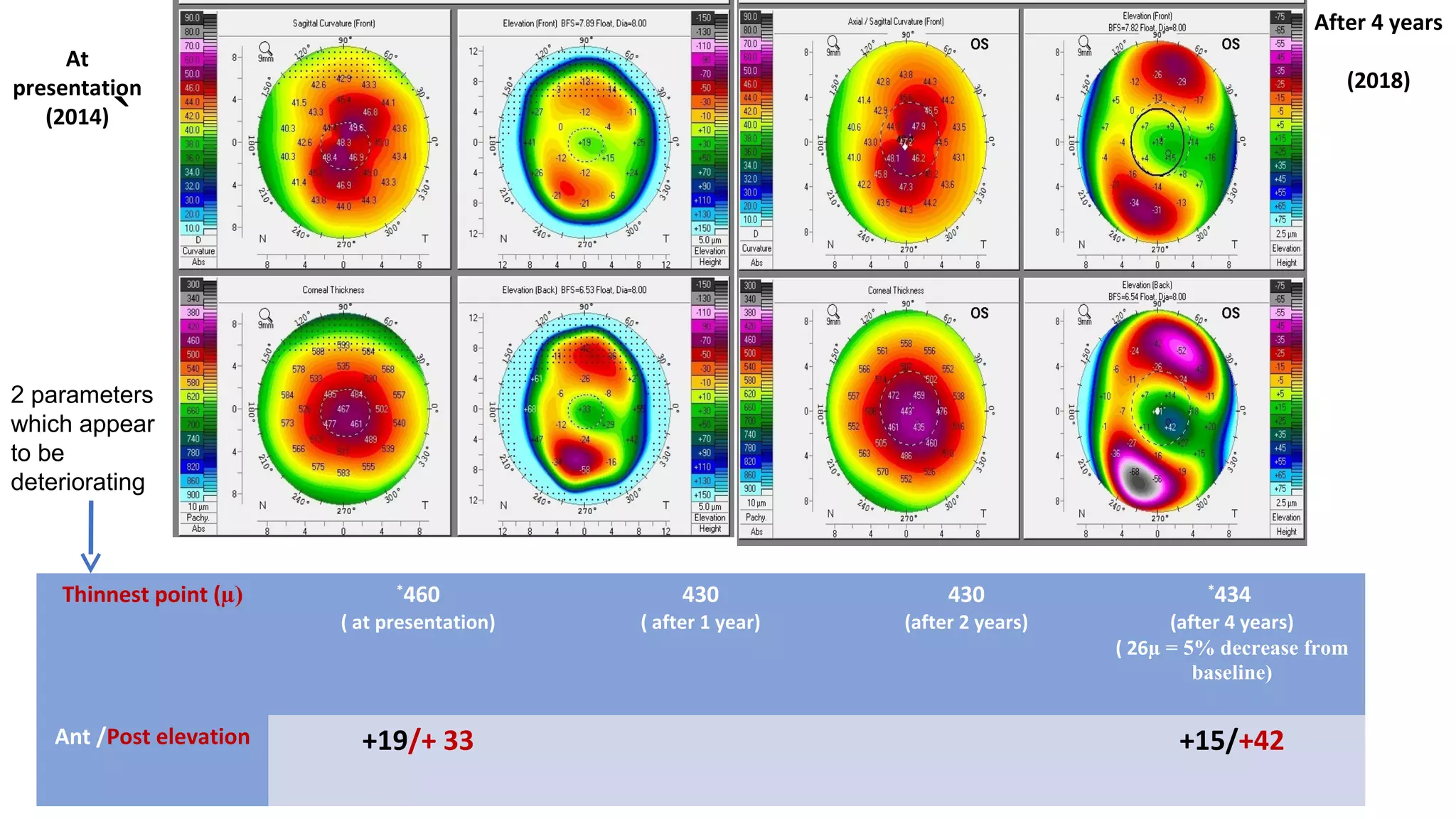
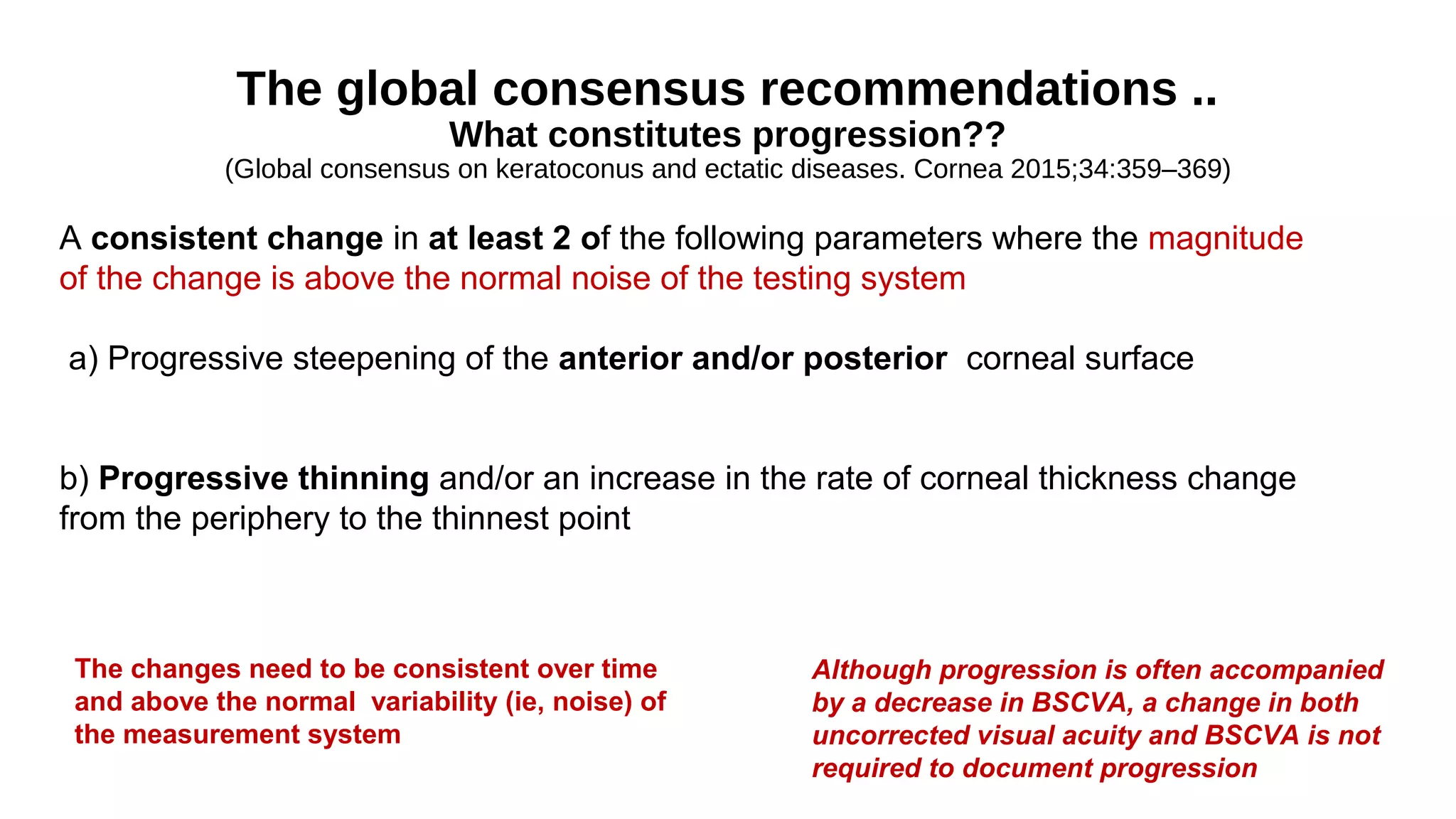
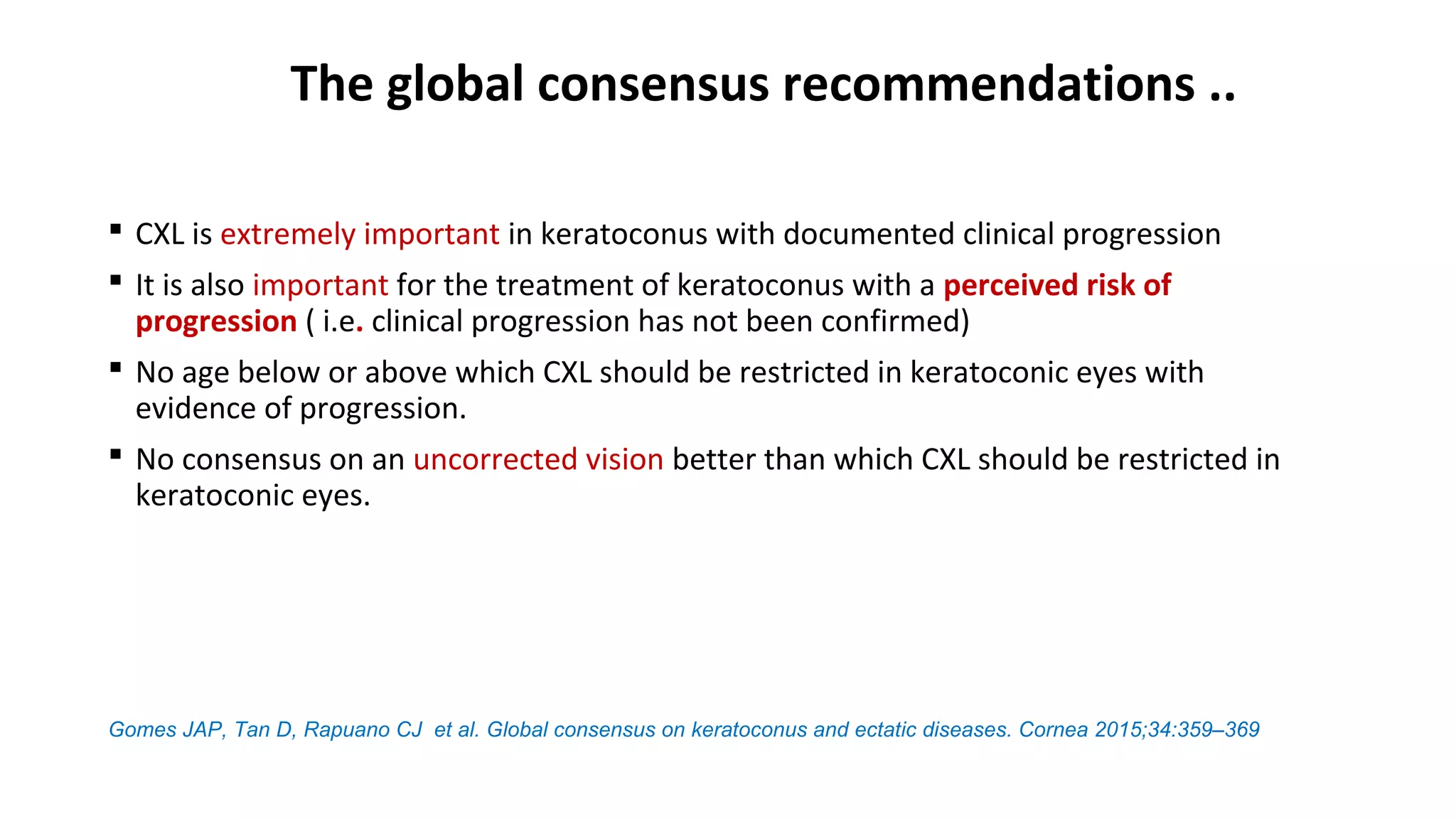
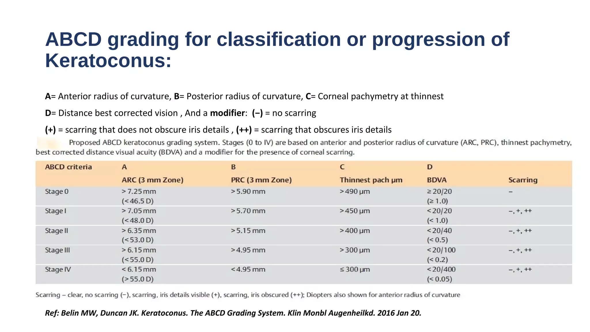
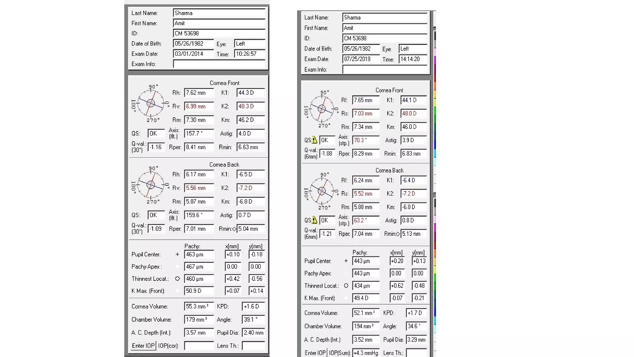
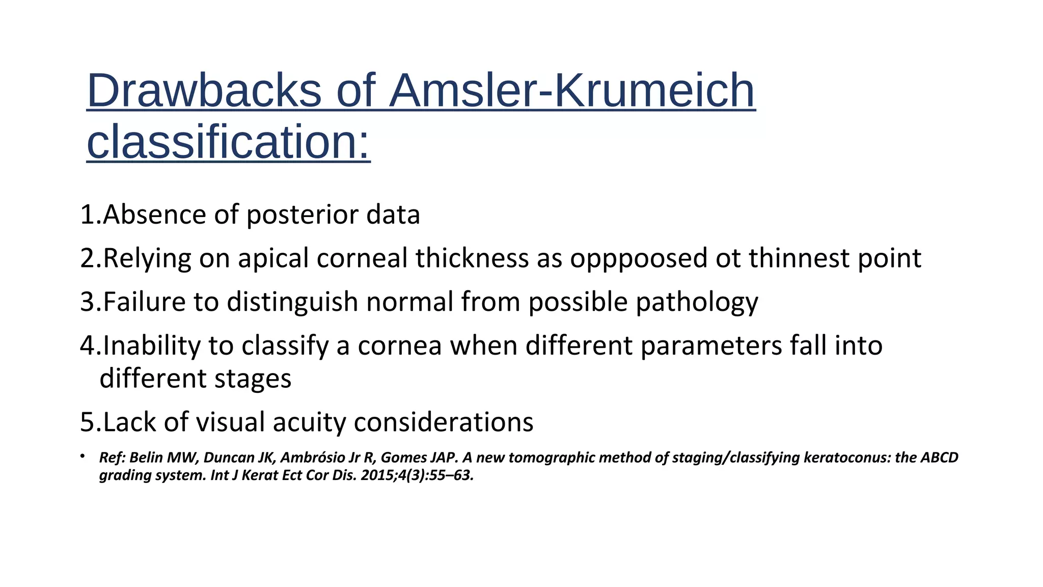
Grand round
13-08-2018
Dr. Sandeep Choudhary
Dr. Monika Balyan
Dr. Chintan Malhotra
Aim
• To discuss the management protocol of a patient with bilateral
asymmetric keratoconus.
• 31Yr/Male
• CO 53698
• Date of presentation : 23 January, 2014
• Diminution of vision in both eyes (RE>LE) since 15 years
• H/O bilateral itching and eye rubbing (affecting both eyes equally)
• No h/o DM/HTN/any other systemic illness
• No family h/o keratoconus
Right eye Left eye
Vision (Unaided) CF <1m 6/36
CDVA CF1m
(-5.00 Dsph / -5.50 Dcyl X 10)
6/9
(-2.50 Dsph / -2.50 Dcyl X 160)
IOP (mm Hg) 12 13
Anterior segment (Shown in coming
slide)
diagram diagram
Posterior segment WNL WNL
Lids and adnexa Papillae present on upper tarsal
conjunctiva
Papillae present on upper tarsal
conjunctiva
OD
OS
Right eye
K1 44.3 D
K2 49.6 D
Km 46.8 D
Thinnest location
thickness
580µ
Astigmatism 5.2 D
Kmax 51.5 D
OD
Unreliable
parameters
because of poor
scan quality
OD OS
On presentation
Left eye
K1 44.3 D
K2 48.3 D
Km 46.2 D
Thinnest location
thickness
460 µ
Astigmatism 4.0 D
Kmax 50.9 D
Anterior /posterior
elevation
+19µ/+33 µ
OS
Diagnosis
Right eye: Stage IV keratoconus
Left eye: Stage I keratoconus Amsler-Krumeich
classification
RIGHT EYE
LEFT EYE
Gomes JA, Rapuano CJ, Belin MW ARJ. Global consensus on keratoconus diagnosis. Group of Panelists for the Global Delphi Panel of Keratoconus and Ectatic Diseases. Cornea.
2015; 34: 359– 369
Management
algorithm
(Thin cornea
Subepith. scarring)
(Very steep cornea
Subepith. scarring)
We opted to
observe
because of low
perceived risk
of progression.
C3R only if
progression
documented on
serial follow
ups
At Presentation
Post Op
4 years since DALK
At presentation
Last follow up
6 months after
DALK
4 years since
presentation
Refraction -0.75 DSph /-
5.50 Dcyl X 1050
6/24
-2.00 DSph /-
1.50
DcylX125=
6/9 (p)
Selective suture removal done during course
of follow up to manage astigmatism
Course of right eye following surgery
(Deep Anterior Lamellar Keratoplasty)
At Presentation
Post Op
4 years since DALK
At presentation
Last follow up
Course of left eye (being observed for progression)
On presentation 1 year since
presentation
2 years since
presentation
4 years since
presentation
Refraction
CDVA
(MRSE)
-2.50 DSph /-
3.50 Dcyl X 155
6/9
(-4.25 D)
-2.50 DSph /-
3.50 Dcyl X
115
6/9
(-4.25 D)
-1.50 DSph /-
4.50 Dcyl X
165
6/9p
(-3.75 D )
-1.00 DSph /-
4.50 DcylX165
6/9
(-3.25 D)
Km (Sim K)
(D)
46.2 45.9 45.6 46.0
Astigmatism
(D)
4.0 4.2 4.1 3.9
Kmax (D) 50.9 50.3 50.1 49.4
No definite criterion has been established
for progression
Clinically
Presence of 1 or more at 2 consecutive
evaluations over 6 months has been
considered progression in literature
An increase (on Pentacam)
A) Kmax ≥ 1.0 D
Sim K ≥ 1.5 D
Astigmatism ≥ 1.0 D
(topographic parameters)
(2) MRSE by 0.5 D
or manifest cylinder by 1.0 D
Ref: Greenstein SA, Fry KL, Bhatt J, Hersh PS. Natural history of corneal haze after collagen crosslinking for keratoconus and corneal
ectasia:Scheimpflug and biomicroscopic analysis. J Cataract Refract Surg 2010; 36:2105–2114.
Stable on basis of these parameters
`
At
presentation
(2014)
Thinnest point (µ) *
460
( at presentation)
430
( after 1 year)
430
(after 2 years)
*
434
(after 4 years)
( 26µ = 5% decrease from
baseline)
Ant /Post elevation +19/+ 33 +15/+42
After 4 years
(2018)
2 parameters
which appear
to be
deteriorating
The global consensus recommendations ..
What constitutes progression??
(Global consensus on keratoconus and ectatic diseases. Cornea 2015;34:359–369)
A consistent change in at least 2 of the following parameters where the magnitude
of the change is above the normal noise of the testing system
a) Progressive steepening of the anterior and/or posterior corneal surface
b) Progressive thinning and/or an increase in the rate of corneal thickness change
from the periphery to the thinnest point
Although progression is often accompanied
by a decrease in BSCVA, a change in both
uncorrected visual acuity and BSCVA is not
required to document progression
The changes need to be consistent over time
and above the normal variability (ie, noise) of
the measurement system
(Cornea 2015;34:285–289)
Thank you
The global consensus recommendations ..
 CXL is extremely important in keratoconus with documented clinical progression
 It is also important for the treatment of keratoconus with a perceived risk of
progression ( i.e. clinical progression has not been confirmed)
 No age below or above which CXL should be restricted in keratoconic eyes with
evidence of progression.
 No consensus on an uncorrected vision better than which CXL should be restricted in
keratoconic eyes.
Gomes JAP, Tan D, Rapuano CJ et al. Global consensus on keratoconus and ectatic diseases. Cornea 2015;34:359–369
ABCD grading for classification or progression of
Keratoconus:
A= Anterior radius of curvature, B= Posterior radius of curvature, C= Corneal pachymetry at thinnest
D= Distance best corrected vision , And a modifier: (−) = no scarring
(+) = scarring that does not obscure iris details , (++) = scarring that obscures iris details
Ref: Belin MW, Duncan JK. Keratoconus. The ABCD Grading System. Klin Monbl Augenheilkd. 2016 Jan 20.
• Keratoconus is a non-inflammatory disease characterised by progressive corneal
thinning and apical protrusion. Typically, it presents in early adulthood and visual
symptoms result from irregular astigmatism and increasing myopia.
• It is reported to have bilateral involvement in over 90% of patients.
• A study in 83 patients demonstrates, quantitatively, the asymmetry of disease
found in patients at the point of initial diagnosis of keratoconus. (1)
• Rubbing the eye is a well-known risk factor, it has been reported that bilateral
keratoconus presents more severely in the side of the dominant hand of
individuals who excessively rub eye. (2)
• Ref: 1. Burns DM, Johnston FM, Frazer DG, Patterson C, Jackson AJ. Keratoconus: An analysis of corneal asymmetry. Br J Ophthalmol. 2004;88:1252–
5.
2. Bral N, Termote K, Unilateral Keratoconus after Chronic Eye Rubbing by the Nondominant Hand. Case Rep Ophthalmol 2017;8:558-561
Parameters to document progression
Progression of keratoconus has been noted to be associated with changes in following:
1. Anterior corneal surface
2: Epithelial thickness
3: BCVA.
4: Corneal thickness
5: Posterior surface
Ref: Belin M. Parameters to Document Progression of Keratoconus. The ability to measure how a patient’s disease changes is key. Cataract & Refractive Surgery Today Europe.
July/August 2014:20–22
Drawbacks of Amsler-Krumeich
classification:
1.Absence of posterior data
2.Relying on apical corneal thickness as opppoosed ot thinnest point
3.Failure to distinguish normal from possible pathology
4.Inability to classify a cornea when different parameters fall into
different stages
5.Lack of visual acuity considerations
• Ref: Belin MW, Duncan JK, Ambrósio Jr R, Gomes JAP. A new tomographic method of staging/classifying keratoconus: the ABCD
grading system. Int J Kerat Ect Cor Dis. 2015;4(3):55–63.

Keratoconus a case of asymmetric keratoconus and management protocol

  • 1.
    Grand round 13-08-2018 Dr. SandeepChoudhary Dr. Monika Balyan Dr. Chintan Malhotra
  • 2.
    Aim • To discussthe management protocol of a patient with bilateral asymmetric keratoconus.
  • 3.
    • 31Yr/Male • CO53698 • Date of presentation : 23 January, 2014 • Diminution of vision in both eyes (RE>LE) since 15 years • H/O bilateral itching and eye rubbing (affecting both eyes equally) • No h/o DM/HTN/any other systemic illness • No family h/o keratoconus
  • 4.
    Right eye Lefteye Vision (Unaided) CF <1m 6/36 CDVA CF1m (-5.00 Dsph / -5.50 Dcyl X 10) 6/9 (-2.50 Dsph / -2.50 Dcyl X 160) IOP (mm Hg) 12 13 Anterior segment (Shown in coming slide) diagram diagram Posterior segment WNL WNL Lids and adnexa Papillae present on upper tarsal conjunctiva Papillae present on upper tarsal conjunctiva
  • 5.
    OD OS Right eye K1 44.3D K2 49.6 D Km 46.8 D Thinnest location thickness 580µ Astigmatism 5.2 D Kmax 51.5 D OD Unreliable parameters because of poor scan quality
  • 6.
    OD OS On presentation Lefteye K1 44.3 D K2 48.3 D Km 46.2 D Thinnest location thickness 460 µ Astigmatism 4.0 D Kmax 50.9 D Anterior /posterior elevation +19µ/+33 µ OS
  • 7.
    Diagnosis Right eye: StageIV keratoconus Left eye: Stage I keratoconus Amsler-Krumeich classification
  • 8.
    RIGHT EYE LEFT EYE GomesJA, Rapuano CJ, Belin MW ARJ. Global consensus on keratoconus diagnosis. Group of Panelists for the Global Delphi Panel of Keratoconus and Ectatic Diseases. Cornea. 2015; 34: 359– 369 Management algorithm (Thin cornea Subepith. scarring) (Very steep cornea Subepith. scarring) We opted to observe because of low perceived risk of progression. C3R only if progression documented on serial follow ups
  • 9.
    At Presentation Post Op 4years since DALK At presentation Last follow up 6 months after DALK 4 years since presentation Refraction -0.75 DSph /- 5.50 Dcyl X 1050 6/24 -2.00 DSph /- 1.50 DcylX125= 6/9 (p) Selective suture removal done during course of follow up to manage astigmatism Course of right eye following surgery (Deep Anterior Lamellar Keratoplasty)
  • 10.
    At Presentation Post Op 4years since DALK At presentation Last follow up Course of left eye (being observed for progression) On presentation 1 year since presentation 2 years since presentation 4 years since presentation Refraction CDVA (MRSE) -2.50 DSph /- 3.50 Dcyl X 155 6/9 (-4.25 D) -2.50 DSph /- 3.50 Dcyl X 115 6/9 (-4.25 D) -1.50 DSph /- 4.50 Dcyl X 165 6/9p (-3.75 D ) -1.00 DSph /- 4.50 DcylX165 6/9 (-3.25 D) Km (Sim K) (D) 46.2 45.9 45.6 46.0 Astigmatism (D) 4.0 4.2 4.1 3.9 Kmax (D) 50.9 50.3 50.1 49.4 No definite criterion has been established for progression Clinically Presence of 1 or more at 2 consecutive evaluations over 6 months has been considered progression in literature An increase (on Pentacam) A) Kmax ≥ 1.0 D Sim K ≥ 1.5 D Astigmatism ≥ 1.0 D (topographic parameters) (2) MRSE by 0.5 D or manifest cylinder by 1.0 D Ref: Greenstein SA, Fry KL, Bhatt J, Hersh PS. Natural history of corneal haze after collagen crosslinking for keratoconus and corneal ectasia:Scheimpflug and biomicroscopic analysis. J Cataract Refract Surg 2010; 36:2105–2114. Stable on basis of these parameters
  • 11.
    ` At presentation (2014) Thinnest point (µ)* 460 ( at presentation) 430 ( after 1 year) 430 (after 2 years) * 434 (after 4 years) ( 26µ = 5% decrease from baseline) Ant /Post elevation +19/+ 33 +15/+42 After 4 years (2018) 2 parameters which appear to be deteriorating
  • 12.
    The global consensusrecommendations .. What constitutes progression?? (Global consensus on keratoconus and ectatic diseases. Cornea 2015;34:359–369) A consistent change in at least 2 of the following parameters where the magnitude of the change is above the normal noise of the testing system a) Progressive steepening of the anterior and/or posterior corneal surface b) Progressive thinning and/or an increase in the rate of corneal thickness change from the periphery to the thinnest point Although progression is often accompanied by a decrease in BSCVA, a change in both uncorrected visual acuity and BSCVA is not required to document progression The changes need to be consistent over time and above the normal variability (ie, noise) of the measurement system
  • 13.
  • 16.
  • 17.
    The global consensusrecommendations ..  CXL is extremely important in keratoconus with documented clinical progression  It is also important for the treatment of keratoconus with a perceived risk of progression ( i.e. clinical progression has not been confirmed)  No age below or above which CXL should be restricted in keratoconic eyes with evidence of progression.  No consensus on an uncorrected vision better than which CXL should be restricted in keratoconic eyes. Gomes JAP, Tan D, Rapuano CJ et al. Global consensus on keratoconus and ectatic diseases. Cornea 2015;34:359–369
  • 18.
    ABCD grading forclassification or progression of Keratoconus: A= Anterior radius of curvature, B= Posterior radius of curvature, C= Corneal pachymetry at thinnest D= Distance best corrected vision , And a modifier: (−) = no scarring (+) = scarring that does not obscure iris details , (++) = scarring that obscures iris details Ref: Belin MW, Duncan JK. Keratoconus. The ABCD Grading System. Klin Monbl Augenheilkd. 2016 Jan 20.
  • 20.
    • Keratoconus isa non-inflammatory disease characterised by progressive corneal thinning and apical protrusion. Typically, it presents in early adulthood and visual symptoms result from irregular astigmatism and increasing myopia. • It is reported to have bilateral involvement in over 90% of patients. • A study in 83 patients demonstrates, quantitatively, the asymmetry of disease found in patients at the point of initial diagnosis of keratoconus. (1) • Rubbing the eye is a well-known risk factor, it has been reported that bilateral keratoconus presents more severely in the side of the dominant hand of individuals who excessively rub eye. (2) • Ref: 1. Burns DM, Johnston FM, Frazer DG, Patterson C, Jackson AJ. Keratoconus: An analysis of corneal asymmetry. Br J Ophthalmol. 2004;88:1252– 5. 2. Bral N, Termote K, Unilateral Keratoconus after Chronic Eye Rubbing by the Nondominant Hand. Case Rep Ophthalmol 2017;8:558-561
  • 21.
    Parameters to documentprogression Progression of keratoconus has been noted to be associated with changes in following: 1. Anterior corneal surface 2: Epithelial thickness 3: BCVA. 4: Corneal thickness 5: Posterior surface Ref: Belin M. Parameters to Document Progression of Keratoconus. The ability to measure how a patient’s disease changes is key. Cataract & Refractive Surgery Today Europe. July/August 2014:20–22
  • 22.
    Drawbacks of Amsler-Krumeich classification: 1.Absenceof posterior data 2.Relying on apical corneal thickness as opppoosed ot thinnest point 3.Failure to distinguish normal from possible pathology 4.Inability to classify a cornea when different parameters fall into different stages 5.Lack of visual acuity considerations • Ref: Belin MW, Duncan JK, Ambrósio Jr R, Gomes JAP. A new tomographic method of staging/classifying keratoconus: the ABCD grading system. Int J Kerat Ect Cor Dis. 2015;4(3):55–63.

Editor's Notes

  • #11 MRSE: Manifest refractive spherical equivalent
  • #13 The changes need to be consistent over time and above the normal variability (ie, noise) of the measurement system (this will vary by system).