
Download as PDF, PPTX
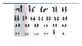
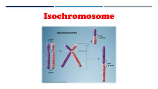
![Isochromosome
Isochromosome (i): Two identical chromosome
arms positioned as mirror images of each other
[i(17q)]
Isochromosome formation leads to both loss and
gain of genetic material
i(17q) consists of two chromosome 17 long arms,
without short arms. Cells with i(17q) generally also
have one normal chromosome 17; thus, they have one
copy of 17p and three copies of 17q.](https://image.slidesharecdn.com/karyotyping-201016130933/85/Karyotyping-70-320.jpg)


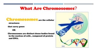
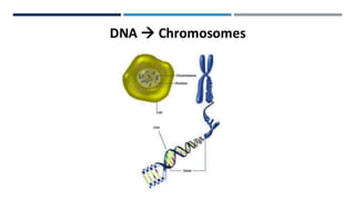
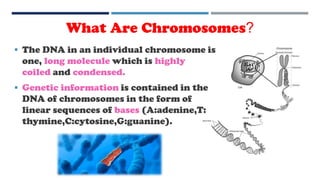
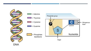
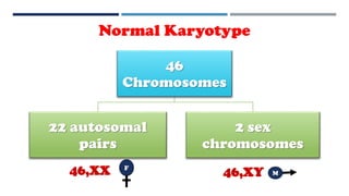
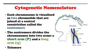
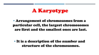
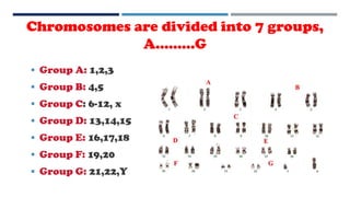
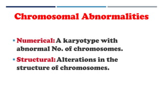
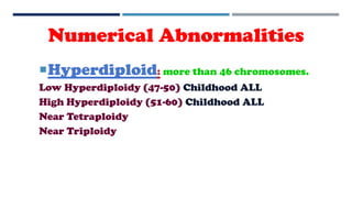
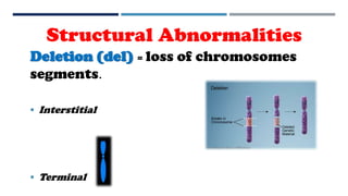
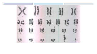
Chromosomes are cellular structures that carry genes. They are composed of DNA and protein and are found in the nucleus of cells. Genetic information is contained in the linear sequence of DNA bases on chromosomes. There are normally 46 chromosomes in humans arranged into 22 paired autosomes and two sex chromosomes. Chromosomal abnormalities can be numerical, involving extra or missing chromosomes, or structural, involving changes in chromosome structure like deletions, translocations, or inversions. Karyotyping allows visualization of chromosomes and identification of any abnormalities.


