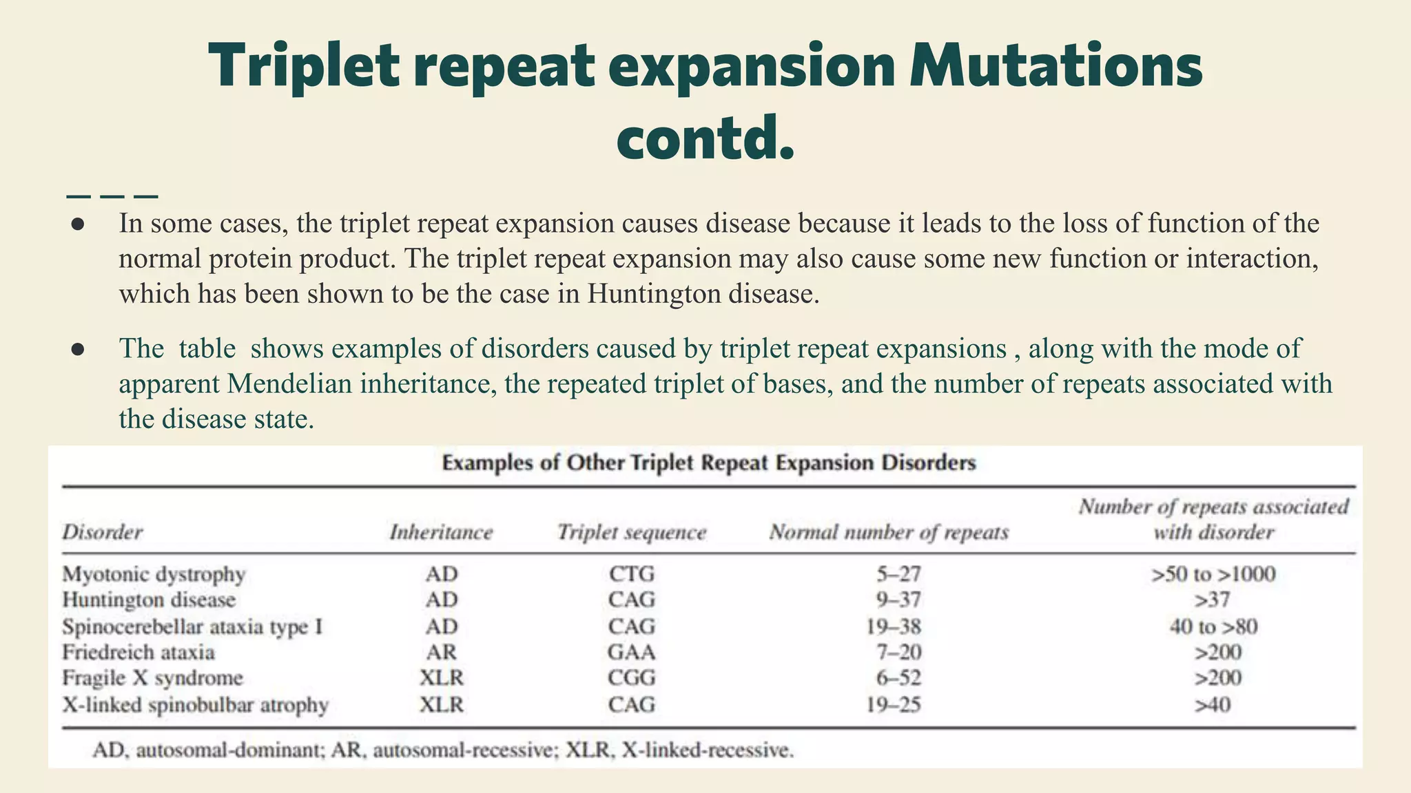
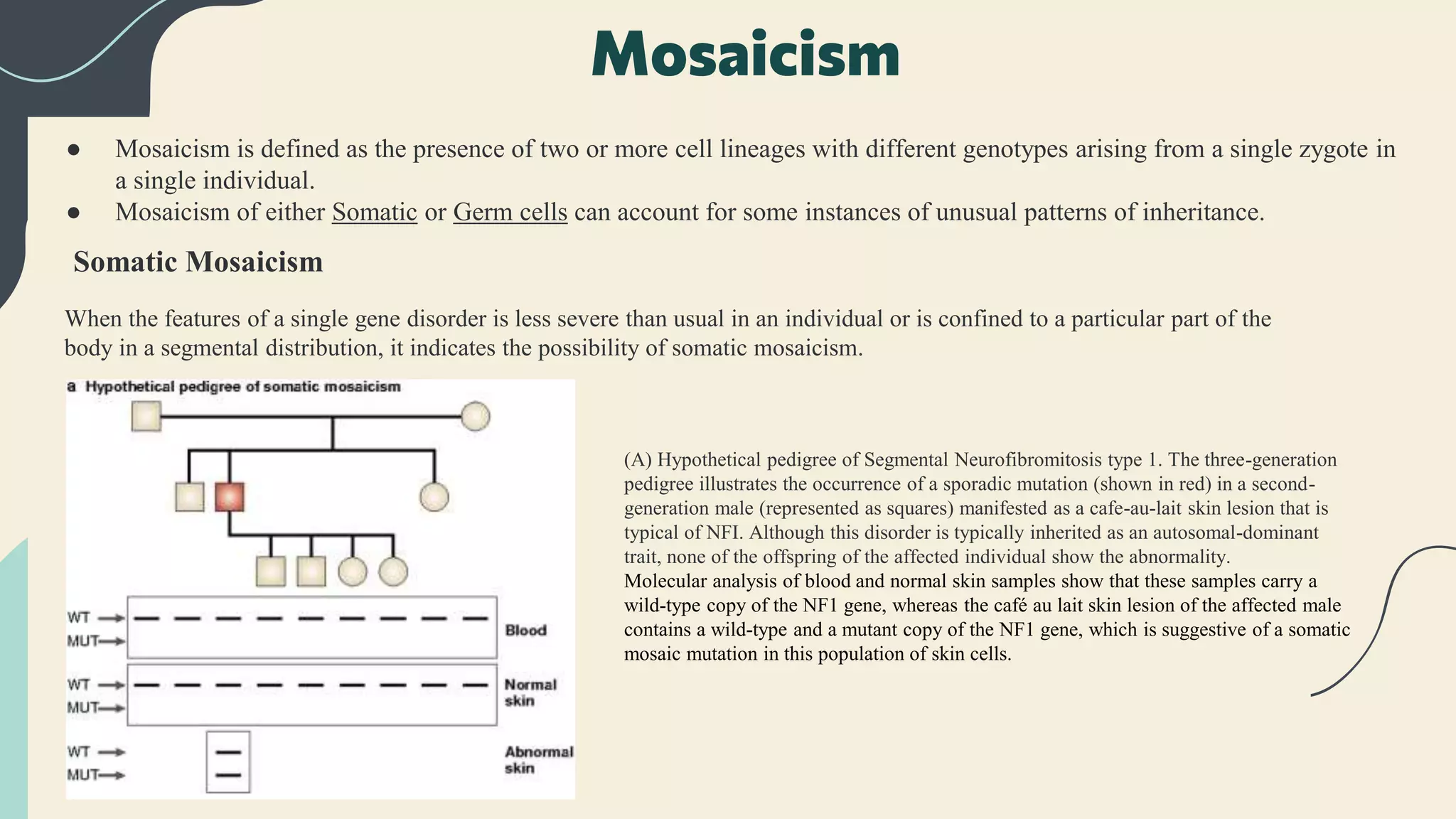
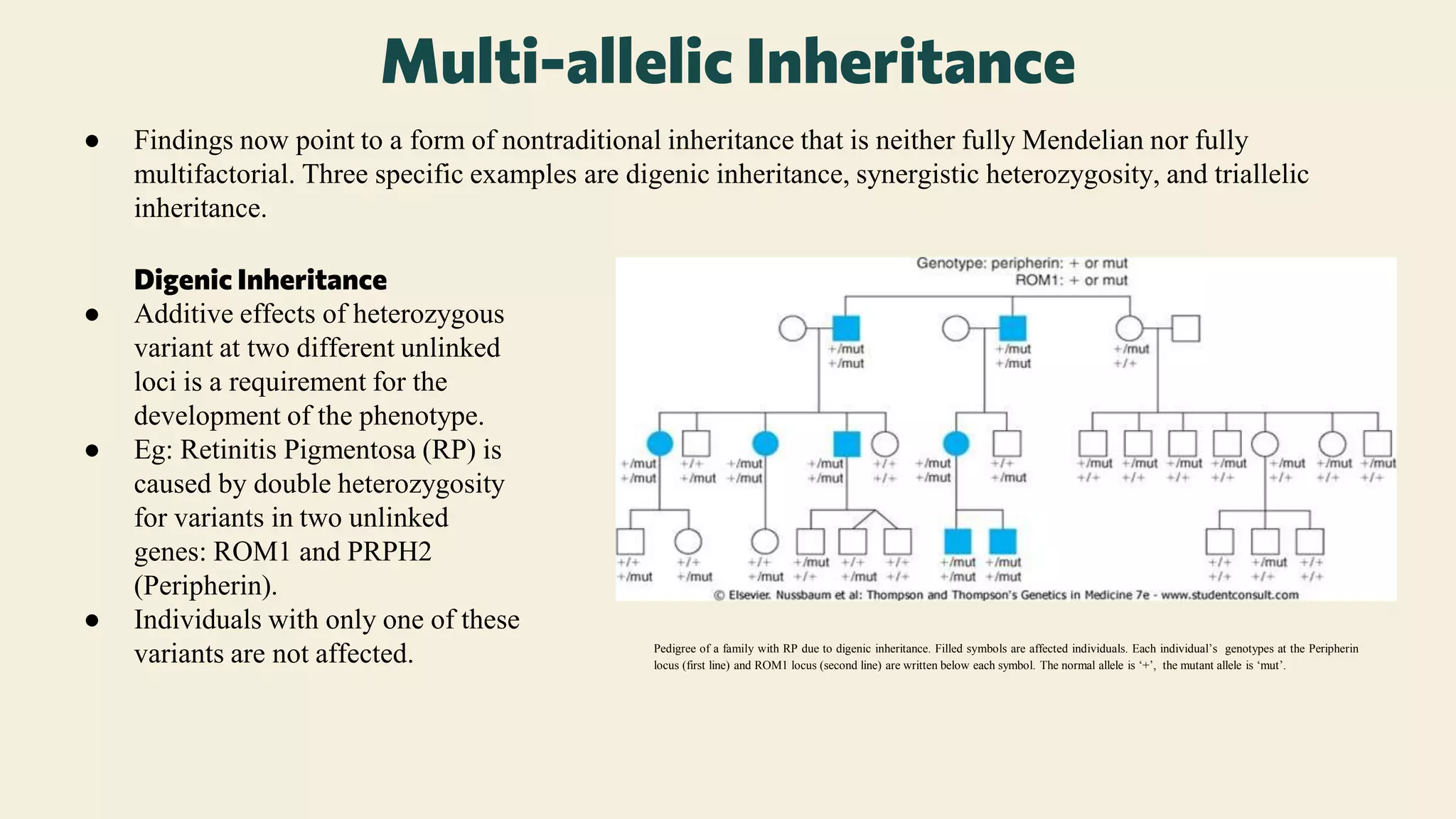
Nontraditional inheritance refers to the pattern of inheritance of a trait or phenotype that occurs predictably, recurrently, and in some cases familially, but does not follow the rules of typical Mendelian autosomal or sex chromosome inheritance.
Key concepts:
1. Introduction
2. Triplet repeat expansion mutations
3. Anticipation
4. Mosaicism
5.Genomic imprinting
6. Uniparental disomy
7. Mitochondrial inheritance
8. Multi-allelic inheritance
3. Introduction
● The “rules” of segregation of alleles originally defined by Gregor Mendel explained much of the
phenomena associated with inheritance and have been dogmatically applied in the field of genetics.
● However, there are situations in which the rules of Mendelian inheritance cannot explain observed
phenomena.
● A variety of molecular mechanisms have been identified that explain certain phenomena that are not
easily explained by traditional Mendelian patterns of inheritance.
● These non-Mendelian mechanisms differ on a molecular basis, but can be described as a group by the
term “non- traditional mechanisms of inheritance” or “non-traditional inheritance.”
● Stated simply, nontraditional inheritance refers to the pattern of inheritance of a trait or phenotype that
occurs predictably, recurrently, and in some cases familially, but does not follow the rules of typical
Mendelian autosomal or sex chromosome inheritance.
● It has become clear in recent years that not all forms of inheritance follow classical mendelian laws
regarding the equal contribution from both parents. These nontraditional modes of inheritance and gene
expression include cytoplasmic inheritance, mosaicism, uniparental disomy, and genomic imprinting.
4. Triplet repeat expansion Mutations
● The first disorder identified as resulting from this form of nontraditional inheritance is Fragile X syndrome
(FRAXA).
● The mental retardation syndrome was inherited in a classic pattern of X-linked inheritance, with carrier mothers
who, might have affected brothers or uncles, passing the trait on to half of their male offspring.
● However, some pedigree patterns demonstrating what came to be known as the Sherman paradox presented
families in which a male appeared to have passed the trait on to his daughters, but he himself was not affected,
even though he might have affected brothers or uncles. This pattern could not be explained by typical X-linked
inheritance.
● The solution to the Sherman paradox became apparent when a repeated sequence of CGG nucleotides, a “triplet
repeat” was found in the FMR1 gene (Xq27.3).
● In an affected individual the sequence is repeated more than 200 times. (normal: 5–44 times)
5. Triplet repeat expansion Mutations
contd.
● Premutations that are alleles that are somewhat larger than normal,are prone to further expansion
(increase in the number of repeats), leading in some cases to the full mutation.
● Occasionally, a female carrier of a premutation may pass on to her son an allele that has not undergone
further expansion. He will not be affected, but can pass on the premutation to his daughters, who then
will have a high risk of having an affected child by passing on a further expanded allele.
● Another interesting observation about FRAXA is that, contrary to Mendelian expectations for an X-
linked recessive disorder, carrier females are often affected.
● In fact, as many as 50% of females who carry a full expansion will have measurable cognitive defects.
This is not because of variation in the triplet repeat expansion, but is instead a result of intraindividual
variation in X-chromosome inactivation.
● Some triplet repeats are more prone to expansion during female meiosis (e.g., fragile X and myotonic
dystrophy) whereas others are more likely to expand when inherited from the father (e.g., Huntington
disease).
6. Triplet repeat expansion Mutations
contd.
● In some cases, the triplet repeat expansion causes disease because it leads to the loss of function of the
normal protein product. The triplet repeat expansion may also cause some new function or interaction,
which has been shown to be the case in Huntington disease.
● The table shows examples of disorders caused by triplet repeat expansions , along with the mode of
apparent Mendelian inheritance, the repeated triplet of bases, and the number of repeats associated with
the disease state.
7. Anticipation
● Anticipation which refers to an observed phenomenon where a genetic disorder appears to become more
severe and/or appear at an earlier age, in successive generations.
● Expansion of the CTG triple repeat in the 3’ untranslated end of the Myotonic Dystrophy gene occurring
predominanly in maternal meiosis appears to be the explanation for severe neonatal form of Myotonic
dystrophy when transmitted by mildly affected mother.
● Fragile X syndrome (CGG repeats) behaves in a similar way with major instability in the expansion
occurring during maternal meiosis.
● CAG repeats in the 5’ end of the Huntington disease gene in paternal meiosis, accounts for the
increased risk of early onset because of further expansion of CAG repeats.
● Another example is inherited Spinocerebellar ataxia group of conditions.
8. Somatic Mosaicism
When the features of a single gene disorder is less severe than usual in an individual or is confined to a particular part of the
body in a segmental distribution, it indicates the possibility of somatic mosaicism.
(A) Hypothetical pedigree of Segmental Neurofibromitosis type 1. The three-generation
pedigree illustrates the occurrence of a sporadic mutation (shown in red) in a second-
generation male (represented as squares) manifested as a cafe-au-lait skin lesion that is
typical of NFI. Although this disorder is typically inherited as an autosomal-dominant
trait, none of the offspring of the affected individual show the abnormality.
Molecular analysis of blood and normal skin samples show that these samples carry a
wild-type copy of the NF1 gene, whereas the café au lait skin lesion of the affected male
contains a wild-type and a mutant copy of the NF1 gene, which is suggestive of a somatic
mosaic mutation in this population of skin cells.
Mosaicism
● Mosaicism is defined as the presence of two or more cell lineages with different genotypes arising from a single zygote in
a single individual.
● Mosaicism of either Somatic or Germ cells can account for some instances of unusual patterns of inheritance.
9. ● In this pedigree, you can see that unaffected parents have one affected son, one affected
daughter, and one unaffected son. Although this disorder is inherited as an autosomal-
dominant trait, the pedigree is more consistent with either autosomal-recessive inheritance or
germ-line mosaicism for a mutation in one of the tuberous sclerosis genes in one of the
parents.
● Molecular analysis of blood samples shows that both parents are homozygous for the wild-type
TSC1 allele, suggesting that they are not typical heterozygous carriers. Blood samples from the
affected children show that both are heterozygous for the mutant TSC1 allele, whereas the
unaffected son carries only the wild-type TSC1 allele.
● Analysis of germ line (sperm) cells from the males in the pedigree shows that the father and
the affected son carry the mutant TSC1 allele.
● Taken together, these results suggest that the father has undergone a germ line mosaic
mutation such that some of his sperm cells carry the mutant TSC1 allele.
Germline Mosaicism
● There have been several clinical descriptions of families with autosomal dominant disorders, such as
Achondroplasia and Osteogenesis imperfecta, and X-linked recessive disorders such as Duchenne
muscular dystrophy and Haemophilia, in which the parents are phenotypically normal and the results of
genetic tests are also normal, but in which mor one of their offsprings have been affected.
● Most favoured explanation is Germline mosaicism in one of the parents.
10. Genomic Imprinting
● The “parent of origin” effect is referred to as genomic imprinting and DNA methylation, occurring mostly at a
CpG site, is the main mechanism by which expression is modified.
● So far at least 80 human genes are known to be imprinted, and the regions are known as differentially methylated
regions (DMRs).
● These DMRS include imprinting control regions (ICRs) that control gene expression across
● some autosomal genes are normally expressed only from one member of the gene pair even though both genes in
the pair have normal base sequence because one allele has been inactivated.
● This exposes at least three different, and fascinating, causes of nontraditional inheritance not because of
variation in the DNA sequence of the genes involved, but instead because of changes affecting the way the genes
are transcribed and expressed.
● Evidence of Genomic imprinting has been observed in two pairs of well known dysmorphic syndromes:
-Prader-Willi syndrome (PWS) and Angelman syndrome (AS)
-Beckwith-Wiedemann syndrome (BWS) and Russell-Silver syndrome (RSS)
11. PWS and AS
● Prader–Willi syndrome (PWS) and Angelman syndrome (AS) exemplify several aspects of nontraditional
inheritance. In the case of PWS and AS, the parent of origin of chromosome 15 affects the expression of some
genes.
● Both of these syndromes can develop as a result of a structural abnormality of the imprinting centre. This
abnormality is known as imprinting mutation.
● An imprinted domain at the 15q11- q13 is the main factor responsible for both the syndromes.
● The genes for both of these disorders are very close together and thus the centre is involved in inactivation of the
genes involved in imprinting
● But, it is the loss of maternal contribution that leads to Angelman syndrome and loss of paternal contribution that
leads to Prader Willi Syndrome.
● In the case of Angelman Syndrome most cases are not inherited but rather caused due to deletion of about 4
million bp in length which results in silencing of maternal gene UBE3A along with the already silenced paternal
gene. This leads to phenotypic syndromes of Angelman syndrome.
● There have also been cases where the maternal gene is lost and two copies of the gene from the father is present.
This is called as uniparental disomy.
12. ● Prader Willi syndrome is caused by loss of genes in the same region on the chromosome 15, namely the loss of
the SNRPN gene which is obtained paternally. The most common mechanism leading to PWS is a small
interstitial deletion on the chromosome 15, thus silencing the gene, inherited from the father..
● This recurrent deletion accounts for about 70–75% of all cases of PWS, and occurs with a frequency as high as 1
in 20,000 liveborn infants (the overall incidence of PWS).
13. ● Uniparental disomy (UPD) occurs when both chromosomes of a pair or areas from one chromosome in
any individual have been inherited from a single parent.
● UPD can be of 2 types: uniparental isodisomy or uniparental heterodisomy.
● When an individual inherits two copies of the same homologue from one parent through an error in
meiosis II, this is called Uniparental isodisomy.
● This is particularly important when the parent is a carrier of an autosomal recessive disorder. If the
offspring of a carrier parent has UPD with isodisomy for a chromosome that carries an abnormal gene,
the abnormal gene will be present in 2 copies, and the phenotype will be that of the autosomal recessive
disorder.
● If an individual inherits two different homologues from one parent through an error in meiosis I, it is
known as Uniparental heterodisomy.
● Examples include some cases of Beckwith-Wiedemann syndrome resulting from UPD for chromosome
11, transient neonatal diabetes resulting from UPD of chromosome 6, some cases of Silver-Russell
syndrome resulting from UPD of chromosome 7.
Uniparental disomy
14. ● UPD most often occurs as a result of a trisomy present at fertilization.
● PWS occurs when there are two copies of the maternally inherited chromosome 15 present. Most
often this results from malsegregation during female meiosis.
● This is nonviable unless there is a second postmitotic event, usually loss of one of the chromosome
15s resulting from malsegregation during mitosis (called “trisomy rescue”).
● This often occurs because of anaphase lag, where one chromosome fails to move along with the
others as the mitotic spindles separate during cell division, and the chromosome is lost into the
cytoplasm of one of the daughter cells.
● It is apparent that there are two possible outcomes from
this event.
-the loss of one of the two chromosomes that came from the same
parent, resulting in a cell that is now back to the normal and
appropriate chromosomal complement, having one chromosome
15 from the father and one from the mother.
-alternatively, the chromosome lost in the trisomy rescue process
may be from the parent who only contributed one chromosome.
This rescues the trisomy, and allows the pregnancy to continue, but
if there are imprinted genes on the involved chromosome they will
have abnormal expression.
15. Mitochondrial Inheritance
● Mitochondrial inheritance appears in a matrilineal pattern.
● The disorder affects both males and females, but are only be
transmitted through females.
● The nucleotide sequence of these mitochondrial genomes is not
identical, so that in any particular ovum there may be a variety
of mutations, none of which are present in every copy of the
mtDNA.
● If a variant occurs in the mtDNA of an individual there will be
two populations of mtDNA, so called heteroplasmy.
● Different developing tissues may acquire different complements of mtDNA mutations, and, depending on the
effect of the mutation and the energy requirements of the tissue in question, there may be selection for one
mtDNA genome over another, leading to accumulation or loss of a particular mutation in a particular tissue type.
This explains range of phenotypic severity in mitochondrial disorders.
● Eg: Most cases of Kearn-Sayre are associated with large deletions of the mitochondrial genome, but some cases
have been reported with point mutations in the same leucine tRNA associated with mitochondrial
encephalomyopathy (MELAS). The fact that two distinct phenotypes may result from defects in the same tRNA
may be a result of tissue heteroplasmy.
16. Multi-allelic Inheritance
● Findings now point to a form of nontraditional inheritance that is neither fully Mendelian nor fully
multifactorial. Three specific examples are digenic inheritance, synergistic heterozygosity, and triallelic
inheritance.
Digenic Inheritance
● Additive effects of heterozygous
variant at two different unlinked
loci is a requirement for the
development of the phenotype.
● Eg: Retinitis Pigmentosa (RP) is
caused by double heterozygosity
for variants in two unlinked
genes: ROM1 and PRPH2
(Peripherin).
● Individuals with only one of these
variants are not affected. Pedigree of a family with RP due to digenic inheritance. Filled symbols are affected individuals. Each individual’s genotypes at the Peripherin
locus (first line) and ROM1 locus (second line) are written below each symbol. The normal allele is ‘+’, the mutant allele is ‘mut’.
17. ● Synergistic heterozygosity is a multigenic inheritance pattern in which mutations in multiple genes in one or
multiple related metabolic pathways lead to sufficient disruption of flux through the pathway, mimicking a
monogenic disorder.
● Although both of these forms of inheritance would lead to Mendelian (recessive) proportions of affected
individuals on pedigree analysis, triallelic inheritance would not.
● Another example of nontraditional inheritance is pn that has been described in an isolated population with a
high rate of Bardet-Biedl syndrome (BBS).
● In studying several families it was found that some affected individuals were homozygous for mutations in a
previously identified BBS gene, whereas there were unaffected family members that were also homozygous for
the same mutations.
● Further investigation revealed that the difference between the affected and the unaffected individuals was that
those affected also had heterozygous mutations of another gene known to be associated with BBS.
● Therefore, in this population, homozygosity for mutation in the first locus was not sufficient to cause the
phenotype, but required a third abnormal allele in a different gene, thus Triallelic inheritance.
18. ● M Turnpenny, P. D., & Ellard, S. (2020). Emery's elements of medical genetics. Philadelphia:
Elsevier/Churchill Livingstone.
● Carlos A. Bacino and Brendan Lee (2020). Nelson Textbook of Pediatrics, Chapter 98, 652-
676.e1
● Chial, H. & Craig, J. (2008) mtDNA and mitochondrial diseases. Nature Education 1(1):217
● Candless S.E., Cassidy S.B. (2006) Nontraditional Inheritance. In: Runge M.S., Patterson C.
(eds) Principles of Molecular Medicine. Humana Press. https://doi.org/10.1007/978-1-59259-
963-9_2
Reference