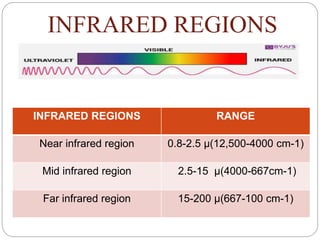
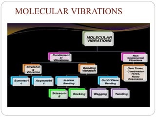
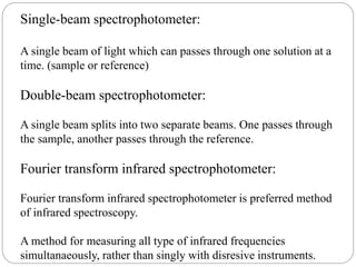
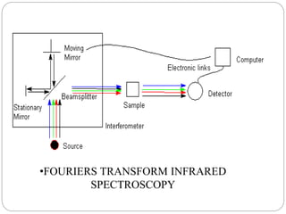
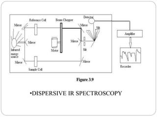
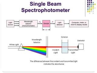
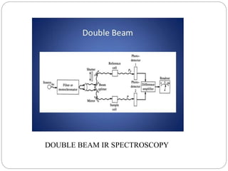
Presentation by : Pawankumar Hanamant Yadav, Rahul Nimba Patil
1st year M. Pharm (sem –l)
Dept. : Pharmaceutics & Pharmacology
Guided by : Prof. Kardile sir
Subject : Modern pharmaceutical analytical Techniques
RAJGAD DNYANPEETH’s
COLLEGE OF PHARMACY , BHOR
•Introduction
• IR Regions
• Principle
• Molecular vibrations
• Fundamental vibrations
• Non-Fundamental vibrations
• Instrumentation
•Applications
CONTENTS:
INTRODUCTION
•Infrared spectroscopy (IR spectroscopy) is the
spectroscopy that deals with the infrared region
of the electromagnetic spectrum, that is light with
a longer wavelength and lower frequency than
visible light
•Infrared Spectroscopy is the analysis of infrared
light interacting with a molecule.
•It is based on absorption spectroscopy
INFRARED REGIONS
INFRARED REGIONS RANGE
Near infrared region 0.8-2.5 µ(12,500-4000 cm-1)
Mid infrared region 2.5-15 µ(4000-667cm-1)
Far infrared region 15-200 µ(667-100 cm-1)
•When the energy in the form of IR is applied and if the of vibration,
the applied IR frequency = Natural frequency of vibration, the
absorption of IR takes place and peak is observed.
•Molecules are excited to the higher energy state from the ground
state when they absorbed IR radiation.
•When a compound is exposed to IR radiation, it selectively absorb
the radiations resulting in vibration of the molecules of the compound
, giving rise to closely packed absorption bands, called as IR
absorption spectrum
•The bands correspond to characteristic functional groups and the
bonds present in a chemical substance. Thus an IR spectrum of a
compound is considered as the fingerprints for its chemical
identification.
PRINCIPLE
MOLECULAR VIBRATIONS
What is a vibration in a molecule?
“Any change in shape of the molecule-
stretching of bonds, bending of bonds, or
internal rotation around single bonds”.
Why we study the molecular vibration?
Because whenever the interaction b/w
electromagnetic waves & matter occur so
change appears in these vibrations.
Molecular vibration divided into 2
main types :
FUNDAMENTAL VIBRATIONS:
Vibrations which appear as band in the spectra.
NON FUNDAMENTAL VIBRATIONS:
Vibrations which appears as a result of fundamental
vibrations.
FUNDAMENTAL VIBRATIONS
STRETCHING
VIB.
1.Streching
vibration Involves a
continuous change
in the inter atomic
distance along the
axis of the bond
b/w 2 atoms.
2.It requires more
energy so appear at
shorter wavelength.
BENDING
VIB.
1.Bending
vibrations are
characterized by a
change in the
angle b/w two
bonds.
2.It requires less
energy so appear
at longer
wavelength.
Stretching vibration is further
divided into
SYMMETRIC VIB.
Inter atomic
distance b/w 2
atoms
increases/decreas
es.
ASYMMETRIC
VIB.
Inter atomic
distance b/w 2
atoms is
alternate/opposite.
BENDING VIBRATION IS DIVIDED INTO:
IN PLANE
BENDING
OUT OF
PLANE
BENDING
•If all the atoms are
on same plane.
•If 2 atoms are on same plane
while the 1 atom is on opposite
plane.
In-Plane Bending further divided
into:
SCISSORING:
When two atoms move away or close
towards each other.
ROCKING:
Change in angle between a group of
atom.
WAGGING:
•Change in angle between the plane of a groups of atoms.
TWISTING:
•Change in angle between the plane of 2 groups of atom.
OUT PLANE BENDING IS FURTHER
DIVIDED INTO
NON FUNDAMENTAL
VIBRATIONS:
Fermi
Resonance :
Interaction
between
fundamental
vibration &
overtones or
combination
tones.
Ex. CO2
OVERTONES:
these are
observed at
twice the
frequency of
strong band.
EX.
Carbonyl
group.
COMBINATION
TONES:
Weak bands that
appear
occasionally at
frequencies that
are
sum/difference of
two or more
fundamental
bands.
Hooke’s law:
It gives relation between frequency of oscillation, atomic mass,
force constant of the bond.
Thus vibrational frequency is
c - velocity of light
F - force constant
Mx - mass of atom x
My – mass of atom y
Since the force constant measures the strength of bond, value of f is
for
Hydrogen bonding:
Effect of hydrogen bonding on IR:
Proton donor group(s-orbital)
Proton acceptor group(p-orbital)
Hydrogen bonding
Examples of proton donor and proton acceptor group
Proton donor group:
Carboxyl, hydroxyl, amine or amide group
Proton acceptor:
Oxygen, Nitrogen, Halogens and unsaturated group
Types of hydrogen bonding:
Intermolecular hydrogen bonding:
Factors affecting on hydrogen bonding:
Temprature
Concentration
Molecular geometry
Relative activity
basicity
Instrumentation:
The main parts of IR spectrometer are as follows:
Radiation source
Sample cells
Monochromators
Detectors
Recorder
Radiation source:
The IR instrumemts require a source of radient energy which
emit IR radiation which must me:
Sufficient intensity
Continues
Stable
Source of IR radiation are as follows:
Globar source:
Rod of silicon carbide.
Heated upto 1300 degree centrigrade.
Produce radiant energy from 1-40 micron.
Nerst glower:
Rod of zirconium and yittrium.
Heated upto 1500 centigrade.
Emits radistion between 0.4-20 micron.
Sample cells:
For gas samples:
The of gas sapmle can be obtained by permitting the sample to
To exapand into an evacuated cell, also called a cuvette.
For solution samples:
Infrared solution cells consist of two windows of pressed salt
sealed. samples that are liquid at room temprature are usually
analysed in pure form or in solution.
The most common solvents are:
Carbon tetrachloride(CCl4)
Carbon disulfide(CS2)
For solid sample:
Solids reduced to small pieces can be examined as a thin paste or
Mull.
The mull is formed by grinding a 2-5 milligrams of the samples
in the sample in the presence of one or two drops of a
hydrocarbons oil(nujol oil).
The resulting mull is then examined as a flim between flat salt
plates.
Another technique is to ground a milligram or less of the
sample with about 100 milligram potassium bromide.
The mixture is then pressed in an evaluate die to produce a
transparent disk.
Monochromator:
Monochromator is used as dispersive element.
Monochromaors consist of mainly 3 parts:
Entrace slit
Dispersive element
Entrance slit
Light enters into the entrance slit then light falls on dispersive
element then dispersive element emit the radiation towards exit
slit then light exit by exit slit.
Two types of monochromators are:
Prism monochromators
Grating monochromators
Prism monochromators:
Used as dispersive element.
Constructed of various metal halide salts.
Sodium chloride is mot commonly used as prism salt.
This salts are subjected to mechanical and thermal instability.
Grating monochromators:
Gratings are made up of some glass, quartz, alkyl hakides.
The mechanism is that diffraction produces reinforcement.
Detectors:
An infrared detector is a detector that reacts to infrared radiation.
There are two types of detectors:
Thermal detectors
Non-thermal detectors
Thermal detectors:
It can be used over a wide range of wavelengths and they
operate at room temperature.
There are 4 types of thermal detectors:
Bolometers detectors
Thermo-couple detectors
Pyro-electric detectors
Golay cells
Bolometrs detectors:
It is derived from greek word (bolometron)
Bolo-for something thrown
Metron-measure
It is use to detect the IR radiation.
Construction:
A bolometer consist of an absorptive element,such as a thin
layer of metal.
Most of bolometers use semiconductors or superconductor
absorptive elements rather than metals.
Thermocouple:
It consists of pair of junction of different metals e.g- 2 pieces
of bismuth fused to either and of a piece of antimony.
Thermocouple detectors are voltage generated devices, which
can be thought of as miniature arrays of thermocouple
junctions.
Pyroelectric detectors:
When an electric field is applied across any dielectric material
electric polarisation takes place.
The magnitude of polarisation is a function of dielectric constant
of that material.
This pyroelectric crystal is sandwiched between 2 electrodes
which are IR transparent.
e.g.- triglycerine sulphate.
Golay cell:
It is made up of metal cylinder and flexible.
Whole chamber is filled with xenon gas.
In chamber temperature increases and gases expand and diaphragm
deforms.
Finally detect as a signal.
Non thermal detectors:
Thin film overlapse over non conducting surface.
Absorption of IR radiation.
Non conducting valance electron negative to higher energy
conducting state.
Electric resistance decrease and voltage drops.
Recorders:
The radiant enrgy received by detector is converted into
measurable electrical signal and is amplified by amplifier.
The amplified signals are recorded and plotted.
It records trransmittance of sample as well as function of wave
number.
It is computer based advanced software version used now a days.
IR absorption spectrophotometers:
These are of two types:
Single-beam spectrophotometer
Double-beam spectrophotometer
Single-beam spectrophotometer:
A single beam of light which can passes through one solution at a
time. (sample or reference)
Double-beam spectrophotometer:
A single beam splits into two separate beams. One passes through
the sample, another passes through the reference.
Fourier transform infrared spectrophotometer:
Fourier transform infrared spectrophotometer is preferred method
of infrared spectroscopy.
A method for measuring all type of infrared frequencies
simultanaeously, rather than singly with disresive instruments.
•FOURIERS TRANSFORM INFRARED
SPECTROSCOPY
•DISPERSIVE IR SPECTROSCOPY
DOUBLE BEAM IR SPECTROSCOPY
Fundamental region:
Group frequency region:
Consisting of absorption bands of the functional groups.
Frequency = 4000-1300cm-1
wavelength = 2.5-8
Fringer print region:
IR spectra is called as “fingerprints” because no other chemical
species will have similar IR spectrum.
Frequency = 1300-650cm-1
Wavelength = 8-15.4
Applications:
Detection of impurities:
Determined by comparing sample spectrum with the spectrumof
pure reference compound.eg: ketone impurity in alcohols.
Protein quantification:
By measuring amide bonds in protein chains, we can accurately
quantifies an intrinsic component of every protein.
Forensic analysis:
It is used in both crimnal and civil cases.eg: identifying polymer
degradation, in determining the blood alcohol content.
Studying progress of reaction:
It is applicable to observe rate of disappearance of characteristic
absorption band in reactants.
Identify molecular structure:
Identification is done based on position of absorption bands in
the spectrum.eg: c=o at 1717cm-1
Identification of substances:
It is use to compare spectrums because no two samples will have
identical IR spectrum.
IR spectroscopy

IR spectroscopy

  • 1.
    Presentation by :Pawankumar Hanamant Yadav, Rahul Nimba Patil 1st year M. Pharm (sem –l) Dept. : Pharmaceutics & Pharmacology Guided by : Prof. Kardile sir Subject : Modern pharmaceutical analytical Techniques RAJGAD DNYANPEETH’s COLLEGE OF PHARMACY , BHOR
  • 3.
    •Introduction • IR Regions •Principle • Molecular vibrations • Fundamental vibrations • Non-Fundamental vibrations • Instrumentation •Applications CONTENTS:
  • 4.
    INTRODUCTION •Infrared spectroscopy (IRspectroscopy) is the spectroscopy that deals with the infrared region of the electromagnetic spectrum, that is light with a longer wavelength and lower frequency than visible light •Infrared Spectroscopy is the analysis of infrared light interacting with a molecule. •It is based on absorption spectroscopy
  • 5.
    INFRARED REGIONS INFRARED REGIONSRANGE Near infrared region 0.8-2.5 µ(12,500-4000 cm-1) Mid infrared region 2.5-15 µ(4000-667cm-1) Far infrared region 15-200 µ(667-100 cm-1)
  • 6.
    •When the energyin the form of IR is applied and if the of vibration, the applied IR frequency = Natural frequency of vibration, the absorption of IR takes place and peak is observed. •Molecules are excited to the higher energy state from the ground state when they absorbed IR radiation. •When a compound is exposed to IR radiation, it selectively absorb the radiations resulting in vibration of the molecules of the compound , giving rise to closely packed absorption bands, called as IR absorption spectrum •The bands correspond to characteristic functional groups and the bonds present in a chemical substance. Thus an IR spectrum of a compound is considered as the fingerprints for its chemical identification. PRINCIPLE
  • 7.
  • 8.
    What is avibration in a molecule? “Any change in shape of the molecule- stretching of bonds, bending of bonds, or internal rotation around single bonds”. Why we study the molecular vibration? Because whenever the interaction b/w electromagnetic waves & matter occur so change appears in these vibrations.
  • 9.
    Molecular vibration dividedinto 2 main types : FUNDAMENTAL VIBRATIONS: Vibrations which appear as band in the spectra. NON FUNDAMENTAL VIBRATIONS: Vibrations which appears as a result of fundamental vibrations.
  • 10.
    FUNDAMENTAL VIBRATIONS STRETCHING VIB. 1.Streching vibration Involvesa continuous change in the inter atomic distance along the axis of the bond b/w 2 atoms. 2.It requires more energy so appear at shorter wavelength. BENDING VIB. 1.Bending vibrations are characterized by a change in the angle b/w two bonds. 2.It requires less energy so appear at longer wavelength.
  • 11.
    Stretching vibration isfurther divided into SYMMETRIC VIB. Inter atomic distance b/w 2 atoms increases/decreas es. ASYMMETRIC VIB. Inter atomic distance b/w 2 atoms is alternate/opposite.
  • 12.
    BENDING VIBRATION ISDIVIDED INTO: IN PLANE BENDING OUT OF PLANE BENDING •If all the atoms are on same plane. •If 2 atoms are on same plane while the 1 atom is on opposite plane.
  • 13.
    In-Plane Bending furtherdivided into: SCISSORING: When two atoms move away or close towards each other. ROCKING: Change in angle between a group of atom.
  • 14.
    WAGGING: •Change in anglebetween the plane of a groups of atoms. TWISTING: •Change in angle between the plane of 2 groups of atom. OUT PLANE BENDING IS FURTHER DIVIDED INTO
  • 15.
    NON FUNDAMENTAL VIBRATIONS: Fermi Resonance : Interaction between fundamental vibration& overtones or combination tones. Ex. CO2 OVERTONES: these are observed at twice the frequency of strong band. EX. Carbonyl group. COMBINATION TONES: Weak bands that appear occasionally at frequencies that are sum/difference of two or more fundamental bands.
  • 16.
    Hooke’s law: It givesrelation between frequency of oscillation, atomic mass, force constant of the bond. Thus vibrational frequency is c - velocity of light F - force constant Mx - mass of atom x My – mass of atom y Since the force constant measures the strength of bond, value of f is for
  • 17.
    Hydrogen bonding: Effect ofhydrogen bonding on IR: Proton donor group(s-orbital) Proton acceptor group(p-orbital) Hydrogen bonding Examples of proton donor and proton acceptor group Proton donor group: Carboxyl, hydroxyl, amine or amide group Proton acceptor: Oxygen, Nitrogen, Halogens and unsaturated group
  • 18.
    Types of hydrogenbonding: Intermolecular hydrogen bonding: Intermolecular hydrogen bonding:
  • 19.
    Factors affecting onhydrogen bonding: Temprature Concentration Molecular geometry Relative activity basicity
  • 20.
    Instrumentation: The main partsof IR spectrometer are as follows: Radiation source Sample cells Monochromators Detectors Recorder
  • 21.
    Radiation source: The IRinstrumemts require a source of radient energy which emit IR radiation which must me: Sufficient intensity Continues Stable Source of IR radiation are as follows: Globar source: Rod of silicon carbide.
  • 22.
    Heated upto 1300degree centrigrade. Produce radiant energy from 1-40 micron. Nerst glower: Rod of zirconium and yittrium. Heated upto 1500 centigrade. Emits radistion between 0.4-20 micron. Sample cells: For gas samples: The of gas sapmle can be obtained by permitting the sample to
  • 23.
    To exapand intoan evacuated cell, also called a cuvette. For solution samples: Infrared solution cells consist of two windows of pressed salt sealed. samples that are liquid at room temprature are usually analysed in pure form or in solution. The most common solvents are: Carbon tetrachloride(CCl4) Carbon disulfide(CS2) For solid sample: Solids reduced to small pieces can be examined as a thin paste or
  • 24.
    Mull. The mull isformed by grinding a 2-5 milligrams of the samples in the sample in the presence of one or two drops of a hydrocarbons oil(nujol oil). The resulting mull is then examined as a flim between flat salt plates. Another technique is to ground a milligram or less of the sample with about 100 milligram potassium bromide. The mixture is then pressed in an evaluate die to produce a transparent disk. Monochromator: Monochromator is used as dispersive element.
  • 25.
    Monochromaors consist ofmainly 3 parts: Entrace slit Dispersive element Entrance slit Light enters into the entrance slit then light falls on dispersive element then dispersive element emit the radiation towards exit slit then light exit by exit slit. Two types of monochromators are: Prism monochromators Grating monochromators
  • 26.
    Prism monochromators: Used asdispersive element. Constructed of various metal halide salts. Sodium chloride is mot commonly used as prism salt. This salts are subjected to mechanical and thermal instability. Grating monochromators: Gratings are made up of some glass, quartz, alkyl hakides. The mechanism is that diffraction produces reinforcement.
  • 27.
    Detectors: An infrared detectoris a detector that reacts to infrared radiation. There are two types of detectors: Thermal detectors Non-thermal detectors Thermal detectors: It can be used over a wide range of wavelengths and they operate at room temperature. There are 4 types of thermal detectors:
  • 28.
    Bolometers detectors Thermo-couple detectors Pyro-electricdetectors Golay cells Bolometrs detectors: It is derived from greek word (bolometron) Bolo-for something thrown Metron-measure It is use to detect the IR radiation.
  • 29.
    Construction: A bolometer consistof an absorptive element,such as a thin layer of metal. Most of bolometers use semiconductors or superconductor absorptive elements rather than metals. Thermocouple: It consists of pair of junction of different metals e.g- 2 pieces of bismuth fused to either and of a piece of antimony. Thermocouple detectors are voltage generated devices, which can be thought of as miniature arrays of thermocouple junctions.
  • 30.
    Pyroelectric detectors: When anelectric field is applied across any dielectric material electric polarisation takes place. The magnitude of polarisation is a function of dielectric constant of that material. This pyroelectric crystal is sandwiched between 2 electrodes which are IR transparent. e.g.- triglycerine sulphate. Golay cell: It is made up of metal cylinder and flexible.
  • 31.
    Whole chamber isfilled with xenon gas. In chamber temperature increases and gases expand and diaphragm deforms. Finally detect as a signal. Non thermal detectors: Thin film overlapse over non conducting surface. Absorption of IR radiation. Non conducting valance electron negative to higher energy conducting state. Electric resistance decrease and voltage drops.
  • 32.
    Recorders: The radiant enrgyreceived by detector is converted into measurable electrical signal and is amplified by amplifier. The amplified signals are recorded and plotted. It records trransmittance of sample as well as function of wave number. It is computer based advanced software version used now a days. IR absorption spectrophotometers: These are of two types: Single-beam spectrophotometer Double-beam spectrophotometer
  • 33.
    Single-beam spectrophotometer: A singlebeam of light which can passes through one solution at a time. (sample or reference) Double-beam spectrophotometer: A single beam splits into two separate beams. One passes through the sample, another passes through the reference. Fourier transform infrared spectrophotometer: Fourier transform infrared spectrophotometer is preferred method of infrared spectroscopy. A method for measuring all type of infrared frequencies simultanaeously, rather than singly with disresive instruments.
  • 34.
  • 35.
  • 37.
    DOUBLE BEAM IRSPECTROSCOPY
  • 38.
    Fundamental region: Group frequencyregion: Consisting of absorption bands of the functional groups. Frequency = 4000-1300cm-1 wavelength = 2.5-8 Fringer print region: IR spectra is called as “fingerprints” because no other chemical species will have similar IR spectrum. Frequency = 1300-650cm-1 Wavelength = 8-15.4
  • 39.
    Applications: Detection of impurities: Determinedby comparing sample spectrum with the spectrumof pure reference compound.eg: ketone impurity in alcohols. Protein quantification: By measuring amide bonds in protein chains, we can accurately quantifies an intrinsic component of every protein. Forensic analysis: It is used in both crimnal and civil cases.eg: identifying polymer degradation, in determining the blood alcohol content.
  • 40.
    Studying progress ofreaction: It is applicable to observe rate of disappearance of characteristic absorption band in reactants. Identify molecular structure: Identification is done based on position of absorption bands in the spectrum.eg: c=o at 1717cm-1 Identification of substances: It is use to compare spectrums because no two samples will have identical IR spectrum.