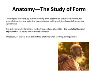
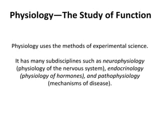
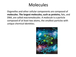
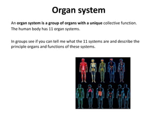
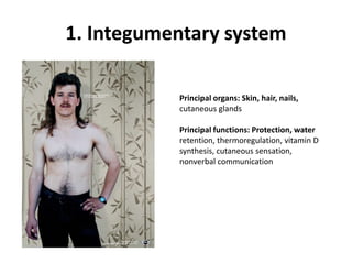
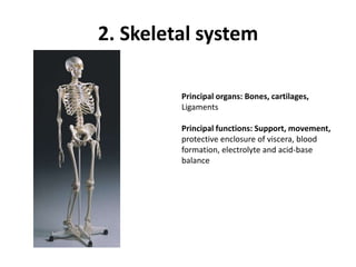
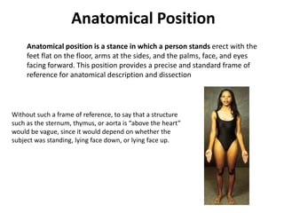
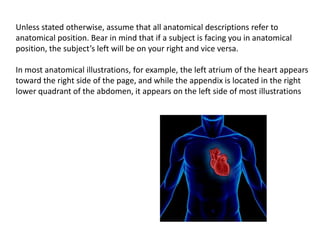
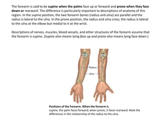
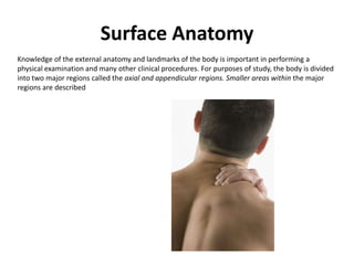
This document provides an overview of anatomy and physiology. It begins by defining anatomy as the study of structure and physiology as the study of function. It describes different methods of studying anatomy, including surface observation, dissection, palpation, auscultation, and percussion. It then discusses the hierarchy of biological complexity from molecules to cells to tissues to organs to organ systems. The document also defines and provides examples of organs, tissues, organelles, and molecules. It lists and describes the 11 major organ systems of the human body. Finally, it introduces some key anatomical concepts and terminology used to describe the human body.
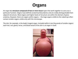

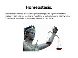


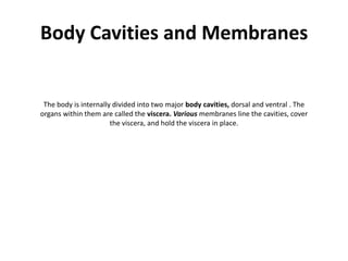
![Anatomical PlanesMany views of the body are based on real or imaginary “slices” called sections or planes. “Section” implies an actual cut or slice to reveal internal anatomy, whereas “plane” implies an imaginary flat surface passing through the body. The three major anatomical planes are sagittal, frontal, and transverseFrontal planeTransverse planeMedian [Mid-Sagittal] plane](https://image.slidesharecdn.com/l1introduction-091002030426-phpapp01/85/L1-Introduction-43-320.jpg)
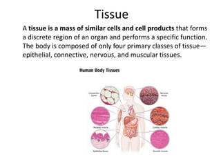
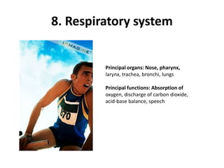
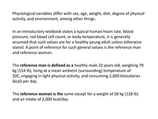
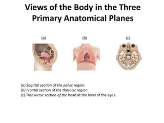
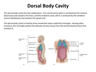
![A sagittal plane passes vertically through the body or an organ and divides it into right and left portions. The sagittal plane that divides the body or organ into equal haves is also called the median (midsagittal) plane. The head and pelvic organs are commonly illustrated on the median planeMedian [Mid-Sagittal] plane](https://image.slidesharecdn.com/l1introduction-091002030426-phpapp01/85/L1-Introduction-44-320.jpg)


![Lesson6 [2 Oth Oct 2008]](https://cdn.slidesharecdn.com/ss_thumbnails/lesson62othoct2008-091022023449-phpapp01-thumbnail.jpg?width=640&height=640&fit=bounds)


