Downloaded 234 times

![Safety: Adverse Effects
• 63% of studies report 1 mild ‘adverse effect’
– Itching, tingling, headache, burning sensation,
discomfort
• However:
– Active tDCS Rate = Sham Rate
– Except:
• Skin reddening (Tx w/ Ketoprofen)
43
Fregni et al. (2014). Clinical Research and Regulatory Affairs, [Early
Online]: 1-14.](https://image.slidesharecdn.com/tdcs-slideshare-170326201006/75/Intro-to-Transcranial-Direct-Curent-Stimulation-tDCS-43-2048.jpg)
![Safety: Serious Adverse Effects
• Review: No “serious adverse events” since
1998 in >10,000 subjects
• 1964 study: “respiratory and motor paralysis”
– Bifrontal anodal electrodes with leg cathode
– 10x intended current strength (likely ~3mA)
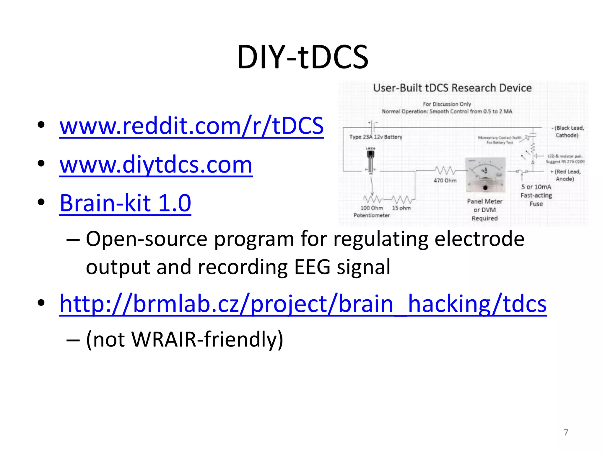
– DIY-tDCS concerns
44
Fregni et al. (2014). Clinical Research and Regulatory Affairs, [Early
Online]: 1-14.
Lippold O. C. J., & Redfearn, J. W. T. (1964). Mental changes resulting
from the passage of small direct currents through the human brain.
110(469): 768-772](https://image.slidesharecdn.com/tdcs-slideshare-170326201006/75/Intro-to-Transcranial-Direct-Curent-Stimulation-tDCS-44-2048.jpg)
![Safety: Physiological Evidence
• No pathological changes in:
– Serum enolase (marker of neuronal damage)
– HRV
– EEG
• 100x the charge density used in humans is
required to cause brain damage in rats
– Discomfort in humans starts at 2-3x
45
Fregni et al. (2014). Clinical Research and Regulatory Affairs, [Early
Online]: 1-14.](https://image.slidesharecdn.com/tdcs-slideshare-170326201006/75/Intro-to-Transcranial-Direct-Curent-Stimulation-tDCS-45-2048.jpg)
![Safety: Standard Parameters
• Current strength <2.5mA
• Duration <60min
• ≤2 sessions per day
• This does not imply going beyond these
parameters is not safe!
47
Fregni et al. (2014). Clinical Research and Regulatory Affairs, [Early
Online]: 1-14.](https://image.slidesharecdn.com/tdcs-slideshare-170326201006/75/Intro-to-Transcranial-Direct-Curent-Stimulation-tDCS-47-2048.jpg)
![Safety: Unknowns
• Long-term usage
• Need for more studies on safety
48
Fregni et al. (2014). Clinical Research and Regulatory Affairs, [Early
Online]: 1-14.](https://image.slidesharecdn.com/tdcs-slideshare-170326201006/75/Intro-to-Transcranial-Direct-Curent-Stimulation-tDCS-48-2048.jpg)
![Future Research Should Explore…
• effects of tDCS on cortical areas outside of M1
– neurotransmitters, receptors, neurons, and
networks are heterogeneous
• interaction of of [Stimulation x Task]
• timing and duration of stimulation (before,
during or after a task)
• multi-electrode stimulation of functional
networks
52
Shin, Y.-I., Foerster, A., & Nitsche, M. A. (2015).
Neuropsychologia, 69: 154-175](https://image.slidesharecdn.com/tdcs-slideshare-170326201006/75/Intro-to-Transcranial-Direct-Curent-Stimulation-tDCS-52-2048.jpg)

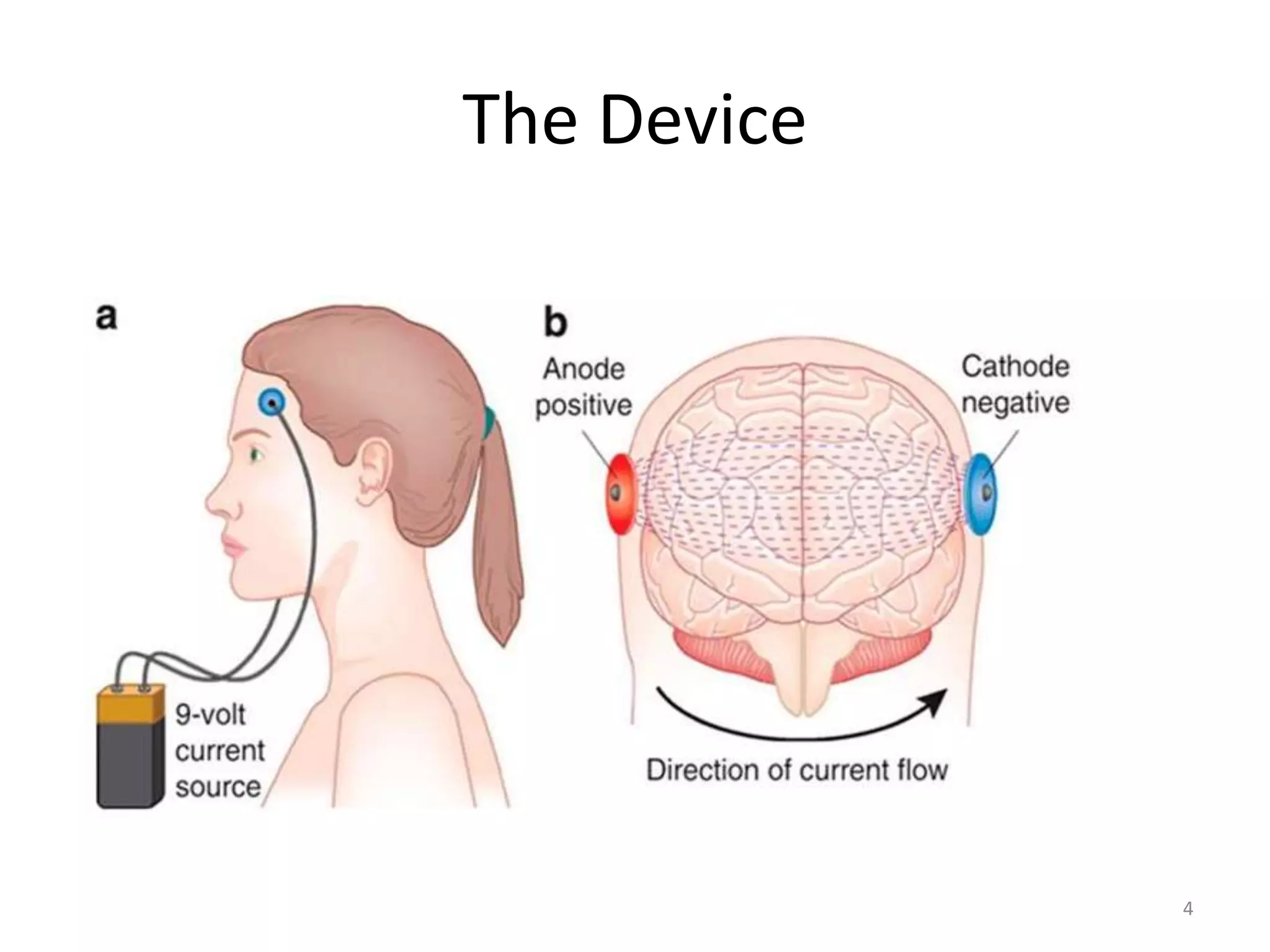
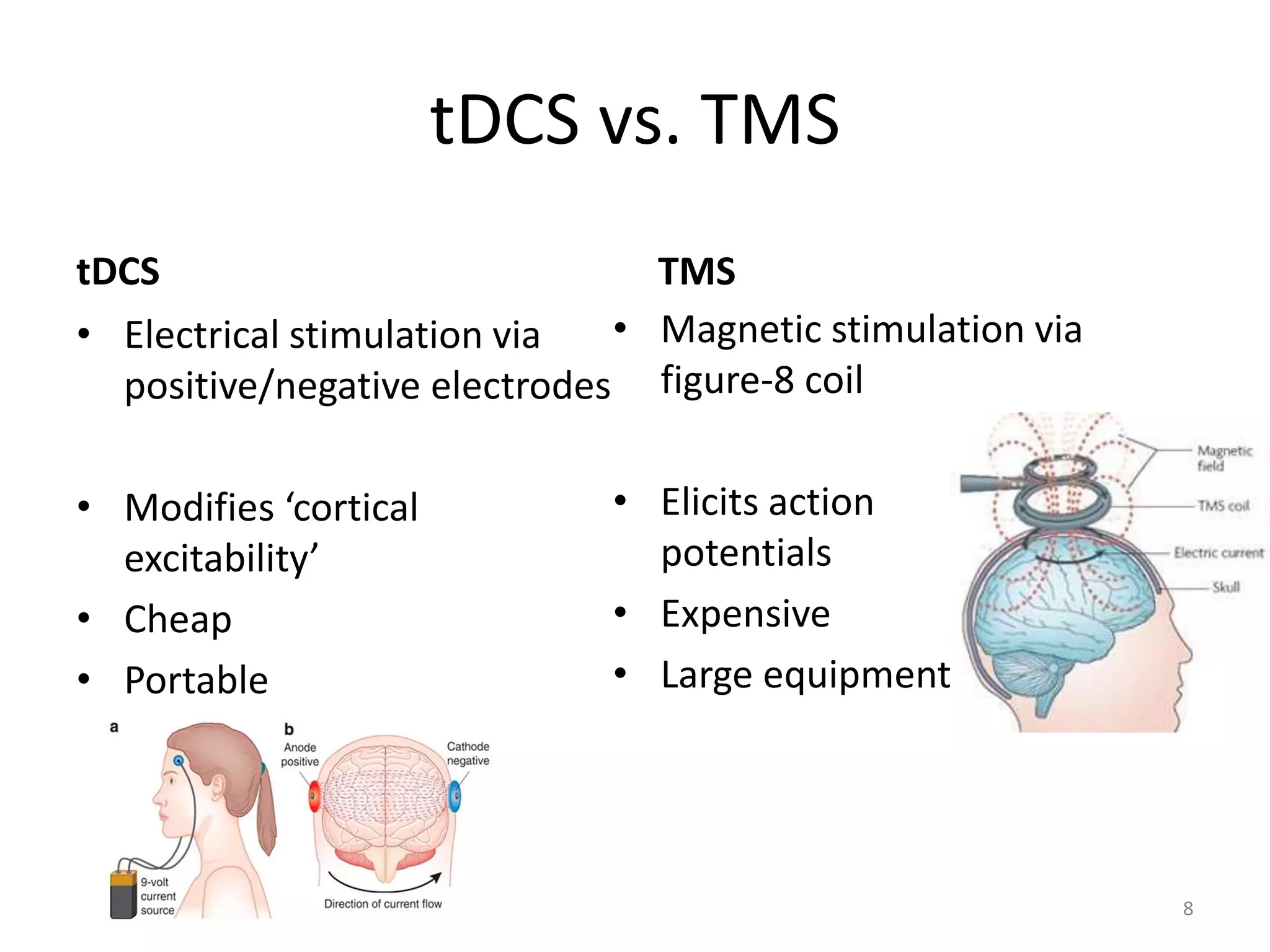
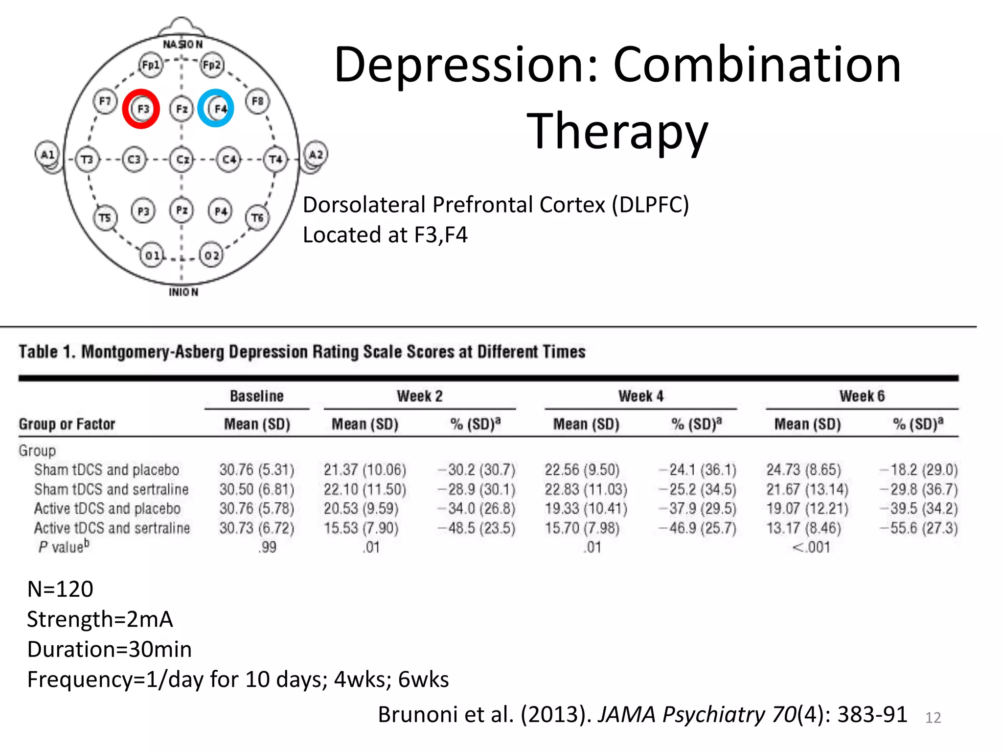
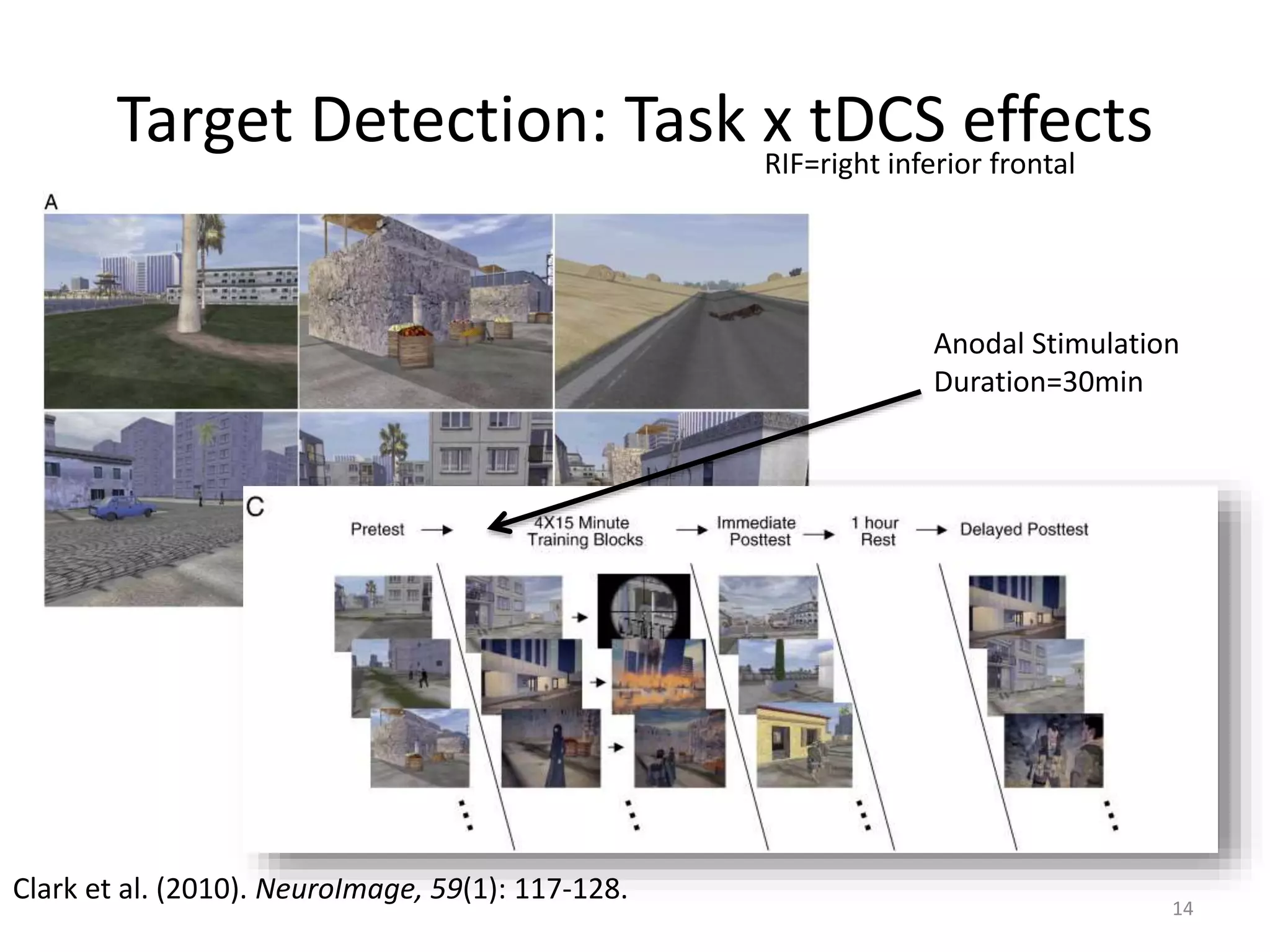
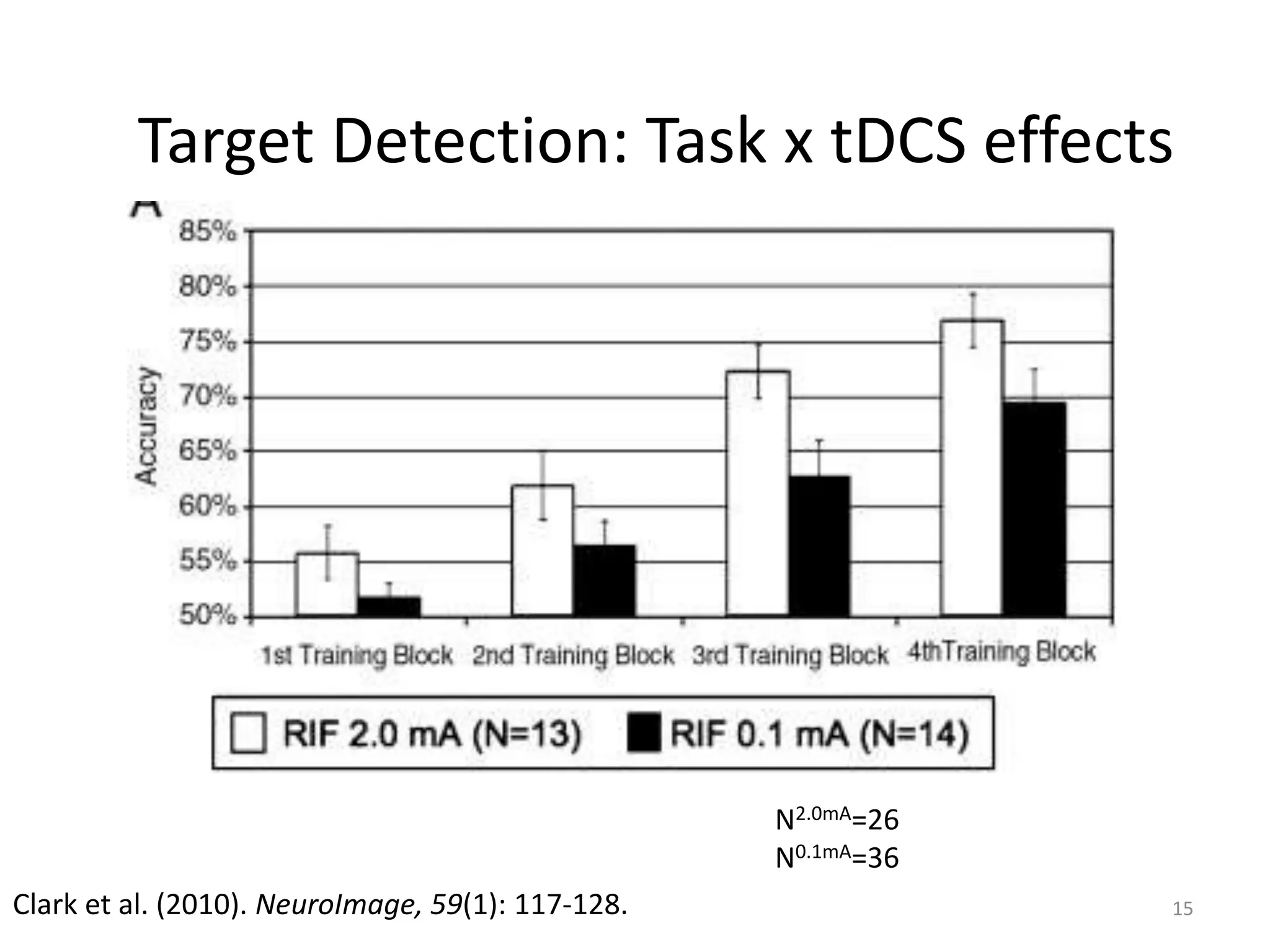
This document summarizes Transcranial Direct Current Stimulation (tDCS). It discusses the history of tDCS, how it works, applications in therapeutic and enhancement contexts, physiological effects and basis, safety considerations, and the future of research. tDCS involves applying a weak electrical current to the brain via electrodes to modulate cortical excitability. It has therapeutic potential for conditions like depression, motor rehabilitation, and is being studied for cognitive enhancement. Research suggests its effects are mediated by changes in neuronal membrane potentials and synaptic plasticity.