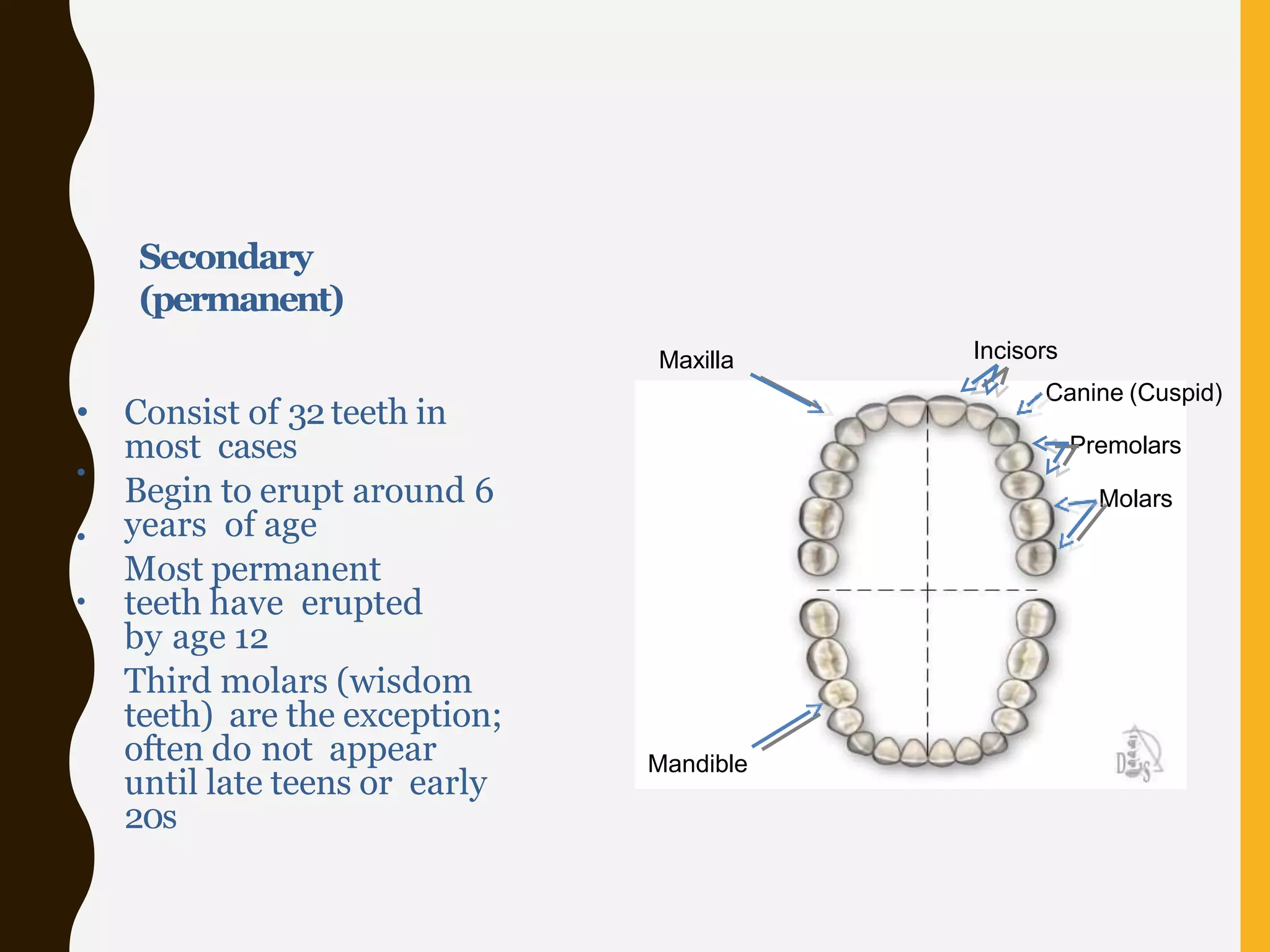
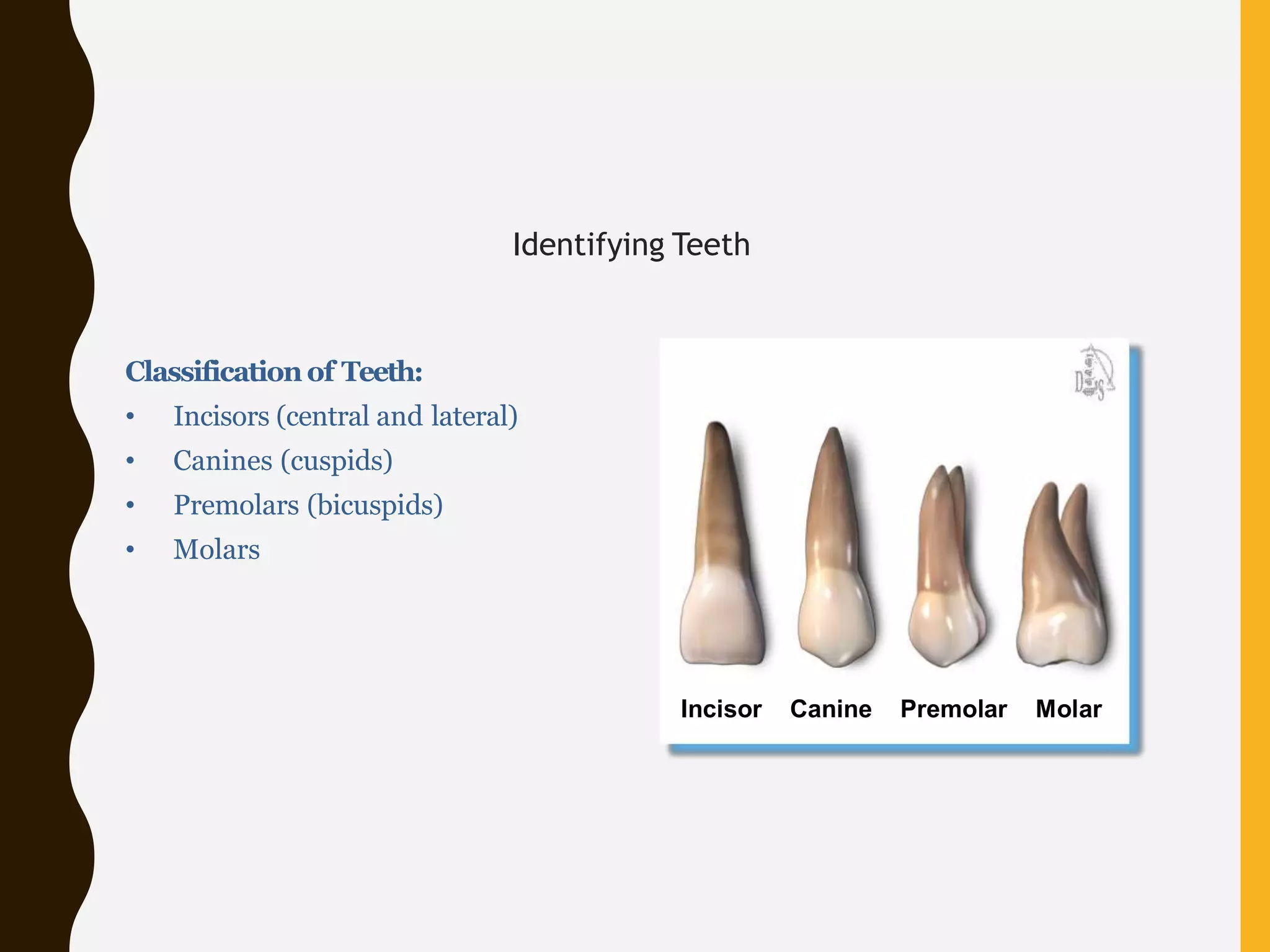
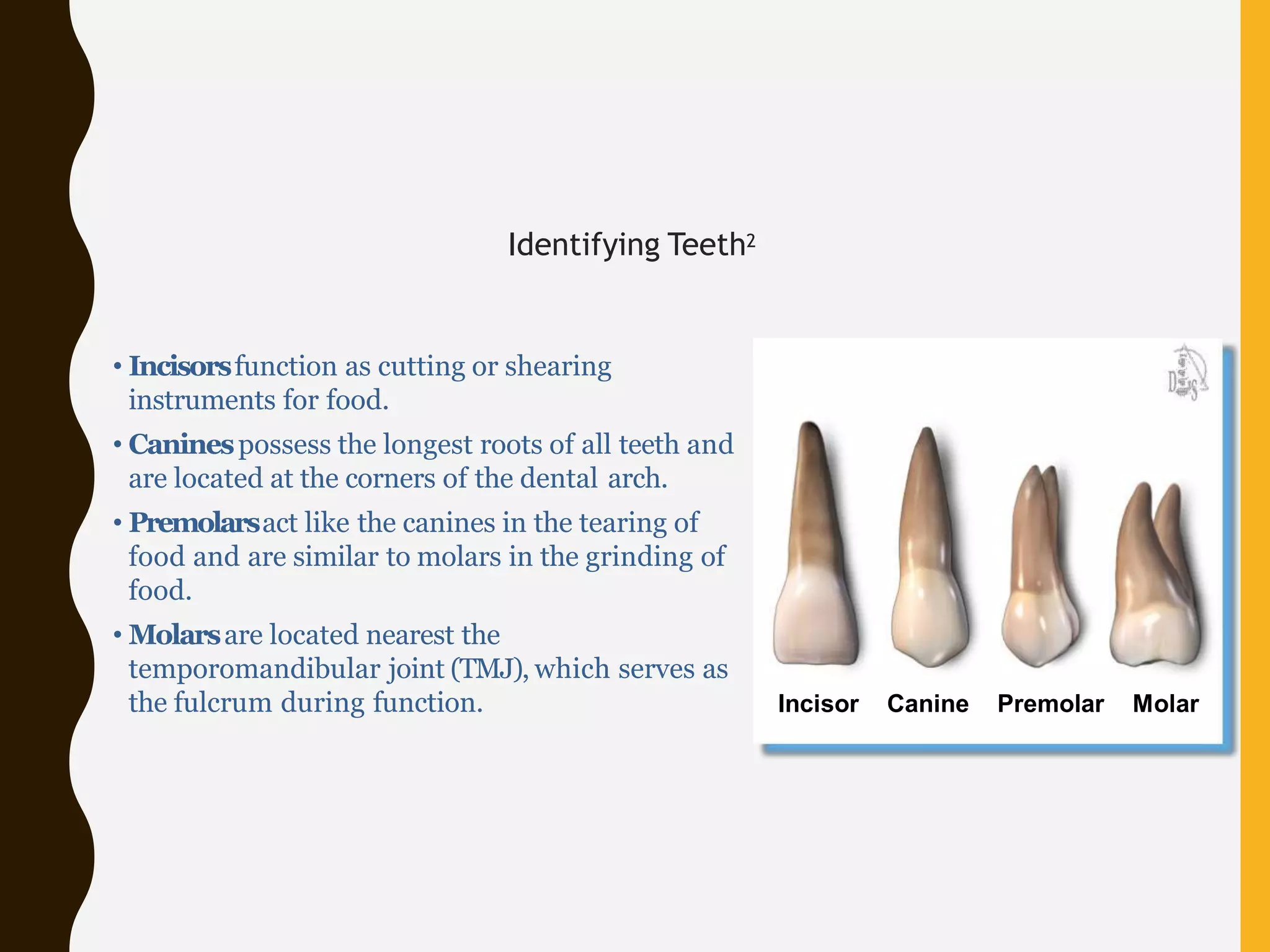
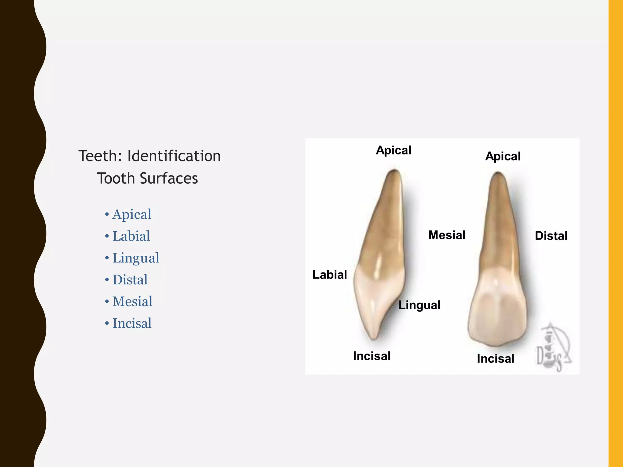
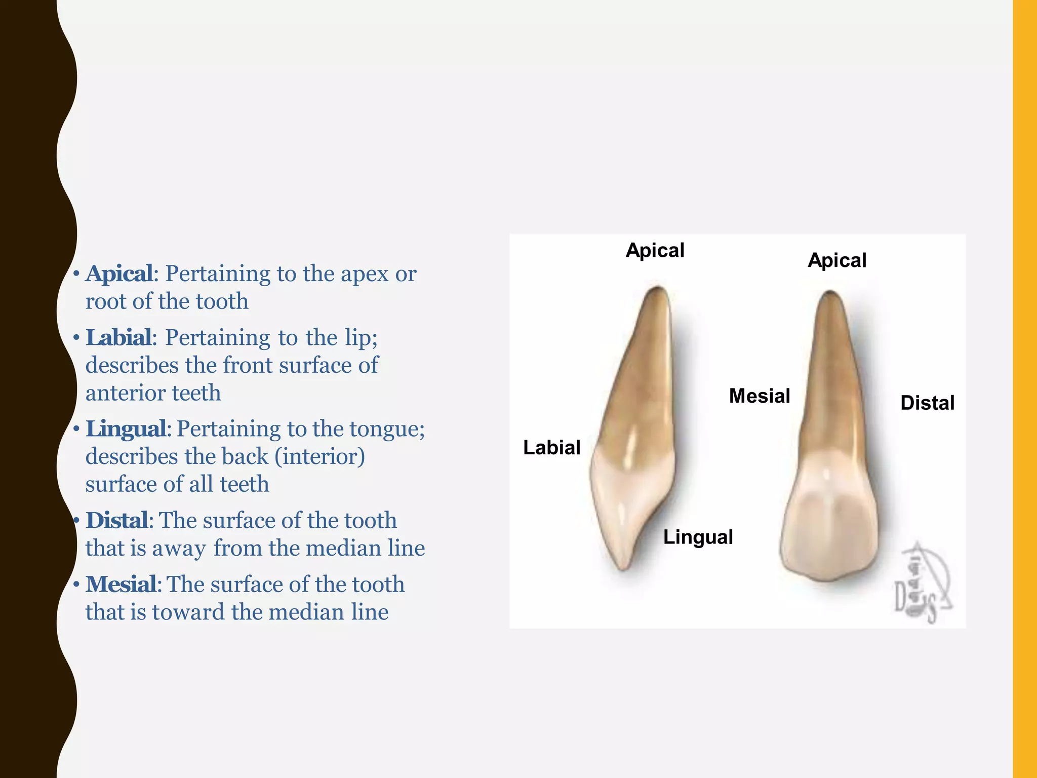
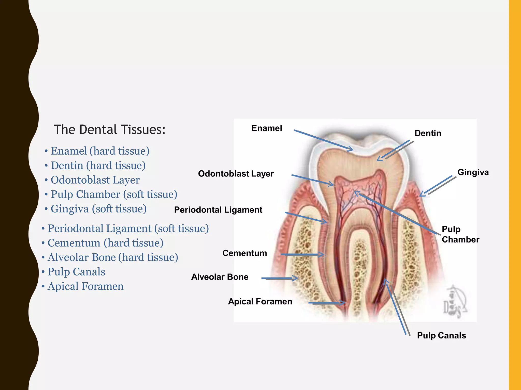
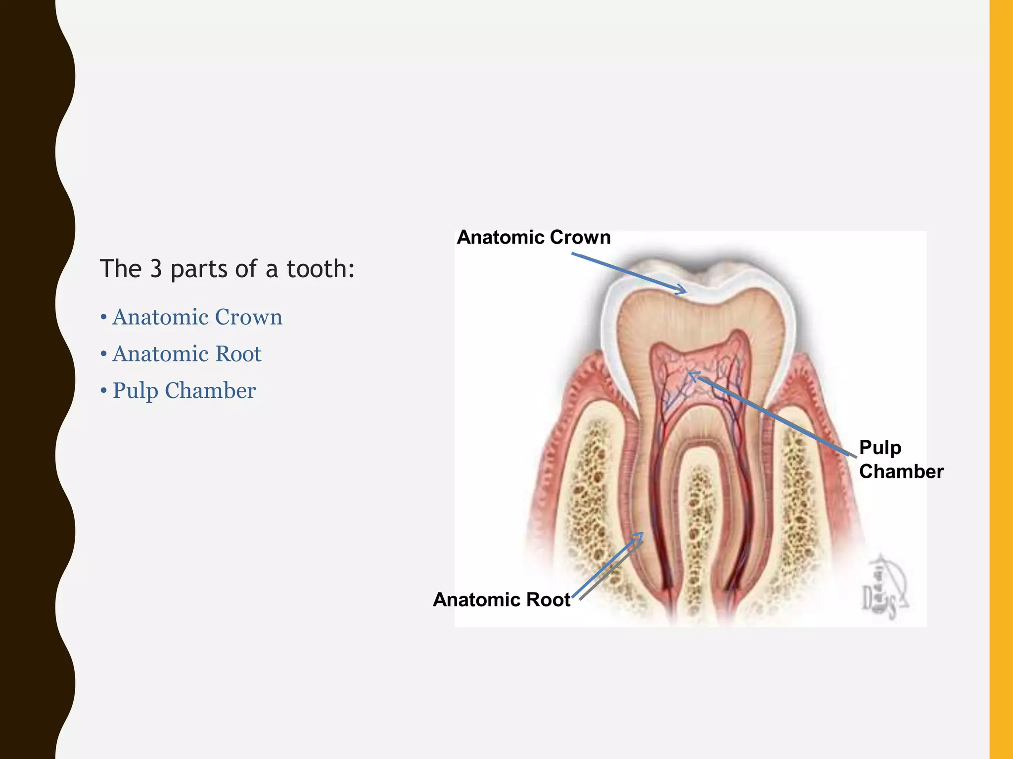
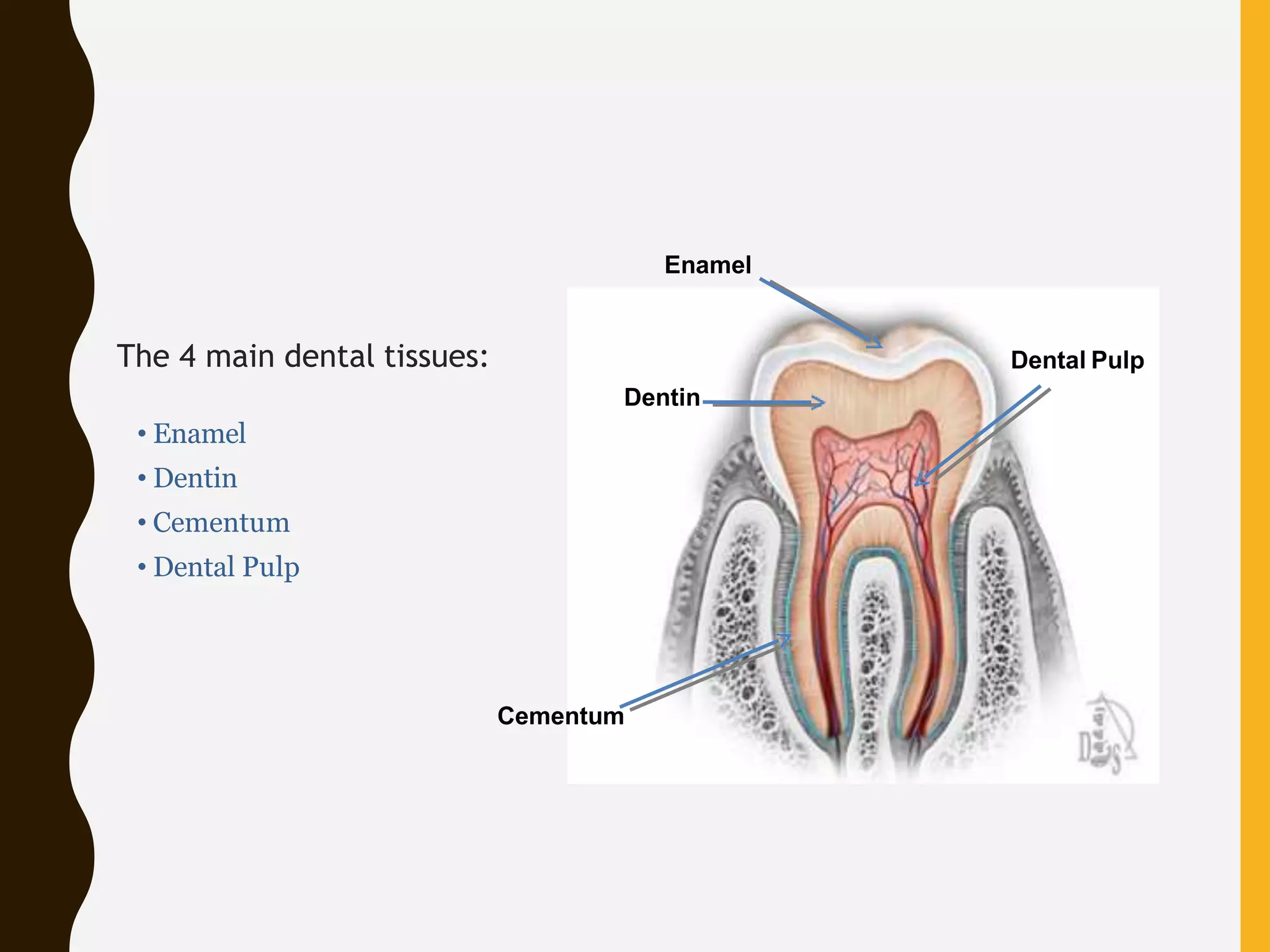
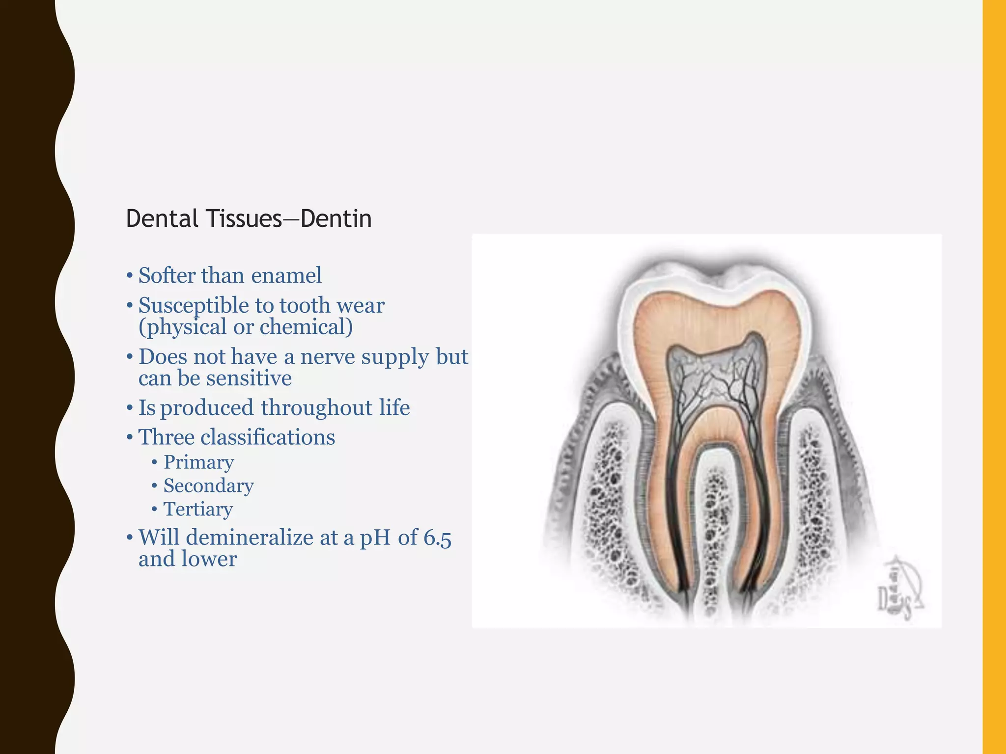
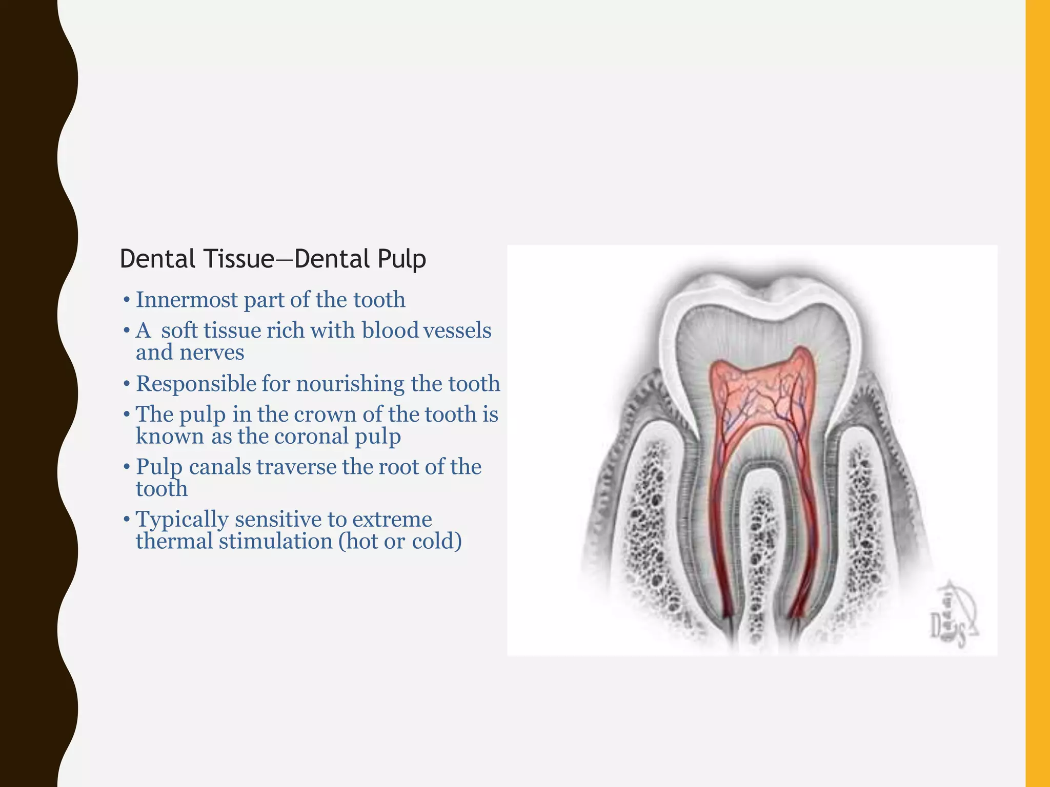
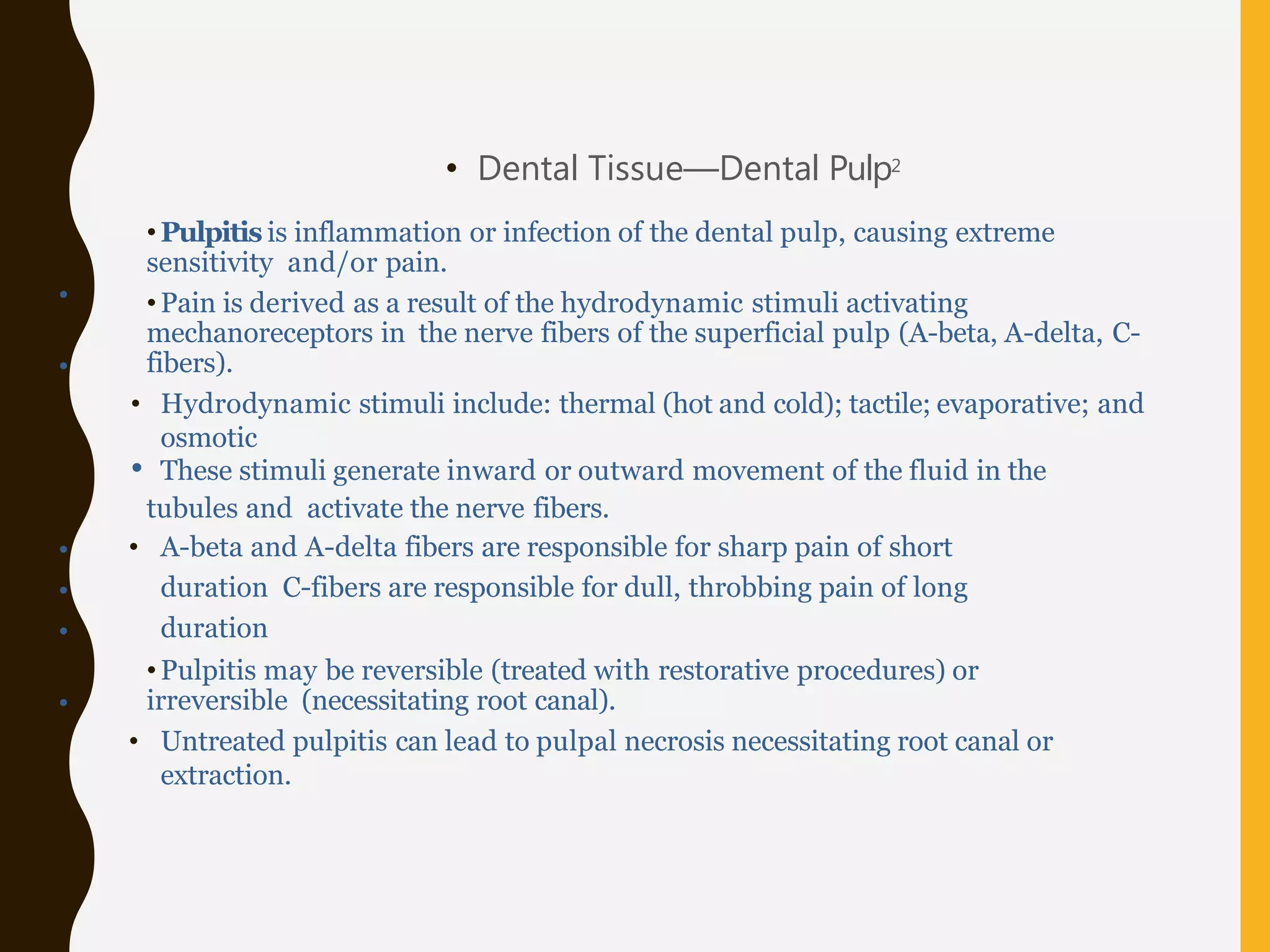
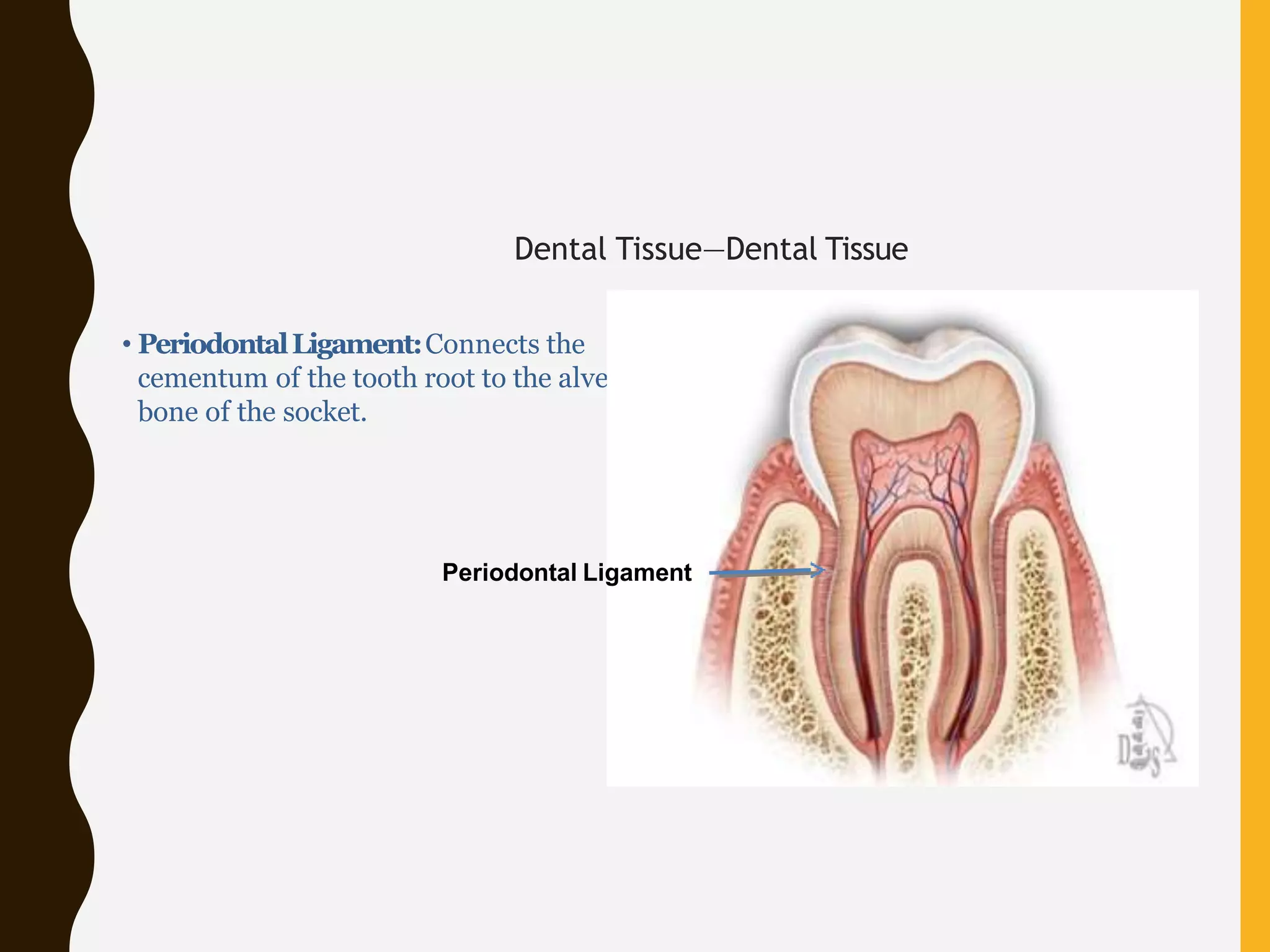
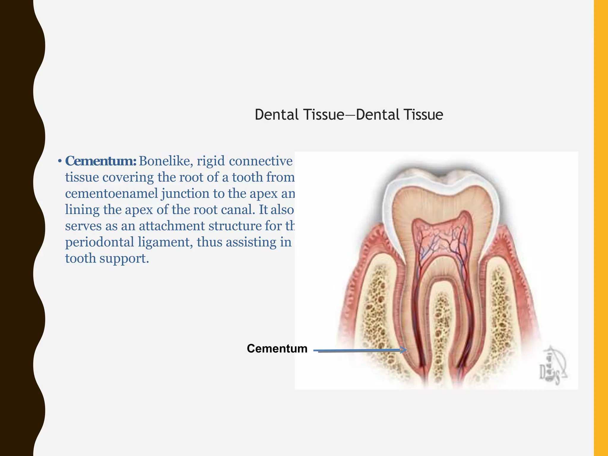
DENTAL ANATOMY&
PHYSIOLOGY
Definition (teeth): There are two
definitions
•Primary (deciduous)
•Secondary (permanent)
Elements
A tooth is made up of three
elements:
•Water
•Organic materials
•Inorganic materials
Primary (deciduous)
•
•
• Consist of 20 teeth
• Begin to form during the
first trimester of
pregnancy
Typically begin
erupting around 6
months
Most children have a
complete primary
dentition by 3years of age
Dentition (teeth): There are two dentitions
Secondary
(permanent)
•
•
•
• Consist of 32 teeth in
most cases
Begin to erupt around 6
years of age
Most permanent
teeth have erupted
by age 12
Third molars (wisdom
teeth) are the exception;
often do not appear
until late teens or early
20s
Mandible
Maxilla Incisors
Canine (Cuspid)
Premolars
Molars
Classification of Teeth:
• Incisors (central and lateral)
• Canines (cuspids)
• Premolars (bicuspids)
• Molars
Identifying Teeth
Incisor Canine Premolar Molar
Identifying Teeth2
Incisor Canine Premolar Molar
• Incisorsfunction as cutting or shearing
instruments for food.
• Caninespossess the longest roots of all teeth and
are located at the corners of the dental arch.
• Premolarsact like the canines in the tearing of
food and are similar to molars in the grinding of
food.
• Molarsare located nearest the
temporomandibular joint (TMJ), which serves as
the fulcrum during function.
• Apical
• Labial
• Lingual
• Distal
• Mesial
• Incisal
Teeth: Identification
Tooth Surfaces
Labial
Apical
Lingual
Distal
Apical
Mesial
Incisal Incisal
• Apical: Pertaining to the apex or
root of the tooth
• Labial: Pertaining to the lip;
describes the front surface of
anterior teeth
• Lingual:Pertaining to the tongue;
describes the back (interior)
surface of all teeth
• Distal: The surface of the tooth
that is away from the median line
• Mesial:The surface of the tooth
that is toward the median line
Labial
Apical
Lingual
Distal
Apical
Mesial
Enamel
Alveolar Bone
Pulp
Chamber
• Periodontal Ligament (soft tissue)
• Cementum (hard tissue)
• Alveolar Bone (hard tissue)
• Pulp Canals
• Apical Foramen
The Dental Tissues:
• Enamel (hard tissue)
• Dentin (hard tissue)
• Odontoblast Layer
• Pulp Chamber (soft tissue)
• Gingiva (soft tissue)
Dentin
Odontoblast Layer Gingiva
Periodontal Ligament
Cementum
Pulp Canals
Apical Foramen
Anatomic Crown
The 3 parts of a tooth:
• Anatomic Crown
• Anatomic Root
• Pulp Chamber
Anatomic Root
Pulp
Chamber
Anatomic Crown
Anatomic Root
Pulp
Chamber
• The anatomiccrownis the
portion of the tooth covered by
enamel.
• The anatomicrootis the lower
two thirds of a tooth.
• The pulp chamberhouses the
dental pulp, an organ of
myelinated and unmyelinated
nerves, arteries, veins, lymph
channels, connective tissue cells,
and various other cells.
• Enamel
• Dentin
• Cementum
• Dental Pulp
The 4 main dental tissues:
Enamel
Dentin
Cementum
Dental Pulp
Dental Tissues—Enamel
• Structure
• Highly calcified and hardest tissue
in the body
• Crystalline in nature
• Enamel rods
• Insensitive—no nerves
• Acid-soluble—will demineralize at a
pH of 5.5 and lower
• Cannot be renewed
• Darkens with age as enamel is lost
• Fluoride and saliva can help with
remineralization
Dental Tissues—Enamel
• Enamel can be lost by:
– Physicalmechanism
• Abrasion (mechanical wear)
• Attrition (tooth-to-tooth contact)
• Abfraction (lesions)
– Chemical dissolution
• Erosion by extrinsic acids (from diet)
• Erosion by intrinsic acids (from the oral
cavity/digestive tract)
• Multifactorial etiology
–Combination of physical and chemical
factors
Dental Tissues—Dentin
• Softer than enamel
• Susceptible to tooth wear
(physical or chemical)
• Does not have a nerve supply but
can be sensitive
• Is produced throughout life
• Three classifications
• Primary
• Secondary
• Tertiary
• Will demineralize at a pH of 6.5
and lower
•
• Primarydentinforms the initial shape of the tooth.
• Secondarydentinis deposited after the formation of the primary dentin on all internal
aspects of the pulp cavity.
Tertiarydentin,or “reparative dentin” is formed by replacement odontoblasts in
response to moderate-level irritants such as attrition, abrasion, erosion, trauma,
moderate-rate dental caries, and some operative procedures.
Dental Tissues—Dentin
Three classifications:
Dental Tissue—Cementum
• Thin layer of mineralized tissue
covering the dentin
• Softer than enamel and dentin
• Anchors the tooth to the alveolar
bone along with the periodontal
ligament
• Not sensitive
Dental Tissue—Dental Pulp
• Innermost part of the tooth
• A soft tissue rich with blood vessels
and nerves
• Responsible for nourishing the tooth
• The pulp in the crown of the tooth is
known as the coronal pulp
• Pulp canals traverse the root of the
tooth
• Typically sensitive to extreme
thermal stimulation (hot or cold)
•
•
•
•
•
•
• Dental Tissue—Dental Pulp2
•Pulpitis is inflammation or infection of the dental pulp, causing extreme
sensitivity and/or pain.
•Pain is derived as a result of the hydrodynamic stimuli activating
mechanoreceptors in the nerve fibers of the superficial pulp (A-beta, A-delta, C-
fibers).
• Hydrodynamic stimuli include: thermal (hot and cold); tactile; evaporative; and
osmotic
• These stimuli generate inward or outward movement of the fluid in the
tubules and activate the nerve fibers.
• A-beta and A-delta fibers are responsible for sharp pain of short
duration C-fibers are responsible for dull, throbbing pain of long
duration
•Pulpitis may be reversible (treated with restorative procedures) or
irreversible (necessitating root canal).
• Untreated pulpitis can lead to pulpal necrosis necessitating root canal or
extraction.
Periodontal Tissues
• Gingiva
• Alveolar Bone
• Periodontal Ligament
• Cementum
Gingiva
Alveolar bone
Cementum
Periodontal Ligament
Dental Tissue—Dental Tissue
• Gingiva: The part of the oral mucosa
overlying the crowns of unerupted teeth
and encircling the necks of erupted teeth,
serving as support structure for
subadjacent tissues.
Gingiva
Dental Tissue—Dental Tissue
• Alveolar Bone:Also called the “alveolar
process”; the thickened ridge of bone
containing the tooth sockets in the
mandible and maxilla.
Alveolar bone
Dental Tissue—Dental Tissue
• PeriodontalLigament:Connects the
cementum of the tooth root to the alveolar
bone of the socket.
Periodontal Ligament
Dental Tissue—Dental Tissue
• Cementum:Bonelike, rigid connective
tissue covering the root of a tooth from the
cementoenamel junction to the apex and
lining the apex of the root canal. It also
serves as an attachment structure for the
periodontal ligament, thus assisting in
tooth support.
Cementum
Oral Cavity/Environment
• Plaque
• Saliva
• pH Values
• Demineralization
• Remineralization
•
•
• is a biofilm
• contains more than 600different
identified species of bacteria
there is harmless and harmful
plaque
salivary pellicle allows the bacteria
to adhere to the tooth surface,
which begins the formation of
plaque
Oral Cavity
Plaque:
•
•
complex mixture of fluids
performs protective functions:
– lubrication—aids swallowing
– mastication
– key role in remineralization
of enamel and dentin
– buffering
Oral Cavity
Saliva:
• mineral salts dissolve into the
surrounding salivary fluid:
•
– enamel at approximate pH of 5.5
or lower
– dentin at approximate pH of 6.5
or lower
erosion or caries can occur
Oral Cavity
Demineralization:7
•
•
•
pH comes back to neutral (7)
saliva-rich calcium and
phosphates
minerals penetrate the damaged
enamel surface and repair it:
– enamel pH is above 5.5
– dentin pH is above 6.5
Oral Cavity
Remineralization:
Intro to dental anatomy

Intro to dental anatomy

  • 1.
  • 2.
    Definition (teeth): Thereare two definitions •Primary (deciduous) •Secondary (permanent)
  • 3.
    Elements A tooth ismade up of three elements: •Water •Organic materials •Inorganic materials
  • 4.
    Primary (deciduous) • • • Consistof 20 teeth • Begin to form during the first trimester of pregnancy Typically begin erupting around 6 months Most children have a complete primary dentition by 3years of age Dentition (teeth): There are two dentitions
  • 5.
    Secondary (permanent) • • • • Consist of32 teeth in most cases Begin to erupt around 6 years of age Most permanent teeth have erupted by age 12 Third molars (wisdom teeth) are the exception; often do not appear until late teens or early 20s Mandible Maxilla Incisors Canine (Cuspid) Premolars Molars
  • 6.
    Classification of Teeth: •Incisors (central and lateral) • Canines (cuspids) • Premolars (bicuspids) • Molars Identifying Teeth Incisor Canine Premolar Molar
  • 7.
    Identifying Teeth2 Incisor CaninePremolar Molar • Incisorsfunction as cutting or shearing instruments for food. • Caninespossess the longest roots of all teeth and are located at the corners of the dental arch. • Premolarsact like the canines in the tearing of food and are similar to molars in the grinding of food. • Molarsare located nearest the temporomandibular joint (TMJ), which serves as the fulcrum during function.
  • 8.
    • Apical • Labial •Lingual • Distal • Mesial • Incisal Teeth: Identification Tooth Surfaces Labial Apical Lingual Distal Apical Mesial Incisal Incisal
  • 9.
    • Apical: Pertainingto the apex or root of the tooth • Labial: Pertaining to the lip; describes the front surface of anterior teeth • Lingual:Pertaining to the tongue; describes the back (interior) surface of all teeth • Distal: The surface of the tooth that is away from the median line • Mesial:The surface of the tooth that is toward the median line Labial Apical Lingual Distal Apical Mesial
  • 10.
    Enamel Alveolar Bone Pulp Chamber • PeriodontalLigament (soft tissue) • Cementum (hard tissue) • Alveolar Bone (hard tissue) • Pulp Canals • Apical Foramen The Dental Tissues: • Enamel (hard tissue) • Dentin (hard tissue) • Odontoblast Layer • Pulp Chamber (soft tissue) • Gingiva (soft tissue) Dentin Odontoblast Layer Gingiva Periodontal Ligament Cementum Pulp Canals Apical Foramen
  • 11.
    Anatomic Crown The 3parts of a tooth: • Anatomic Crown • Anatomic Root • Pulp Chamber Anatomic Root Pulp Chamber
  • 12.
    Anatomic Crown Anatomic Root Pulp Chamber •The anatomiccrownis the portion of the tooth covered by enamel. • The anatomicrootis the lower two thirds of a tooth. • The pulp chamberhouses the dental pulp, an organ of myelinated and unmyelinated nerves, arteries, veins, lymph channels, connective tissue cells, and various other cells.
  • 13.
    • Enamel • Dentin •Cementum • Dental Pulp The 4 main dental tissues: Enamel Dentin Cementum Dental Pulp
  • 14.
    Dental Tissues—Enamel • Structure •Highly calcified and hardest tissue in the body • Crystalline in nature • Enamel rods • Insensitive—no nerves • Acid-soluble—will demineralize at a pH of 5.5 and lower • Cannot be renewed • Darkens with age as enamel is lost • Fluoride and saliva can help with remineralization
  • 15.
    Dental Tissues—Enamel • Enamelcan be lost by: – Physicalmechanism • Abrasion (mechanical wear) • Attrition (tooth-to-tooth contact) • Abfraction (lesions) – Chemical dissolution • Erosion by extrinsic acids (from diet) • Erosion by intrinsic acids (from the oral cavity/digestive tract) • Multifactorial etiology –Combination of physical and chemical factors
  • 16.
    Dental Tissues—Dentin • Softerthan enamel • Susceptible to tooth wear (physical or chemical) • Does not have a nerve supply but can be sensitive • Is produced throughout life • Three classifications • Primary • Secondary • Tertiary • Will demineralize at a pH of 6.5 and lower
  • 17.
    • • Primarydentinforms theinitial shape of the tooth. • Secondarydentinis deposited after the formation of the primary dentin on all internal aspects of the pulp cavity. Tertiarydentin,or “reparative dentin” is formed by replacement odontoblasts in response to moderate-level irritants such as attrition, abrasion, erosion, trauma, moderate-rate dental caries, and some operative procedures. Dental Tissues—Dentin Three classifications:
  • 18.
    Dental Tissue—Cementum • Thinlayer of mineralized tissue covering the dentin • Softer than enamel and dentin • Anchors the tooth to the alveolar bone along with the periodontal ligament • Not sensitive
  • 19.
    Dental Tissue—Dental Pulp •Innermost part of the tooth • A soft tissue rich with blood vessels and nerves • Responsible for nourishing the tooth • The pulp in the crown of the tooth is known as the coronal pulp • Pulp canals traverse the root of the tooth • Typically sensitive to extreme thermal stimulation (hot or cold)
  • 20.
    • • • • • • • Dental Tissue—DentalPulp2 •Pulpitis is inflammation or infection of the dental pulp, causing extreme sensitivity and/or pain. •Pain is derived as a result of the hydrodynamic stimuli activating mechanoreceptors in the nerve fibers of the superficial pulp (A-beta, A-delta, C- fibers). • Hydrodynamic stimuli include: thermal (hot and cold); tactile; evaporative; and osmotic • These stimuli generate inward or outward movement of the fluid in the tubules and activate the nerve fibers. • A-beta and A-delta fibers are responsible for sharp pain of short duration C-fibers are responsible for dull, throbbing pain of long duration •Pulpitis may be reversible (treated with restorative procedures) or irreversible (necessitating root canal). • Untreated pulpitis can lead to pulpal necrosis necessitating root canal or extraction.
  • 21.
    Periodontal Tissues • Gingiva •Alveolar Bone • Periodontal Ligament • Cementum Gingiva Alveolar bone Cementum Periodontal Ligament
  • 22.
    Dental Tissue—Dental Tissue •Gingiva: The part of the oral mucosa overlying the crowns of unerupted teeth and encircling the necks of erupted teeth, serving as support structure for subadjacent tissues. Gingiva
  • 23.
    Dental Tissue—Dental Tissue •Alveolar Bone:Also called the “alveolar process”; the thickened ridge of bone containing the tooth sockets in the mandible and maxilla. Alveolar bone
  • 24.
    Dental Tissue—Dental Tissue •PeriodontalLigament:Connects the cementum of the tooth root to the alveolar bone of the socket. Periodontal Ligament
  • 25.
    Dental Tissue—Dental Tissue •Cementum:Bonelike, rigid connective tissue covering the root of a tooth from the cementoenamel junction to the apex and lining the apex of the root canal. It also serves as an attachment structure for the periodontal ligament, thus assisting in tooth support. Cementum
  • 26.
    Oral Cavity/Environment • Plaque •Saliva • pH Values • Demineralization • Remineralization
  • 27.
    • • • is abiofilm • contains more than 600different identified species of bacteria there is harmless and harmful plaque salivary pellicle allows the bacteria to adhere to the tooth surface, which begins the formation of plaque Oral Cavity Plaque:
  • 28.
    • • complex mixture offluids performs protective functions: – lubrication—aids swallowing – mastication – key role in remineralization of enamel and dentin – buffering Oral Cavity Saliva:
  • 29.
    • mineral saltsdissolve into the surrounding salivary fluid: • – enamel at approximate pH of 5.5 or lower – dentin at approximate pH of 6.5 or lower erosion or caries can occur Oral Cavity Demineralization:7
  • 30.
    • • • pH comes backto neutral (7) saliva-rich calcium and phosphates minerals penetrate the damaged enamel surface and repair it: – enamel pH is above 5.5 – dentin pH is above 6.5 Oral Cavity Remineralization: