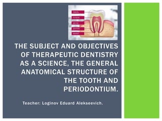
SlideShare a Scribd company logo
1 of 42
Teacher: Loginov Eduard Alekseevich.
THE SUBJECT AND OBJECTIVES
OF THERAPEUTIC DENTISTRY
AS A SCIENCE, THE GENERAL
ANATOMICAL STRUCTURE OF
THE TOOTH AND
PERIODONTIUM.
 Therapeutic dentistry is a science that studies the features of
diagnosis, clinic, treatment, prevention and rehabilitation of
patients with diseases of the hard tissues of the teeth,
periodontal, and oral mucosa.
 The purpose of therapeutic dentistry:
Effective prevention, diagnosis and conservative treatment of
diseases of the teeth, periodontal and oral mucosa.
The subject of therapeutic dentistry: teeth, periodontal and oral
mucosa and other organs of the oral cavity.
THERAPEUTIC DENTISTRY
 Prevention and treatment of
caries and its complications.
 Prevention and treatment of
non-carious lesions.
 Treatment and prevention of
periodontal diseases.
 Prevention and treatment of
diseases of the periodontal and
oral mucosa.
 Dispensary examination of
dental patients.
THE OBJECTIVES OF THERAPEUTIC
DENTISTRY
in-depth study of the etiology,
pathogenesis and clinic of the most
common dental diseases;
study of diagnostic methods for the
most important clinical syndromes in
major dental diseases;
mastering the basic practical skills
necessary for the examination and
treatment of dental patients of various
ages;
mastering the basic principles of
treatment and prevention of dental
diseases;
to form the methodological and
methodological foundations of clinical
thinking and rational action of the
doctor.
DISCIPLINE OBJECTIVES
 The teeth are part of the human chewing-speech apparatus
and are solid formations located in the oral cavity and
performing the function of biting off, holding and grinding
food. They are also involved in the formation of the face and
the pronunciation of some sounds.
TEETH (DENTIS)
Clinical crown height - from the free
edge of the gums to the incisal edge
or occlusal surface of the tooth.
Clinical Root Length - starts at the
clinical crown and ends at the root
apex.
The teeth are fixed in the dental
alveoli by the dental ligament -
periodont.
ANATOMICAL STRUCTURE OF THE
TOOTH(DENS)
Crown
Tooth neck
Root
 Anatomical crown - the part of the tooth that is covered with
enamel (1)
 Clinical crown - the part of the tooth that is visible in the
mouth and protrudes above the gum (2)
CROWN OF THE TOOTH
TOOTH NECK
The anatomical
formation, which is the
place of transition of the
crown to the root of the
tooth, corresponds to the
enamel-cement border.
 Tooth root (radix dentis) - is
attached to the walls of the
alveoli by bundles of
collagen fibers surrounding
the tooth root and located
between the cement and
the walls of the alveoli.
 This connective tissue is
called the periodontium, it
is similar to the periosteum
and forms the
articulationes
dentoaveolares.
TOOTH ROOT
 The dentogingival fibers (fibrae
dentogingivales) go from the cement of
the root to the connective tissue of the
gums.
 Interdental fibers (fibrae interdentales)
go from the cement of the tooth through
the interdental septum to the cement of
the adjacent tooth.
 Cemento-alveolar fibers (fibrae
cementoalveolares) go from the cement
to the walls of the dental alveoli.
The totality of the formations
surrounding the root of the tooth,
including the gum, periodontium, bone
tissue of the dental alveoli, the
corresponding section of the alveolar
process and cement form the
periodontium (parodontium), which is
the supporting-retaining apparatus of
the tooth.
PERIODONTAL STRUCTURE
Periodontium -
located between
the cement of the
root and the bone
tissue of the
alveoli, contains
blood, lymph
vessels and nerve
fibers.
Periosteum
covering the
alveolar
process
Root cement
Gum
Alveolar bone
tissue
Bone tissue of
the tooth socket
PERIODONTAL STRUCTURE. GUM
1 – marginal gingiva
2 - gingival groove
3 - attached gingiva
4 - gingival groove
The mucous membrane covering the alveolar ridge
of the jaw and the neck of the tooth, which is
tightly attached to them (attached gum). The
marginal part of the gum is freely located at the
neck of the tooth and has no attachment to it
(unattached gum).
GUM
The space formed by the tooth and
unattached gums.
Gingival sulcus
A recess located at the transition of the
free gum to the attached one.
Gingival
groove
Gingival
papilla
it is the part of the gum that fills the
interdental space.
Support-holding Shock-absorbing
Pressure
distribution
Uniting teeth
into a dentition.
Sensory (tactile,
pain perception,
pressure)
Reflex Plastic Trophic
Barrier
Adaptation to
functional and
topographic
changes
Promote
physiological
changes in the
tooth
Ability to repair
tissue after
traumatic injury
Participation in
growth, teething,
changing teeth
Renewal of
periodontal
tissues
FUNCTIONS OF PERIODONT
TOOTH TISSUE
in structure it is similar to coarse-fibrous bone, differs in the absence of
cells and greater hardness. It is represented by the processes of
odontoblasts, cells that are located in the peripheral parts of the dental
pulp. There are numerous dentinal tubules (tubuli dentinales), in which
the dentinal processes of odontoblasts are located. There is an outer
and an inner (is a zone of constant growth) layer of dentin.
formed by enamel prisms (prismae enameli), which have a
polygonal shape, run radially relative to the longitudinal axis of
the tooth.
formed by a basic substance impregnated with salts and
containing collagen fibers. Cementocytes are located in the area
of the apex of the tooth, interroot sections in special cavities.
Dentine(dentin
um)
Cement
(cementum)
Enamel
(enamelum)-
 Hard mineralized tissue that covers the outside of
the crown of the tooth and protects the dentin and
pulp from external stimuli. The thickness of the
enamel layer is maximum in the area of the
tubercles and minimum in the area of the neck.
Enamel is the hardest tissue in the human body.
Consists of inorganic (96 - 99%) and only 1 - 4% -
of organic substances (proteins and water).
 The main structural formation of enamel is an
enamel prism with a diameter of 4 - 6 microns.
The number of prisms is several million. Enamel
prisms, concentrating in beams (10 - 20), form S-
shaped bends. Between the prisms there is an
interprismatic substance, which is 0.5 - 5.0% of
the enamel volume.
 The main structural unit of the prism is
hydroxyapatite crystals - Ca10 (PO4) 6 (OH) 2.
 Enamel functions - protective, trophic (dental
liquor).
ENAMEL(ENAMELUM)
1 - enamel prism
2 - interprism substance
 Calcified tooth tissue, which forms its
bulk and shape. Dentin contains 70%
inorganic (hydroxyapatite), 20% organic
(collagen), 10% water. Dentin consists of
a calcified intercellular substance
(represented by collagen fibers),
penetrated by dentinal tubules (thin
tubules that penetrate dentin from the
pulp to the periphery). Predentin is the
inner (unusual) part of dentin adjacent
to the layer of odontoblasts. Predentin is
the dentin growth zone. Dentin is
subdivided into: Primary - formed before
the eruption of the tooth; Secondary -
formed after the eruption of a tooth;
Tertiary - formed in response to
irritation. Dentin functions: trophic,
sensory, protective.
DENTINE(DENTINIM)
 Calcified tooth tissue. Covers the root and neck
of the tooth. Contains 50-60% inorganic
substances (hydroxyapatite), 30-40% organic
(collagen). The thickness of the cement is not
the same: it is thinner in the area of the neck
(20 - 50 µm) and thicker in the area of the root
apex (100 - 150 µm). Cement is subdivided into
acellular (primary) and cellular (secondary). The
primary cement adheres to the dentin, covering
the lateral surfaces of the root. Secondary
cement covers the apical third of the root and
the bifurcation area of the roots of multi-rooted
teeth. It is located on top of the acellular
cementum, but sometimes directly adjacent to
the dentin. Cellular cement consists of cells
(cementocytes and cementoblasts) and
intercellular substance. Cement functions:
protective, retaining, reparative.
CEMENT
 In the area of the root apex, the canals
end in an apical (apical) foramen. Pulp
(pulpa dentis) - Loose fibrous-connective
tissue with a significant content of
cellular elements. Distinguish between
the pulp of the vessels, the pulp of the
crown (pulpa coronalis) and the pulp of
the root (pulpa radicularis).
TOOTH CAVITY
There is a cavity inside the tooth that follows the
outer contours of the tooth - the cavity of the tooth
(cavitas dentis) or the cavity of the pulp (cavitas
pulparis), which includes: cavity of the crown (cavitas
coronae) root canal (canalis radicis dentis)
FORMS OF CAVITIES OF PERMANENT
TEETH
 Loose connective tissue that fills the tooth
cavity. At the apical foramen, the pulp
gradually passes into the periodontal tissue.
The pulp consists of intercellular substance
and cells. The intercellular substance is
represented by collagen and pre-collagen
fibers (elastic fibers are absent in the pulp)
and the main substance, which has a
gelatinous consistency. There are three cell
layers in the pulp: peripheral, intermediate
and central. The peripheral layer is
represented by highly differentiated cells -
odontoblasts. The intermediate
(subodontoblastic) layer is stellate cells with
processes - pulpocytes. The central layer
consists of star-shaped process cells,
collagen fibers, nerve elements, and blood
vessels.
PULP
PULP FUNCTIONS
protective
function
trophic
function
sensory
function
 Blood supply: upper jaw teeth from the maxillary artery
system, the teeth of the lower jaw - from the lower alveolar
artery, the branches of the lingual, facial and superficial
temporal arteries play only an auxiliary role in the blood
supply to the teeth.
 Venous outflow - from the teeth - in the same name and
enters the pterygoid plexus. The veins of the teeth form
anastomoses with the veins of the orbit, and the pterygoid
plexus with the pharyngeal and vertebral plexuses and
through them with the venous sinuses of the skull.
ARTERIES AND VEINS OF TEETH
 There are 4 surfaces in each
tooth: 1) facing the vestibule of
the mouth, facies vestibularis,
which in the front teeth is in
contact with the mucous
membrane of the lip (facies
labialis), in the back - with the
mucous membrane of the cheek
(facies buccalis); 2) the chewing
surface or the surface of the
occlusion with the teeth of the
opposite row, facies occlusalis;
The incisors and canines at the
ends facing the similar teeth of
the opposite jaw have an incisal
edge (margoincisalis).
TOOTH SURFACES
3) facing into the oral
cavity, to the tongue,
facies lingualis;
4) in contact with adjacent teeth of their row,
facies contactus. Distinguish between the mesial
surface (facies mesialis) - directed towards the
middle of the dental arch, and the distal (facies
distalis) - facing in the direction from the middle of
the dental arch.
TOOTH SURFACES
ELEMENTS OF THE RELIEF OF THE
CROWN OF THE TEETH
 1 - tooth tubercle,
 2 - elements of the edge
of the tooth,
 3 - edge comb,
 4 - transverse scallop,
 5 - top of the tip,
 6 - belt,
 7 - occlusal gap,
 8 - occlusal fossa,
 9 - roller,
 10 - triangular scallops
ELEMENTS OF THE RELIEF OF THE
CROWN OF THE TEETH
 Teeth are grouped according to morphological and functional
characteristics.
TEETH GROUPS
By the shape of the crown and the function of the teeth
incisors canine
Premolars
(permanent
teeth)
Molars (large
molars)
Teeth are distinguished by the number of roots
single-rooted two-root three-root
By position in the dental arch
front teeth
(incisors)
angled teeth
(canines)
posterior teeth
(premolars and
molars)
TEETH GROUPS
Incisors (dens incisivus)
•front teeth, 4 on each jaw. Their function is to cut and bite off food.
Canine (dens caninus)
•2 on each jaw, serve to tear off food
Premolars (dens premolaris)
•4 on each jaw in a permanent bite, in the milk bite they are not. Serve for crushing,
coarse grinding of food
Molars (dens molaris)
•6 teeth on each jaw in a permanent bite and 4 in a milk bite. Designed for chopping
and grinding food
ROOT NAMES OF MULTI-ROOTED TEETH
1 - buccal
root
2 - palatine
root
3 - buccal-
distal root
4 - buccal-
medial root
5 - distal
root
6 - medial
root
 They allow you to determine the belonging of the tooth to the
upper or lower jaw and the side of the jaw (right or left). There
are three main features:
SIGNS OF TOOTH BELONGING
Crown angle
sign
Crown
curvature
sign
Root
deviation
sign
CROWN ANGLE SIGN
The angle of the tooth crown formed by the
medial surface and the occlusion surface
(chewing surface or incisal edge) is less
than the angle formed by the distal surface
and closing surface. The sign is
determined when viewed from the
vestibular side.
CROWN CURVATURE SIGN
On the vestibular surface, the medial part of the crown is more convex
than the lateral one. The sign is determined when viewed from the side
of the closure.
ROOT DEVIATION SIGN
Curvature of the entire root or its apex in the distal direction (in relation to the
longitudinal axis of the tooth). The sign is determined by examining the tooth
from the vestibular or oral sides.
 The genetically determined sequence of
the location of each tooth and groups of
teeth is called the dentition. The dentition
in its natural position, located according
to the curvature of the jaw, is called the
dental arch. Distinguish: upper dental
arch, arcus dentalis superior and lower
dental arch, arcus dentalis inferior. Each
complete arch (arch) contains 10
deciduous teeth or 16 permanent teeth.
DENTAL ARCH (DENTAL ROW)
FIELDS OF TEETH CONTACT
 The upper dentition has the shape of a semi-ellipse,
the lower one is in the shape of a parabola. In
addition to the dental arch, the alveolar and basal
(apical) arch.
 The alveolar arch is a line drawn along the crest of
the alveolar ridge.
 The basal arch runs along the apex of the roots. Since
in the upper jaw, the crowns of the teeth are inclined
outward, and the roots - inward, the dental arch of the
upper jaw is wider than the basal one. Accordingly, on
the lower jaw, the opposite is true. For this reason,
with complete loss of teeth, the lower jaw protrudes
forward.
ALVEOLAR ARCH, DENTAL ARCH AND
BASAL ARCH
TYPES OF BITE
 There are temporary, removable and permanent bites.
Temporary bite is
represented by 20 teeth
There are temporary
and permanent teeth in
the mixed dentition
Permanent bite
includes 32
permanent teeth
 Research in recent years shows that both dental and somatic diseases
have common risk factors. A number of studies note that various factors
such as social (stress), behavioral (smoking, psychological personality
traits), environmental factors significantly affect the development of
dental and periodontal diseases, as well as the development of such
chronic non-infectious diseases as chronic obstructive pulmonary disease. ,
arterial hypertension, ischemic heart disease. A disease such as diabetes
mellitus is characterized by severe metabolic disorders, which gradually
lead to damage to all organs, forming the polymorbid status of patients.
Periodontitis (inflammation of the tissues surrounding the tooth) is a
characteristic early manifestation of diabetes mellitus. In almost 100% of
cases, patients with diabetes mellitus have problems with periodontal
tissues of varying severity, which are distinguished by a vivid clinical
picture and an aggressive course. The relationship between periodontal
diseases and human cardiovascular pathology has also been proven. It was
found that microbial agents that are on the tooth plaque and are in the
periodontal pocket are also present in the affected coronary vessels, which
lead to extensive heart attacks. Therefore, dental treatment is important
not only in order to look beautiful and chew food correctly, but also to be
healthy. Timely visits to the dentist, regular check-ups and quality
treatment will help maintain the health of the gums and teeth and the
body as a whole!
THE RELATIONSHIP OF SOMATIC AND
DENTAL DISEASES
 Dental illnesses can lead to general illness. The carious
process is accompanied by the development of bacterial flora
in the affected teeth and surrounding tissues. The constant
presence of focal infection depletes the immune system, can
maintain allergies, and cause dysfunction of the
gastrointestinal tract.
THE RELATIONSHIP OF SOMATIC AND
DENTAL DISEASES
THANK YOU FOR ATTENTION!

More Related Content

Similar to 1 тема Стоматология Лечебное дело на англ.pptx

Dental anatomy educational teaching resource
Dental anatomy educational teaching resourceDental anatomy educational teaching resource
Dental anatomy educational teaching resourcescarydentist
 
Dentin with emphasis on applied physiology and pathology
Dentin with emphasis on applied physiology and pathology Dentin with emphasis on applied physiology and pathology
Dentin with emphasis on applied physiology and pathology Nadeem Aashiq
 
Dentin / rotary endodontic courses by indian dental academy
Dentin / rotary endodontic courses by indian dental academyDentin / rotary endodontic courses by indian dental academy
Dentin / rotary endodontic courses by indian dental academyIndian dental academy
 
Anatomy and dev of occlusion/ oral surgery courses
Anatomy and dev of occlusion/ oral surgery courses  Anatomy and dev of occlusion/ oral surgery courses
Anatomy and dev of occlusion/ oral surgery courses Indian dental academy
 
Introduction to ( Dental Terminology).pptx
Introduction to  ( Dental Terminology).pptxIntroduction to  ( Dental Terminology).pptx
Introduction to ( Dental Terminology).pptxSamah Saker
 
Development of periodontium
Development of periodontiumDevelopment of periodontium
Development of periodontiumFatima Gilani
 
An overview on the Pulp Dentin Complex .pptx
An overview on the Pulp Dentin Complex .pptxAn overview on the Pulp Dentin Complex .pptx
An overview on the Pulp Dentin Complex .pptxperiovista
 
Tooth Structures, Periodontium, and Caries Classifications Powerpoint
Tooth Structures, Periodontium, and Caries Classifications PowerpointTooth Structures, Periodontium, and Caries Classifications Powerpoint
Tooth Structures, Periodontium, and Caries Classifications PowerpointKatieHenkel1
 

Similar to 1 тема Стоматология Лечебное дело на англ.pptx (20)

Dental anatomy educational teaching resource
Dental anatomy educational teaching resourceDental anatomy educational teaching resource
Dental anatomy educational teaching resource
 
Dentistry 2023.pptx
Dentistry 2023.pptxDentistry 2023.pptx
Dentistry 2023.pptx
 
Dentin with emphasis on applied physiology and pathology
Dentin with emphasis on applied physiology and pathology Dentin with emphasis on applied physiology and pathology
Dentin with emphasis on applied physiology and pathology
 
Anatomy of tooth
Anatomy of toothAnatomy of tooth
Anatomy of tooth
 
Basics of dentistry
Basics of dentistryBasics of dentistry
Basics of dentistry
 
Dentin
DentinDentin
Dentin
 
Dentin / rotary endodontic courses by indian dental academy
Dentin / rotary endodontic courses by indian dental academyDentin / rotary endodontic courses by indian dental academy
Dentin / rotary endodontic courses by indian dental academy
 
Anatomy and dev of occlusion/ oral surgery courses
Anatomy and dev of occlusion/ oral surgery courses  Anatomy and dev of occlusion/ oral surgery courses
Anatomy and dev of occlusion/ oral surgery courses
 
Dentin2
Dentin2Dentin2
Dentin2
 
Histology of dentin
Histology of dentinHistology of dentin
Histology of dentin
 
Introduction to ( Dental Terminology).pptx
Introduction to  ( Dental Terminology).pptxIntroduction to  ( Dental Terminology).pptx
Introduction to ( Dental Terminology).pptx
 
Gingiva
GingivaGingiva
Gingiva
 
Development of periodontium
Development of periodontiumDevelopment of periodontium
Development of periodontium
 
An overview on the Pulp Dentin Complex .pptx
An overview on the Pulp Dentin Complex .pptxAn overview on the Pulp Dentin Complex .pptx
An overview on the Pulp Dentin Complex .pptx
 
1 normal periodontium.ppt
1 normal periodontium.ppt1 normal periodontium.ppt
1 normal periodontium.ppt
 
Periodontium
Periodontium Periodontium
Periodontium
 
L1 tooth development-r
L1 tooth development-rL1 tooth development-r
L1 tooth development-r
 
Tooth Structures, Periodontium, and Caries Classifications Powerpoint
Tooth Structures, Periodontium, and Caries Classifications PowerpointTooth Structures, Periodontium, and Caries Classifications Powerpoint
Tooth Structures, Periodontium, and Caries Classifications Powerpoint
 
Teeth
TeethTeeth
Teeth
 
Gingiva
GingivaGingiva
Gingiva
 

Recently uploaded

How to Make a Pirate ship Primary Education.pptx
How to Make a Pirate ship Primary Education.pptxHow to Make a Pirate ship Primary Education.pptx
How to Make a Pirate ship Primary Education.pptxmanuelaromero2013
 
Kisan Call Centre - To harness potential of ICT in Agriculture by answer farm...
Kisan Call Centre - To harness potential of ICT in Agriculture by answer farm...Kisan Call Centre - To harness potential of ICT in Agriculture by answer farm...
Kisan Call Centre - To harness potential of ICT in Agriculture by answer farm...Krashi Coaching
 
Micromeritics - Fundamental and Derived Properties of Powders
Micromeritics - Fundamental and Derived Properties of PowdersMicromeritics - Fundamental and Derived Properties of Powders
Micromeritics - Fundamental and Derived Properties of PowdersChitralekhaTherkar
 
Solving Puzzles Benefits Everyone (English).pptx
Solving Puzzles Benefits Everyone (English).pptxSolving Puzzles Benefits Everyone (English).pptx
Solving Puzzles Benefits Everyone (English).pptxOH TEIK BIN
 
Software Engineering Methodologies (overview)
Software Engineering Methodologies (overview)Software Engineering Methodologies (overview)
Software Engineering Methodologies (overview)eniolaolutunde
 
Incoming and Outgoing Shipments in 1 STEP Using Odoo 17
Incoming and Outgoing Shipments in 1 STEP Using Odoo 17Incoming and Outgoing Shipments in 1 STEP Using Odoo 17
Incoming and Outgoing Shipments in 1 STEP Using Odoo 17Celine George
 
Measures of Central Tendency: Mean, Median and Mode
Measures of Central Tendency: Mean, Median and ModeMeasures of Central Tendency: Mean, Median and Mode
Measures of Central Tendency: Mean, Median and ModeThiyagu K
 
Science 7 - LAND and SEA BREEZE and its Characteristics
Science 7 - LAND and SEA BREEZE and its CharacteristicsScience 7 - LAND and SEA BREEZE and its Characteristics
Science 7 - LAND and SEA BREEZE and its CharacteristicsKarinaGenton
 
Employee wellbeing at the workplace.pptx
Employee wellbeing at the workplace.pptxEmployee wellbeing at the workplace.pptx
Employee wellbeing at the workplace.pptxNirmalaLoungPoorunde1
 
The basics of sentences session 2pptx copy.pptx
The basics of sentences session 2pptx copy.pptxThe basics of sentences session 2pptx copy.pptx
The basics of sentences session 2pptx copy.pptxheathfieldcps1
 
Arihant handbook biology for class 11 .pdf
Arihant handbook biology for class 11 .pdfArihant handbook biology for class 11 .pdf
Arihant handbook biology for class 11 .pdfchloefrazer622
 
18-04-UA_REPORT_MEDIALITERAСY_INDEX-DM_23-1-final-eng.pdf
18-04-UA_REPORT_MEDIALITERAСY_INDEX-DM_23-1-final-eng.pdf18-04-UA_REPORT_MEDIALITERAСY_INDEX-DM_23-1-final-eng.pdf
18-04-UA_REPORT_MEDIALITERAСY_INDEX-DM_23-1-final-eng.pdfssuser54595a
 
PSYCHIATRIC History collection FORMAT.pptx
PSYCHIATRIC   History collection FORMAT.pptxPSYCHIATRIC   History collection FORMAT.pptx
PSYCHIATRIC History collection FORMAT.pptxPoojaSen20
 
Introduction to AI in Higher Education_draft.pptx
Introduction to AI in Higher Education_draft.pptxIntroduction to AI in Higher Education_draft.pptx
Introduction to AI in Higher Education_draft.pptxpboyjonauth
 
URLs and Routing in the Odoo 17 Website App
URLs and Routing in the Odoo 17 Website AppURLs and Routing in the Odoo 17 Website App
URLs and Routing in the Odoo 17 Website AppCeline George
 
Grant Readiness 101 TechSoup and Remy Consulting
Grant Readiness 101 TechSoup and Remy ConsultingGrant Readiness 101 TechSoup and Remy Consulting
Grant Readiness 101 TechSoup and Remy ConsultingTechSoup
 

Recently uploaded (20)

How to Make a Pirate ship Primary Education.pptx
How to Make a Pirate ship Primary Education.pptxHow to Make a Pirate ship Primary Education.pptx
How to Make a Pirate ship Primary Education.pptx
 
Kisan Call Centre - To harness potential of ICT in Agriculture by answer farm...
Kisan Call Centre - To harness potential of ICT in Agriculture by answer farm...Kisan Call Centre - To harness potential of ICT in Agriculture by answer farm...
Kisan Call Centre - To harness potential of ICT in Agriculture by answer farm...
 
Micromeritics - Fundamental and Derived Properties of Powders
Micromeritics - Fundamental and Derived Properties of PowdersMicromeritics - Fundamental and Derived Properties of Powders
Micromeritics - Fundamental and Derived Properties of Powders
 
Solving Puzzles Benefits Everyone (English).pptx
Solving Puzzles Benefits Everyone (English).pptxSolving Puzzles Benefits Everyone (English).pptx
Solving Puzzles Benefits Everyone (English).pptx
 
Software Engineering Methodologies (overview)
Software Engineering Methodologies (overview)Software Engineering Methodologies (overview)
Software Engineering Methodologies (overview)
 
Código Creativo y Arte de Software | Unidad 1
Código Creativo y Arte de Software | Unidad 1Código Creativo y Arte de Software | Unidad 1
Código Creativo y Arte de Software | Unidad 1
 
Incoming and Outgoing Shipments in 1 STEP Using Odoo 17
Incoming and Outgoing Shipments in 1 STEP Using Odoo 17Incoming and Outgoing Shipments in 1 STEP Using Odoo 17
Incoming and Outgoing Shipments in 1 STEP Using Odoo 17
 
Measures of Central Tendency: Mean, Median and Mode
Measures of Central Tendency: Mean, Median and ModeMeasures of Central Tendency: Mean, Median and Mode
Measures of Central Tendency: Mean, Median and Mode
 
Science 7 - LAND and SEA BREEZE and its Characteristics
Science 7 - LAND and SEA BREEZE and its CharacteristicsScience 7 - LAND and SEA BREEZE and its Characteristics
Science 7 - LAND and SEA BREEZE and its Characteristics
 
Employee wellbeing at the workplace.pptx
Employee wellbeing at the workplace.pptxEmployee wellbeing at the workplace.pptx
Employee wellbeing at the workplace.pptx
 
The basics of sentences session 2pptx copy.pptx
The basics of sentences session 2pptx copy.pptxThe basics of sentences session 2pptx copy.pptx
The basics of sentences session 2pptx copy.pptx
 
Arihant handbook biology for class 11 .pdf
Arihant handbook biology for class 11 .pdfArihant handbook biology for class 11 .pdf
Arihant handbook biology for class 11 .pdf
 
18-04-UA_REPORT_MEDIALITERAСY_INDEX-DM_23-1-final-eng.pdf
18-04-UA_REPORT_MEDIALITERAСY_INDEX-DM_23-1-final-eng.pdf18-04-UA_REPORT_MEDIALITERAСY_INDEX-DM_23-1-final-eng.pdf
18-04-UA_REPORT_MEDIALITERAСY_INDEX-DM_23-1-final-eng.pdf
 
PSYCHIATRIC History collection FORMAT.pptx
PSYCHIATRIC   History collection FORMAT.pptxPSYCHIATRIC   History collection FORMAT.pptx
PSYCHIATRIC History collection FORMAT.pptx
 
Introduction to AI in Higher Education_draft.pptx
Introduction to AI in Higher Education_draft.pptxIntroduction to AI in Higher Education_draft.pptx
Introduction to AI in Higher Education_draft.pptx
 
Staff of Color (SOC) Retention Efforts DDSD
Staff of Color (SOC) Retention Efforts DDSDStaff of Color (SOC) Retention Efforts DDSD
Staff of Color (SOC) Retention Efforts DDSD
 
TataKelola dan KamSiber Kecerdasan Buatan v022.pdf
TataKelola dan KamSiber Kecerdasan Buatan v022.pdfTataKelola dan KamSiber Kecerdasan Buatan v022.pdf
TataKelola dan KamSiber Kecerdasan Buatan v022.pdf
 
URLs and Routing in the Odoo 17 Website App
URLs and Routing in the Odoo 17 Website AppURLs and Routing in the Odoo 17 Website App
URLs and Routing in the Odoo 17 Website App
 
Model Call Girl in Bikash Puri Delhi reach out to us at 🔝9953056974🔝
Model Call Girl in Bikash Puri  Delhi reach out to us at 🔝9953056974🔝Model Call Girl in Bikash Puri  Delhi reach out to us at 🔝9953056974🔝
Model Call Girl in Bikash Puri Delhi reach out to us at 🔝9953056974🔝
 
Grant Readiness 101 TechSoup and Remy Consulting
Grant Readiness 101 TechSoup and Remy ConsultingGrant Readiness 101 TechSoup and Remy Consulting
Grant Readiness 101 TechSoup and Remy Consulting
 

1 тема Стоматология Лечебное дело на англ.pptx

  • 1. Teacher: Loginov Eduard Alekseevich. THE SUBJECT AND OBJECTIVES OF THERAPEUTIC DENTISTRY AS A SCIENCE, THE GENERAL ANATOMICAL STRUCTURE OF THE TOOTH AND PERIODONTIUM.
  • 2.  Therapeutic dentistry is a science that studies the features of diagnosis, clinic, treatment, prevention and rehabilitation of patients with diseases of the hard tissues of the teeth, periodontal, and oral mucosa.  The purpose of therapeutic dentistry: Effective prevention, diagnosis and conservative treatment of diseases of the teeth, periodontal and oral mucosa. The subject of therapeutic dentistry: teeth, periodontal and oral mucosa and other organs of the oral cavity. THERAPEUTIC DENTISTRY
  • 3.  Prevention and treatment of caries and its complications.  Prevention and treatment of non-carious lesions.  Treatment and prevention of periodontal diseases.  Prevention and treatment of diseases of the periodontal and oral mucosa.  Dispensary examination of dental patients. THE OBJECTIVES OF THERAPEUTIC DENTISTRY
  • 4. in-depth study of the etiology, pathogenesis and clinic of the most common dental diseases; study of diagnostic methods for the most important clinical syndromes in major dental diseases; mastering the basic practical skills necessary for the examination and treatment of dental patients of various ages; mastering the basic principles of treatment and prevention of dental diseases; to form the methodological and methodological foundations of clinical thinking and rational action of the doctor. DISCIPLINE OBJECTIVES
  • 5.  The teeth are part of the human chewing-speech apparatus and are solid formations located in the oral cavity and performing the function of biting off, holding and grinding food. They are also involved in the formation of the face and the pronunciation of some sounds. TEETH (DENTIS)
  • 6. Clinical crown height - from the free edge of the gums to the incisal edge or occlusal surface of the tooth. Clinical Root Length - starts at the clinical crown and ends at the root apex. The teeth are fixed in the dental alveoli by the dental ligament - periodont. ANATOMICAL STRUCTURE OF THE TOOTH(DENS) Crown Tooth neck Root
  • 7.  Anatomical crown - the part of the tooth that is covered with enamel (1)  Clinical crown - the part of the tooth that is visible in the mouth and protrudes above the gum (2) CROWN OF THE TOOTH
  • 8. TOOTH NECK The anatomical formation, which is the place of transition of the crown to the root of the tooth, corresponds to the enamel-cement border.
  • 9.  Tooth root (radix dentis) - is attached to the walls of the alveoli by bundles of collagen fibers surrounding the tooth root and located between the cement and the walls of the alveoli.  This connective tissue is called the periodontium, it is similar to the periosteum and forms the articulationes dentoaveolares. TOOTH ROOT
  • 10.  The dentogingival fibers (fibrae dentogingivales) go from the cement of the root to the connective tissue of the gums.  Interdental fibers (fibrae interdentales) go from the cement of the tooth through the interdental septum to the cement of the adjacent tooth.  Cemento-alveolar fibers (fibrae cementoalveolares) go from the cement to the walls of the dental alveoli. The totality of the formations surrounding the root of the tooth, including the gum, periodontium, bone tissue of the dental alveoli, the corresponding section of the alveolar process and cement form the periodontium (parodontium), which is the supporting-retaining apparatus of the tooth.
  • 11. PERIODONTAL STRUCTURE Periodontium - located between the cement of the root and the bone tissue of the alveoli, contains blood, lymph vessels and nerve fibers. Periosteum covering the alveolar process Root cement Gum Alveolar bone tissue Bone tissue of the tooth socket
  • 12. PERIODONTAL STRUCTURE. GUM 1 – marginal gingiva 2 - gingival groove 3 - attached gingiva 4 - gingival groove The mucous membrane covering the alveolar ridge of the jaw and the neck of the tooth, which is tightly attached to them (attached gum). The marginal part of the gum is freely located at the neck of the tooth and has no attachment to it (unattached gum). GUM The space formed by the tooth and unattached gums. Gingival sulcus A recess located at the transition of the free gum to the attached one. Gingival groove Gingival papilla it is the part of the gum that fills the interdental space.
  • 13. Support-holding Shock-absorbing Pressure distribution Uniting teeth into a dentition. Sensory (tactile, pain perception, pressure) Reflex Plastic Trophic Barrier Adaptation to functional and topographic changes Promote physiological changes in the tooth Ability to repair tissue after traumatic injury Participation in growth, teething, changing teeth Renewal of periodontal tissues FUNCTIONS OF PERIODONT
  • 14. TOOTH TISSUE in structure it is similar to coarse-fibrous bone, differs in the absence of cells and greater hardness. It is represented by the processes of odontoblasts, cells that are located in the peripheral parts of the dental pulp. There are numerous dentinal tubules (tubuli dentinales), in which the dentinal processes of odontoblasts are located. There is an outer and an inner (is a zone of constant growth) layer of dentin. formed by enamel prisms (prismae enameli), which have a polygonal shape, run radially relative to the longitudinal axis of the tooth. formed by a basic substance impregnated with salts and containing collagen fibers. Cementocytes are located in the area of the apex of the tooth, interroot sections in special cavities. Dentine(dentin um) Cement (cementum) Enamel (enamelum)-
  • 15.  Hard mineralized tissue that covers the outside of the crown of the tooth and protects the dentin and pulp from external stimuli. The thickness of the enamel layer is maximum in the area of the tubercles and minimum in the area of the neck. Enamel is the hardest tissue in the human body. Consists of inorganic (96 - 99%) and only 1 - 4% - of organic substances (proteins and water).  The main structural formation of enamel is an enamel prism with a diameter of 4 - 6 microns. The number of prisms is several million. Enamel prisms, concentrating in beams (10 - 20), form S- shaped bends. Between the prisms there is an interprismatic substance, which is 0.5 - 5.0% of the enamel volume.  The main structural unit of the prism is hydroxyapatite crystals - Ca10 (PO4) 6 (OH) 2.  Enamel functions - protective, trophic (dental liquor). ENAMEL(ENAMELUM) 1 - enamel prism 2 - interprism substance
  • 16.  Calcified tooth tissue, which forms its bulk and shape. Dentin contains 70% inorganic (hydroxyapatite), 20% organic (collagen), 10% water. Dentin consists of a calcified intercellular substance (represented by collagen fibers), penetrated by dentinal tubules (thin tubules that penetrate dentin from the pulp to the periphery). Predentin is the inner (unusual) part of dentin adjacent to the layer of odontoblasts. Predentin is the dentin growth zone. Dentin is subdivided into: Primary - formed before the eruption of the tooth; Secondary - formed after the eruption of a tooth; Tertiary - formed in response to irritation. Dentin functions: trophic, sensory, protective. DENTINE(DENTINIM)
  • 17.  Calcified tooth tissue. Covers the root and neck of the tooth. Contains 50-60% inorganic substances (hydroxyapatite), 30-40% organic (collagen). The thickness of the cement is not the same: it is thinner in the area of the neck (20 - 50 µm) and thicker in the area of the root apex (100 - 150 µm). Cement is subdivided into acellular (primary) and cellular (secondary). The primary cement adheres to the dentin, covering the lateral surfaces of the root. Secondary cement covers the apical third of the root and the bifurcation area of the roots of multi-rooted teeth. It is located on top of the acellular cementum, but sometimes directly adjacent to the dentin. Cellular cement consists of cells (cementocytes and cementoblasts) and intercellular substance. Cement functions: protective, retaining, reparative. CEMENT
  • 18.  In the area of the root apex, the canals end in an apical (apical) foramen. Pulp (pulpa dentis) - Loose fibrous-connective tissue with a significant content of cellular elements. Distinguish between the pulp of the vessels, the pulp of the crown (pulpa coronalis) and the pulp of the root (pulpa radicularis). TOOTH CAVITY There is a cavity inside the tooth that follows the outer contours of the tooth - the cavity of the tooth (cavitas dentis) or the cavity of the pulp (cavitas pulparis), which includes: cavity of the crown (cavitas coronae) root canal (canalis radicis dentis)
  • 19. FORMS OF CAVITIES OF PERMANENT TEETH
  • 20.  Loose connective tissue that fills the tooth cavity. At the apical foramen, the pulp gradually passes into the periodontal tissue. The pulp consists of intercellular substance and cells. The intercellular substance is represented by collagen and pre-collagen fibers (elastic fibers are absent in the pulp) and the main substance, which has a gelatinous consistency. There are three cell layers in the pulp: peripheral, intermediate and central. The peripheral layer is represented by highly differentiated cells - odontoblasts. The intermediate (subodontoblastic) layer is stellate cells with processes - pulpocytes. The central layer consists of star-shaped process cells, collagen fibers, nerve elements, and blood vessels. PULP
  • 22.  Blood supply: upper jaw teeth from the maxillary artery system, the teeth of the lower jaw - from the lower alveolar artery, the branches of the lingual, facial and superficial temporal arteries play only an auxiliary role in the blood supply to the teeth.  Venous outflow - from the teeth - in the same name and enters the pterygoid plexus. The veins of the teeth form anastomoses with the veins of the orbit, and the pterygoid plexus with the pharyngeal and vertebral plexuses and through them with the venous sinuses of the skull. ARTERIES AND VEINS OF TEETH
  • 23.  There are 4 surfaces in each tooth: 1) facing the vestibule of the mouth, facies vestibularis, which in the front teeth is in contact with the mucous membrane of the lip (facies labialis), in the back - with the mucous membrane of the cheek (facies buccalis); 2) the chewing surface or the surface of the occlusion with the teeth of the opposite row, facies occlusalis; The incisors and canines at the ends facing the similar teeth of the opposite jaw have an incisal edge (margoincisalis). TOOTH SURFACES
  • 24. 3) facing into the oral cavity, to the tongue, facies lingualis; 4) in contact with adjacent teeth of their row, facies contactus. Distinguish between the mesial surface (facies mesialis) - directed towards the middle of the dental arch, and the distal (facies distalis) - facing in the direction from the middle of the dental arch. TOOTH SURFACES
  • 25.
  • 26.
  • 27. ELEMENTS OF THE RELIEF OF THE CROWN OF THE TEETH  1 - tooth tubercle,  2 - elements of the edge of the tooth,  3 - edge comb,  4 - transverse scallop,  5 - top of the tip,  6 - belt,  7 - occlusal gap,  8 - occlusal fossa,  9 - roller,  10 - triangular scallops
  • 28. ELEMENTS OF THE RELIEF OF THE CROWN OF THE TEETH
  • 29.  Teeth are grouped according to morphological and functional characteristics. TEETH GROUPS By the shape of the crown and the function of the teeth incisors canine Premolars (permanent teeth) Molars (large molars) Teeth are distinguished by the number of roots single-rooted two-root three-root By position in the dental arch front teeth (incisors) angled teeth (canines) posterior teeth (premolars and molars)
  • 30. TEETH GROUPS Incisors (dens incisivus) •front teeth, 4 on each jaw. Their function is to cut and bite off food. Canine (dens caninus) •2 on each jaw, serve to tear off food Premolars (dens premolaris) •4 on each jaw in a permanent bite, in the milk bite they are not. Serve for crushing, coarse grinding of food Molars (dens molaris) •6 teeth on each jaw in a permanent bite and 4 in a milk bite. Designed for chopping and grinding food
  • 31. ROOT NAMES OF MULTI-ROOTED TEETH 1 - buccal root 2 - palatine root 3 - buccal- distal root 4 - buccal- medial root 5 - distal root 6 - medial root
  • 32.  They allow you to determine the belonging of the tooth to the upper or lower jaw and the side of the jaw (right or left). There are three main features: SIGNS OF TOOTH BELONGING Crown angle sign Crown curvature sign Root deviation sign
  • 33. CROWN ANGLE SIGN The angle of the tooth crown formed by the medial surface and the occlusion surface (chewing surface or incisal edge) is less than the angle formed by the distal surface and closing surface. The sign is determined when viewed from the vestibular side.
  • 34. CROWN CURVATURE SIGN On the vestibular surface, the medial part of the crown is more convex than the lateral one. The sign is determined when viewed from the side of the closure.
  • 35. ROOT DEVIATION SIGN Curvature of the entire root or its apex in the distal direction (in relation to the longitudinal axis of the tooth). The sign is determined by examining the tooth from the vestibular or oral sides.
  • 36.  The genetically determined sequence of the location of each tooth and groups of teeth is called the dentition. The dentition in its natural position, located according to the curvature of the jaw, is called the dental arch. Distinguish: upper dental arch, arcus dentalis superior and lower dental arch, arcus dentalis inferior. Each complete arch (arch) contains 10 deciduous teeth or 16 permanent teeth. DENTAL ARCH (DENTAL ROW)
  • 37. FIELDS OF TEETH CONTACT
  • 38.  The upper dentition has the shape of a semi-ellipse, the lower one is in the shape of a parabola. In addition to the dental arch, the alveolar and basal (apical) arch.  The alveolar arch is a line drawn along the crest of the alveolar ridge.  The basal arch runs along the apex of the roots. Since in the upper jaw, the crowns of the teeth are inclined outward, and the roots - inward, the dental arch of the upper jaw is wider than the basal one. Accordingly, on the lower jaw, the opposite is true. For this reason, with complete loss of teeth, the lower jaw protrudes forward. ALVEOLAR ARCH, DENTAL ARCH AND BASAL ARCH
  • 39. TYPES OF BITE  There are temporary, removable and permanent bites. Temporary bite is represented by 20 teeth There are temporary and permanent teeth in the mixed dentition Permanent bite includes 32 permanent teeth
  • 40.  Research in recent years shows that both dental and somatic diseases have common risk factors. A number of studies note that various factors such as social (stress), behavioral (smoking, psychological personality traits), environmental factors significantly affect the development of dental and periodontal diseases, as well as the development of such chronic non-infectious diseases as chronic obstructive pulmonary disease. , arterial hypertension, ischemic heart disease. A disease such as diabetes mellitus is characterized by severe metabolic disorders, which gradually lead to damage to all organs, forming the polymorbid status of patients. Periodontitis (inflammation of the tissues surrounding the tooth) is a characteristic early manifestation of diabetes mellitus. In almost 100% of cases, patients with diabetes mellitus have problems with periodontal tissues of varying severity, which are distinguished by a vivid clinical picture and an aggressive course. The relationship between periodontal diseases and human cardiovascular pathology has also been proven. It was found that microbial agents that are on the tooth plaque and are in the periodontal pocket are also present in the affected coronary vessels, which lead to extensive heart attacks. Therefore, dental treatment is important not only in order to look beautiful and chew food correctly, but also to be healthy. Timely visits to the dentist, regular check-ups and quality treatment will help maintain the health of the gums and teeth and the body as a whole! THE RELATIONSHIP OF SOMATIC AND DENTAL DISEASES
  • 41.  Dental illnesses can lead to general illness. The carious process is accompanied by the development of bacterial flora in the affected teeth and surrounding tissues. The constant presence of focal infection depletes the immune system, can maintain allergies, and cause dysfunction of the gastrointestinal tract. THE RELATIONSHIP OF SOMATIC AND DENTAL DISEASES
  • 42. THANK YOU FOR ATTENTION!