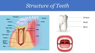
Teeth are made up of three main elements - water, organic materials, and inorganic materials. The tooth is composed of a crown, neck, and root. The crown is covered by the hardest substance in the body, enamel, and below that is dentin. The root is embedded in the jawbone. Tooth decay occurs when bacteria in the mouth produce acids from sugars that dissolve tooth enamel over time. The most common type of tooth decay is dental caries, or cavities, which can damage tooth structures and lead to pain, loss of teeth, and infection if left untreated.


![Dentin
Dentin is the substance between enamel
or
cementum and the pulp chamber.
It is secreted by the odontoblasts of the dental pulp[13].
The formation of dentin is known as dentinogenesis.
The porous, yellow-hued material is made up of 70%
inorganic materials, 20% organic materials, and 10% water
by weight[14].](https://image.slidesharecdn.com/teeth-200919171251/85/Teeth-12-320.jpg)
![Cementum
Cementum is a specialized bone like substance covering the
root of a tooth.[13]
It is approximately 45% inorganic material
(mainly hydroxyapatite), 33% organic material
(mainly collagen) and 22% water.
Cementum is excreted by cementoblasts within the root of the
tooth and is thickest at the root apex.
Its coloration is yellowish and it is softer than dentin and
enamel.](https://image.slidesharecdn.com/teeth-200919171251/85/Teeth-13-320.jpg)
![Dental pulp
The dental pulp is the central part of the tooth filled with soft
connective tissue.[14]
This tissue contains blood vessels and nerves that enter the
tooth from a hole at the apex of the root.[21]
Along the border between the dentin and the pulp are
odontoblasts, which initiate the formation of dentin.[14]
Other cells in the pulp include fibroblasts,
preodontoblasts, macrophages and T lymphocytes.[22]
The pulp is commonly called "the nerve" of the tooth.](https://image.slidesharecdn.com/teeth-200919171251/85/Teeth-14-320.jpg)
![Development
Tooth development is the complex process by which teeth form
from embryonic cells, grow, and erupt into the mouth.
Although many diverse species have teeth, their development is largely the same
as in humans.
For human teeth to have a healthy oral environment, enamel, dentin, cementum,
and the periodontium must all develop during appropriate stages of fetal
development.
Primary teeth start to form in the development of the embryo between the sixth
and eighth weeks, and permanent teeth begin to form in the twentieth week.[23]
If teeth do not start to develop at or near these times, they will not develop at all.](https://image.slidesharecdn.com/teeth-200919171251/85/Teeth-15-320.jpg)

![Supporting structures
The periodontium is the supporting structure of a tooth, helping to attach the
tooth to surrounding tissues and to allow sensations of touch and
pressure.[30]
It consists of the cementum, periodontal ligaments, alveolar bone,
and gingiva. Of these, cementum is the only one that is a part of a tooth.
Periodontal ligaments connect the alveolar bone to the cementum. Alveolar
bone surrounds the roots of teeth to provide support and creates what is
commonly called an alveolus, or "socket".
Lying over the bone is the gingiva or gum, which is readily visible in the
mouth.](https://image.slidesharecdn.com/teeth-200919171251/85/Teeth-17-320.jpg)
![Periodontal ligaments
The periodontal ligament is a specialized connective tissue that attaches the
cementum of a tooth to the alveolar bone.
This tissue covers the root of the tooth within the bone.
Each ligament has a width of 0.15–0.38mm, but this size decreases over
time.[31] The functions of the periodontal ligaments include attachment of the
tooth to the bone, support for the tooth, formation and resorption of bone
during tooth movement, sensation, and eruption.[27]
The cells of the periodontal ligaments include osteoblasts, osteoclasts,
fibroblasts, macrophages, cementoblasts, and epithelial cell rests of
Malassez.[32] Consisting of mostly Type I and III collagen, the fibers are
grouped in bundles and named according to their location.
The groups of fibers are named alveolar crest, horizontal, oblique, periapical,
and interradicular fibers.[33]](https://image.slidesharecdn.com/teeth-200919171251/85/Teeth-18-320.jpg)
![Alveolar bone
The alveolar bone is the bone of the jaw which forms the alveolus around
teeth.[35]
Like any other bone in the human body, alveolar bone is modified throughout
life.
Osteoblasts create bone and osteoclasts destroy it, especially if force is placed
on a tooth.[30]
As is the case when movement of teeth is attempted through orthodontics, an
area of bone under compressive force from a tooth moving toward it has a
high osteoclast level, resulting in bone resorption.
An area of bone receiving tension from periodontal ligaments attached to a
tooth moving away from it has a high number of osteoblasts, resulting in bone
formation.](https://image.slidesharecdn.com/teeth-200919171251/85/Teeth-19-320.jpg)
![Gingiva
The gingiva ("gums") is the mucosal tissue that overlays the jaws.
There are three different types of epithelium associated with the gingiva:
gingival, junctional, and sulcular epithelium.
These three types form from a mass of epithelial cells known as the
epithelial cuff between the tooth and the mouth.[36]
The gingival epithelium is not associated directly with tooth attachment
and is visible in the mouth.
The junctional epithelium, composed of the basal
lamina and hemidesmosomes, forms an attachment to the tooth.[27]
The sulcular epithelium is nonkeratinized stratified squamous tissue on the
gingiva which touches but is not attached to the tooth.[37]](https://image.slidesharecdn.com/teeth-200919171251/85/Teeth-20-320.jpg)

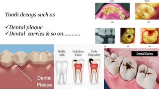
![Plaque
Plaque is a biofilm consisting of large quantities of
various bacteria that form on teeth.[38]
If not removed regularly, plaque buildup can lead
to periodontal problems such as gingivitis.
Given time, plaque can mineralize along the gingiva,
forming tartar.
The microorganisms that form the biofilm are almost
entirely bacteria (mainly streptococcus and anaerobes), with the
composition varying by location in the mouth.[39]
Streptococcus mutans is the most important bacterium associated
with dental caries.](https://image.slidesharecdn.com/teeth-200919171251/85/Teeth-23-320.jpg)
![ Certain bacteria in the mouth live off the remains of foods,
especially sugars and starches. In the absence of oxygen they
produce lactic acid, which dissolves the calcium and phosphorus in
the enamel.[13][40]
This process, known as "demineralization", leads to tooth
destruction. Saliva gradually neutralizes the acids which cause the
pH of the tooth surface to rise above the critical pH, typically
considered to be 5.5. This causes 'remineralization', the return of the
dissolved minerals to the enamel.
If there is sufficient time between the intake of foods then the impact
is limited and the teeth can repair themselves.
Saliva is unable to penetrate through plaque, however, to neutralize
the acid produced by the bacteria](https://image.slidesharecdn.com/teeth-200919171251/85/Teeth-24-320.jpg)
![ Dental caries (cavities), described as "tooth decay", is an infectious disease which
damages the structures of teeth.[41]
The disease can lead to pain, tooth loss, and infection. Dental caries has a long
history, with evidence showing the disease was present in the Bronze, Iron,
and Middle ages but also prior to the neolithic period.[42]
The largest increases in the prevalence of caries have been associated with diet
changes.[43]
Today, caries remains one of the most common diseases throughout the world. In the
United States, dental caries is the most common chronic childhood disease, being at
least five times more common than asthma.[44] Countries that have experienced an
overall decrease in cases of tooth decay continue to have a disparity in the
distribution of the disease.[45]
Among children in the United States and Europe, 60–80% of cases of dental caries
occur in 20% of the population.[46]
Tooth carries](https://image.slidesharecdn.com/teeth-200919171251/85/Teeth-25-320.jpg)
![ Tooth decay is caused by certain types of acid-producing bacteria
which cause the most damage in the presence
of fermentable carbohydrates such as sucrose, fructose,
and glucose.[47][48]
The resulting acidic levels in the mouth affect teeth because a tooth's
special mineral content causes it to be sensitive to low pH.
Depending on the extent of tooth destruction, various treatments
can be used to restore teeth to proper form, function, and aesthetics,
but there is no known method to regenerate large amounts of tooth
structure. Instead, dental health organizations advocate
preventative and prophylactic measures, such as regular oral
hygiene and dietary modifications, to avoid dental caries.[49]](https://image.slidesharecdn.com/teeth-200919171251/85/Teeth-26-320.jpg)
![Discoloration of teeth may result from bacteria stains, tobacco, tea, coffee, foods with an
abundance of chlorophyll, restorative materials, and medications.[67] Stains from bacteria may
cause colors varying from green to black to orange. Green stains also result from foods with
chlorophyll or excessive exposure to copper or nickel. Amalgam, a common dental restorative
material, may turn adjacent areas of teeth black or gray. Long term use of chlorhexidine, a
mouthwash, may encourage extrinsic stain formation near the gingiva on teeth. This is usually
easy for a hygienist to remove. Systemic disorders also can cause tooth
discoloration. Congenital erythropoietic porphyria causes porphyrins to be deposited in teeth,
causing a red-brown coloration. Blue discoloration may occur with alkaptonuria and rarely
with Parkinson's disease. Erythroblastosis fetalis and biliary atresia are diseases which may
cause teeth to appear green from the deposition of biliverdin. Also, trauma may change a tooth
to a pink, yellow, or dark gray color. Pink and red discolorations are also associated in
patients with lepromatous leprosy. Some medications, such as tetracycline antibiotics, may
become incorporated into the structure of a tooth, causing intrinsic staining of the teeth.
Discoloration](https://image.slidesharecdn.com/teeth-200919171251/85/Teeth-27-320.jpg)
![Alteration and eruption
Tooth eruption may be altered by some environmental factors.
When eruption is prematurely stopped, the tooth is said to
be impacted.
The most common cause of tooth impaction is lack of space in the
mouth for the tooth.[68]
Other causes may be tumors, cysts, trauma, and thickened bone or
soft tissue.
Tooth ankylosis occurs when the tooth has already erupted into the
mouth but the cementum or dentin has fused with the alveolar bone.
This may cause a person to retain their primary tooth instead of
having it replaced by a permanent one.](https://image.slidesharecdn.com/teeth-200919171251/85/Teeth-28-320.jpg)
![Abnormalities
Tooth abnormalities may be categorized according to whether
they have environmental or developmental causes.[56]
While environmental abnormalities may appear to have an
obvious cause, there may not appear to be any known cause for
some developmental abnormalities.
Environmental forces may affect teeth during development,
destroy tooth structure after development, discolor teeth at any
stage of development, or alter the course of tooth eruption.
Developmental abnormalities most commonly affect the
number, size, shape, and structure of teeth.](https://image.slidesharecdn.com/teeth-200919171251/85/Teeth-29-320.jpg)
![•Anodontia is the total lack of tooth development.
•Hyperdontia is the presence of a higher-than-normal number of teeth.
•Hypodontia is the lack of development of one or more teeth.
• Oligodontia may be used to describe the absence of 6 or more teeth.
Some systemic disorders which may result in hyperdontia include Apert
syndrome, cleidocranial dysostosis, Crouzon syndrome, Ehlers–Danlos
syndrome, Gardner's syndrome, and Sturge–Weber syndrome.[69] Some
systemic disorders which may result in hypodontia include Crouzon
syndrome, Ectodermal dysplasia, Ehlers–Danlos syndrome, and Gorlin
syndrome.[70]
Abnormalities in number](https://image.slidesharecdn.com/teeth-200919171251/85/Teeth-30-320.jpg)

![Abnormalities in shape
•Gemination occurs when a developing tooth incompletely splits into the
formation of two teeth.
•Fusion is the union of two adjacent teeth during development.
•Concrescence is the fusion of two separate teeth only in their cementum.
•Accessory cusps are additional cusps on a tooth and may manifest as a
Talon cusp, Cusp of Carabelli, or Dens evaginatus.
•Dens invaginatus, also called Dens in dente, is a deep invagination in a tooth
causing the appearance of a tooth within a tooth.
•Ectopic enamel is enamel found in an unusual location, such as the root of a tooth.
•Taurodontism is a condition where the body of the tooth and pulp chamber is enlarged, and is
associated with Klinefelter syndrome, Tricho-dento-osseous syndrome, Triple X syndrome,
and XYY syndrome.[71]
•Hypercementosis is excessive formation of cementum, which may result from trauma,
inflammation, acromegaly, rheumatic fever, and Paget's disease of bone.[71]
•A dilaceration is a bend in the root which may have been caused by trauma to the tooth during
formation.
•Supernumerary roots is the presence of a greater number of roots on a tooth than expected](https://image.slidesharecdn.com/teeth-200919171251/85/Teeth-32-320.jpg)
![Abnormalities in structures
•Amelogenesis imperfecta is a condition in which enamel does not form
properly or at all.[75]
•Dentinogenesis imperfecta is a condition in which dentin does not form
properly and is sometimes associated with osteogenesis imperfecta.[76]
•Dentin dysplasia is a disorder in which the roots and pulp of teeth may
be affected.
•Regional odontodysplasia is a disorder affecting enamel, dentin, and
pulp and causes the teeth to appear "ghostly" on radiographs.[77]
•Diastema is a condition in which there is a gap between two teeth
caused by the imbalance in the relationship between the jaw and the size
of teeth.[78]](https://image.slidesharecdn.com/teeth-200919171251/85/Teeth-33-320.jpg)


![ The purpose of cleaning teeth is to remove plaque, which consists
mostly of bacteria.[50]
Healthcare professionals recommend regular brushing twice a
day (in the morning and in the evening, or after meals) in order to
prevent formation of plaque and tartar.[49]
A toothbrush is able to remove most plaque, except in areas
between teeth.
As a result, flossing is also considered a necessity to maintain oral
hygiene. When used correctly, dental floss removes plaque from
between teeth and at the gum line, where periodontal disease often
begins and could develop caries.](https://image.slidesharecdn.com/teeth-200919171251/85/Teeth-36-320.jpg)
![ Fluoride therapy is often recommended to protect against dental caries.
Water fluoridation and fluoride supplements decrease the incidence of dental
caries.
Fluoride helps prevent dental decay by binding to the hydroxyapatite
crystals in enamel.[52]
The incorporated fluoride makes enamel more resistant to demineralization
and thus more resistant to decay.[27]
Topical fluoride, such as a fluoride toothpaste or mouthwash, is also
recommended to protect teeth surfaces.
Many dentists include application of topical fluoride solutions as part of
routine cleanings.
Protective treatments](https://image.slidesharecdn.com/teeth-200919171251/85/Teeth-37-320.jpg)