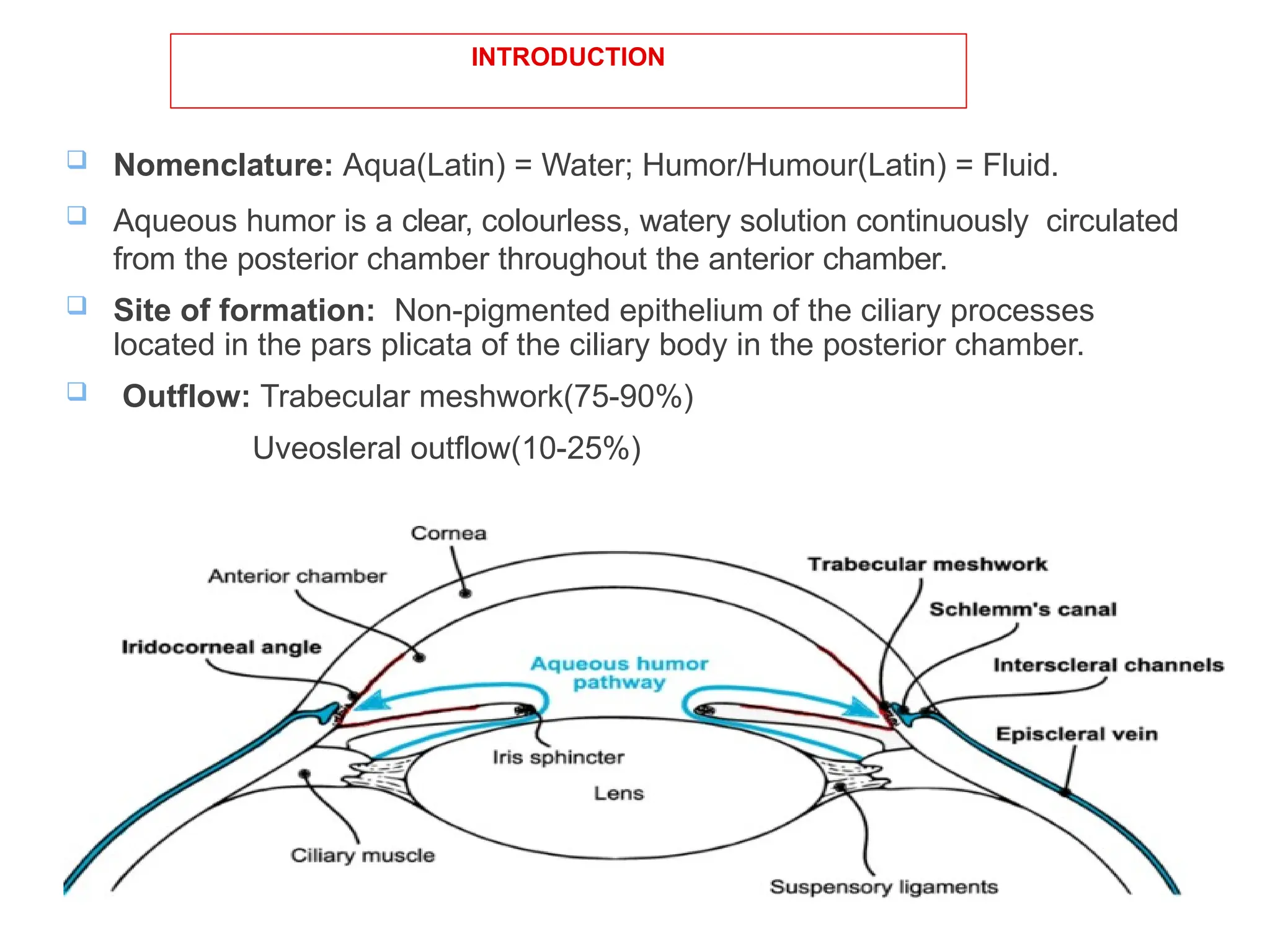
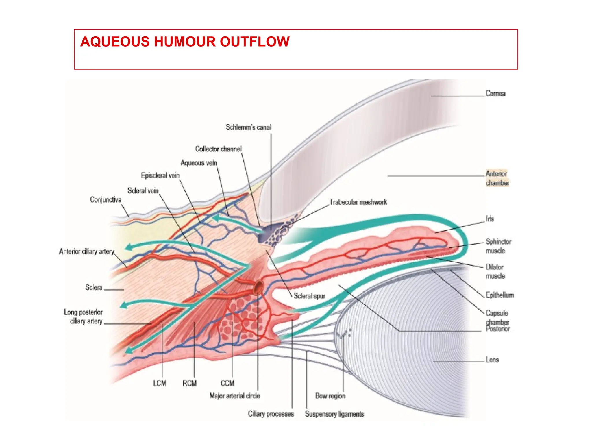
AQUEOUS HUMOUR
DYNAMICS
PRESENTED BY;
DR. RAHUL GUPTA
1ST
YEAR RESIDENT
DEPT. OPHTHALMOLOGY
DHULIKHEL HOSPITAL
DATE: 13TH
SEPT. 2023
LAYOUT
 INTRODUCTION
 HISTORY
 EMBROYOLOGY
 FUNCTION
 PHYSIOCHEMICAL PROPERTIES
 BIOCHEMICAL PROPERTIES
 ANATOMY ( ANTERIOR CHAMBER, ANGLE
STRUCTURE, POSTERIOR CHAMBER)
 FORMATION OF AQUEOUS HUMOR
 OUTFLOW OF AQUEOUS HUMOR
INTRODUCTION
 Nomenclature: Aqua(Latin) = Water; Humor/Humour(Latin) = Fluid.
 Aqueous humor is a clear, colourless, watery solution continuously circulated
from the posterior chamber throughout the anterior chamber.
 Site of formation: Non-pigmented epithelium of the ciliary processes
located in the pars plicata of the ciliary body in the posterior chamber.
 Outflow: Trabecular meshwork(75-90%)
Uveosleral outflow(10-25%)
HISTORY
• Until the early Twentienth century, aqueous humor was
regarded as stagnant fluid.
• Since that time, however it has been shown to be
continously formed and drained, and the associated
anatomic drainage portals(schlemn’s canal, collector
channels, aqueous veins, ciliary muscles interstices)
has been described.
• Boerhavve first describe the presence of aqueous
veins.
• Ascher described a clear fluid in veins of the episclera
and demonstrated by means of external compression
with glass rod that these veins were interconnected
with veins containing blood.
• Goldmann demonstrated that these vessels contained
aqueous humor by injecting fluorescein intravenously
and observing the dye entering the anterior chamber
and subsequently the aqueous veins.
EMBROYOLOGY
• SCLERAL SPUR: At approximately 4 months, the inner
aspect of the anterior sclera(inner limbus) develops a
fibrous, wedge shaped protrusion, called scleral spur.
- By the middle of 7th month the anterior ends of longitudinal
ciliary muscle fibres have established their insertion into
scleral spur.
• Draining Vascular Plexus: The deep and intrascleral
vascular plexus, the aqueous veins and the collector
channels that transverse the sclera at limbal region are
first recognizable at about 12 WOG.
- these vessels differenciates from primitive mesoderm.
• Anterior chamber and aqueous outflow pathway:
Development of anterior chamber depends on structures
which borders it.
• Angle of anterior chamber: Approximately 7 WOG,
angle is occupied by nest of loosely organised
undifferentiated mesenchymal cells that are destined to
develop into the trabecular meshwork.
• Trabecular Meshwork: At 4th MOG the primordium of
the trabecular meshwork is recognizable as an
approximately triangular or wedgeshape structure that
consist of undifferentiated mesenchymal cells of neural
crest origin.
• Schlemn’s Canal: It develops from small plexus of
venous canaliculi by the end of 3rd MOG. These
channels are derived from mesodermal mesenchyme
and initially they functions as blood vessels.
FUNCTIONS OF THE AQUEOUS
HUMOUR
 Maintains Intra-ocular pressure.
 Nutritive function
 Supplies glucose and oxygen to the cornea
 Supplies oxygen, glucose, amino acids and potassium to the lens.
 Excretory function
 Removes lactic acid and carbon dioxide from the cornea.
 Removes lactate, pyruvate and sodium from the lens.
 Provides optically clear medium for passage of light.
 Cornea-aqueous interface acts as diverging lens of low power.
 Helps to clear inflammatory products and cells from the anterior chamber.
PHYSIOCHEMICAL PROPERTIES
 Volume: 0.31ml(anterior chamber: 0.25 ml, posterior
chamber: 0.06ml)
 Refractive index: 1.336
 Viscosity : 1.025- 1.040
 Osmotic pressure: slightly hyperosmotic then plasma by 3 to
5 mosm/l
 PH: Slightly acidic(7.2)
 Rate of formation: 2.3 microliter/min
BIOCHEMICAL COMPOSITION
 Water: 99.9 %
 Proteins;
 5-16 mg /100 ml as compared to plasma( 6-7gm/100ml)
 Immunoglobulins : IgG and IgM but no IgD and IgA
 Amino acids: aqueous /plasma : 0.08 to 3.14
 Non colloidal constituents(Na, K, Ca, Mg, Cl, HCO3,
Lactate, Pyruvate, Ascorbate, Urea, Glucose )
ANATOMY OF THE ANTERIOR CHAMBER
• Volume : 0.25ml
 Anterior boundary : Posterior
surface of cornea
 Posterior boundary
 Anterior surface of the lens in the
pupillary area
 Anterior surface of the iris in the
periphery
 2.5 to 3mm deep in the center in
normal adults
 Communicates with the posterior
chamber through the pupil
SHAFFER’S SYSTEM OF GRADING THE ANGLE WIDTH
SL = Schwalbe’s line, TM = Trabecular meshwork, SS = Scleral spur, CBB = Ciliary body band
VAN HERICK METHOD OF ANGLE
GRADING
Estimate of Angle Width From Anterior Chamber Depth at the Peripheri.
ANGLE DEPTH
Grade 4 anterior chamber depth = corneal thickness
Grade 3 anterior chamber depth = ¼ to ½ corneal thickness
Grade 2 anterior chamber depth = ¼ corneal thickness
Grade 1 anterior chamber depth = Slit like (extremely shallow)
Closed absent peripheral anterior chamber
ANATOMY OF THE POSTERIOR CHAMBER
 Volume: 0.06ml
 Anterior boundary: The
posterior surface of the iris and
part of ciliary body
 Posterior Boundary: The lens
and its zonules.
 Laterally by the ciliary body.
3 different zones:
1) PREZONULAR SPACE (Posterior chamber
proper)
anterior to the zonular fibers and posterior to the iris
2) ZONULAR SPACE (Circumlental or ciliolental
space, canal of Hanover)
Bounded
 centrally equator of the lens
 peripherally by the ciliary process
 anteriorly by the posterior surface of anterior
zonular fibres
 posteriorly by anterior surface of posterior
zonular fibres.
3) RETROZONULAR SPACE( Canal of Petit)
Between the anterior surface of vitreous and the
posterior surface of zonules.
FORMATION OF AQUEOUS HUMOUR
Site of formation:
Nonpigmented epithelium of the ciliary processes
Processes:
 Active secretion(70%).
 Ultrafiltration(10%).
 Diffusion(20%)
Basic anatomy of ciliary body:
CILIARY PROCESSES-
ULTRASTRUCTURE
70-80 radial ridges from the pars
plicata
• Each process is 2mm long,
0.5mm wide and 0.9mm in
height
• Contains central core of
stroma with capillaries
surrounded by double layer
of epithelium.
• Endothelium of the capillaries
is thin and has tiny fenestrae.
Permeable to proteins.
The outer pigmented epithelium
 Contains cuboidal cells.
 Has Carbonic anhydrase enzyme.
 Basal portion has numerous mitochondria
suggesting participation in active metabolic process.
 Separated from stroma by Basement membrane
which is continuation of the Bruch’s membrane
The inner non-pigmented epithelium
Contains columnar cells
 Features of cells involved in fluid transport
 Extensive foldings
 Numerous mitochondria
 Well developed Endoplasmic reticulum
 Tight junction
 Na-K-ATPase(lateral infoldings).
 Impermeable to proteins.
Separated from the aqueous by basement
membrane
Potential space between the 2 layers of
epithelium is called as the ciliary channel
INTERCELLULAR JUNCTIONS
 GAP JUNCTIONS
 PUNCTA ADHERENTIA AND
DESMOSOMES
 TIGHT JUNCTIONS(ZONULA
OCCLUDENTAE)
VASCULAR SUPPLY
Ciliary processes have the richest
blood supply in the whole
body(154microl/min)
 Formation of aqueous humor
removes only 4-8% of the total
plasma in ciliary processes.
 Hence modest reduction in the rate
of plasma flow would not decrease
aqueous formation but profound
vasoconstriction will however have
an effect.
MECHANISM OF FORMATION OF THE
AQUEOUS
 The formation of aqueous humor is a complex process that
involves ultrafiltration and diffusional exchange of water
and solutes with plasma from blood flowing through the ciliary
processes.
 Active transport of substances from dialysate of plasma to
posterior chamber occurs.
 The fluid is further changed by diffusional exchange and
active transport of substances out of the eye.
Each process involved in aqueous humour
formation are:
 Ultrafiltration
 Active transport
 Diffusion
ULTRAFILTRATION
• The process by which fluids and its
solutes crosses semipermeable
membrane under a pressure
gradient (e.g. capillary blood
pressure) is called ultrafiltration.
• As blood passes through the
capillaries of the ciliary
processes ,about 4% of the plasma
filters through the fenestrations in
the capillary wall into the interstitial
spaces between the capillaries and
the ciliary epithelium.
Fluid movement is;
Favored by the hydrostatic pressure
difference between the capillary
pressure and the interstitial fluid
pressure.
Resisted by difference between
oncotic pressure of plasma and
aqueous humour.
• Colloid concentration in the
interstitial space gradually rises due
to non permeability of ciliary
epithelium to colloids .
• Increase concentration of colloids
retards the movement of water from
stroma from posterior chamber but
favors movement of water from
plasma to stroma.
• Active process takes place to
transfer fluid from stroma to
posterior chamber.
• Active process requiring
energy.
• Movement of substances
against the
concentration gradient.
• Carried out by globular
proteins in the membrane.
• Na, Cl and bicarbonate
• Na-K-ATPase inhibitor
Ouabain inhibits aqueous
formation by about 70-
80%.
ACTIVE SECRETION
DIFFUSION
• Diffusion is a movement of a substance across a membrane along its
concentration gradient.
• As aqueous humour passes from posterior to anterior chamber-
diffusional exchange with the surrounding tissues takes place -anterior
chamber aqueous resembles plasma more closely than the posterior
aqueous humour.
 Passive process in which substances flow across a biological
membrane along its concentration gradient.
 Occurs in the lipid portion of membrane.
 Governed by the Fick’s law of diffusion:
Rate of movement=K(C1-C2)
where K:Constant that depends on nature, permeability of
membrane and nature of solute and solvent.
C1-C2:concentration gradient across the membrane.
COMPOSITION OF AQUEOUS HUMOUR
SUBSTANCE
(Nm/kg H2O)
Anterior chamber
aqueous
Plasma
Na 163 176
Cl 126 117
HCO3 22 26
pH 7.21 7.4
Ascorbate 0.92 0.06
Protein 0.02% 7%
BLOOD AQUEOUS BARRIER
The blood aqueous barrier is formed by tight junctions
(zonula occludens and zonula adherans) between the cells
of inner nonpigmented epithelium of ciliary body the
tight junctions of the iris capillary endothelial cells.
 It is not an absolute barrier
 Allows entry of particles depending upon molecular size,
lipid solubility and electrical charge
 So water soluble, low mol wt polar solutes can breach this
barrier
FACTORS THAT BREAK DOWN
BLOOD AQUEOUS BARRIER
FACTORS AFFECTING AQUEOUS
HUMOUR FORMATION
CONDITION EFFECT ON AQUEOUS
FORMATION
Hypothermia Decrease
Hyperthermia Increase
Acidosis Decrease
Alkalosis Increase
DM, Retinal detachment, ocular
inflammation, cyclodestructive procedures,
choroidal detachment, cyclodialysis
Decrease
FACTORS AFFECTING COMPOSITION
OF AQUEOUS HUMOUR
1. Blood aqueous barrier
2. Hemodynamic factors affecting stromal pool
3. Diffusional exchange at iris
4. Metabolites
5. Rate of Aquous drainage
6. Quality of aqueous.
EFFECT OF PHARMACOLOGICAL AGENTS
DRUG EFFECT
Beta adrenergic agonists Increase
Pilocarpine Slightly increase
Beta adrenergic blockers, Alpha
adrenergic agonists, Carbonic
anhydrase inhibitor
Decrease
Halothane, Ouabain, Barbiturates,
Ketamine
Decrease
AQUEOUS HUMOUR OUTFLOW
Anatomy of outflow system
 Trabecular meshwork:
Triangular shape with its
apex at schwalbe line
and base at scleral spur.
 Sieve like structure at
the angle of anterior
chamber through which
90% of the aqueous
leaves the eye.
 It consists of three
portions.
A) Uveal meshwork:
 Innermost part of trabecular
meshwork extending from ciliary
body and iris root to the
schwalbe line.
 Trabeculae are cord like and is
2-3 layers thick.
 Openings vary from 25-
75micron
 Larger middle portion which
extends from scleral spur to
Schwalbe’s line.
 Consists of sheets of
trabeculae perforated by
elliptical openings (5-
50micron)
 Openings are progressively
smaller as they approach
Schlemm’s canal.
B) Corneoscleral
meshwork;
Outermost portion of trabecular
meshwork which links Corneoscleral
meshwork with the endothelium of the
Schlemm's canal.
• It has 3 layers.
 The Juxtacanalicular tissue offers the
major proportion of maximum
resistance to aqueous outflow.
C) Juxtacanalicular
(endothelial) meshwork;
Schlemm’s canal
Endothelial lined oval channel present circumferentially in the
scleral sulcus
 The endothelial cells of the
inner wall are irregular,
spindle shaped and
contains giant vacuoles
whereas outer are smooth
and flat.
 The outer wall contains the
opening of collector channels
which leave schlemm’s canal
at oblique angles and connect
directly or indirectly with
episcleral veins.
COLLECTOR CHANNEL
• Also called intrascleral aqueous
vessels and are 25-35 in no; and
leave the Schlemm’s canal at
oblique angles to terminate
ultimately into episcleral veins.
• Lined by endothelium similar to
outer wall of Schlemm’s canal.
• Direct system: drained by about
8 larger vessels directly into
episcleral vein.
• Indirect system: Fine inter-
connecting channels before
eventually going into episcleral
vein.
EPISCLERAL AND CONJUNCTIVAL
VEINS
• Schlems canal is connected to episcleral
and conjunctival veins by a complex
system of intrascleral channels.
AQUEOUS DRAINAGE
1.Trabecular(conventional)
route:
 70-90%
Trabecular meshwork –juxtacanalicular
tissue-inner wall of Schlemm’s canal-
collector channels –episcleral veins
Bulk flow pressure sensitive route
hence increasing pressure head will
increase outflow
Trabecular flow can be increased by
drugs, laser trabeculoplasty.
2.Uveoscleral(unconventional) route:
 Accounts for 10% of aqueous outflow.
 Aqueous passes across the face of ciliary body into the
suprachoroidal space and is drained by the venous
circulation in the ciliary body, choroid and sclera.
 Some aqueous drains through the iris.
POSSIBLE MECHANISMS OF AQUEOUS
TRANSPORT
 Vacuolation theory
 Leaky endothelial cells
 Sonderman’s channels
 Contractile microfilaments
 Pores in endothelial cells
Vacuolation Theory
Factors affecting aqueous outflow
FACTORS EFFECT
Age Decrease
Hormones
 corticosteroids
 progesterone
 relaxin
 HCG
Decrease
Increase
Increase
Increase
Ciliary muscles tone Increase
AC depth Increase with increasing depth
DRUGS
 Parasympathomimetic( pilocarpine)
 Parasympatholytic(atropine ,
homatropine, tropicamide,
cyclopentolate )
 Bradykinin
 C-AMP
 Substance P
Increase
Decrease
Increase
Increase
Decrease
FACTORS EFFECT
SURGERY
 ALT(argon laser trabeculoplasty
 Filtering surgery
 cyclodialysis
 cataract extraction
 PK
Increase
Increase
Increase
Decrease (temporary effect)
NEURAL REGULATION
 IIIrd nerve stimulation increase
 IIIrd nerve ablation
decrease
 sympathetic stimulation increase
Increase
Decrease
Increase
Particulate matter
 RBC/WBC
 Pigment
Decrease
Decrease
Different methods for physical
measurement of aqueous production
• Tonography
• Suction Cup
• Perfusion
INTRAOCULAR PRESSURE
• It is the pressure exerted by the intraocular contents
on the coats of eyeball
• Normal IOP is maintained by dynamic equilibrium
between aqueous humor formation, aqueous humor
outflow and episcleral venous pressure.
• maintains shape and optical integrity of eyeball.
• Normal IOP = 10-21 mm Hg.
Factors exerting Short-term influence in
IOP
• Diurnal variation
• Postural variation
• Exertional Influences
• Lid and eye movement
• Intraocular conditions
• Systemic conditions
• Environmental conditions
• General Anaesthesia
• food and drugs.
• Genetics
• Age
• Gender
• Refractive Error
• Ethinicity
Factors exerting Long-term influence in
IOP
Some clinical Corelations
• Vascular pressure in episcleral venous system is
around 9 mmHg and IOP varies from 10-21mmHg,
with mean value =15mmHg.
• The pressure gradient from anterior chamber to
episcleral veins is explained by resistance in flow in
different structures in conventional pathway.
• In older age there is increase in resistace to structures
leads to Primary Open Angle Glaucoma(Chronic
simple Glaucoma).
Angle closure Glaucoma:
• occurs when the peripheral iris is in contact with the trabecular
meshwork, either intermittently or permanently (synechial closure).
• Specific mechanisms leading to angle closure can be divided into 2
categories:
• Mechanisms that push the iris from behind. The most common
reason is relative pupillary block, but other reasons include plateau
iris syndrome, enlarged or anteriorly displaced lens, and malignant
glaucoma.
• Mechanisms that pull the iris into contact with the Trabecular
Meshwork. Examples include contraction of inflammatory
membrane as in uveitis, fibrovascular tissue as in iris
neovascularization, or corneal endothelium as in iridocorneal
endothelial syndrome.
Conclusion
• The aqueous humor fills the anterior and posterior
chamber of the eye.
• It is one of the fundamental components in ensuring
the optical physics and health of eye are properly
maintained.
• Continous production of aqueous humor is critical to
the eye’s shape and size.
• It is drained by three routes: Trabecular meshwork,
uveoscleral meshwork, and iris.
Ophthalmology presentation in aqueous humor dynamics.pptx

Ophthalmology presentation in aqueous humor dynamics.pptx

  • 1.
    AQUEOUS HUMOUR DYNAMICS PRESENTED BY; DR.RAHUL GUPTA 1ST YEAR RESIDENT DEPT. OPHTHALMOLOGY DHULIKHEL HOSPITAL DATE: 13TH SEPT. 2023
  • 2.
    LAYOUT  INTRODUCTION  HISTORY EMBROYOLOGY  FUNCTION  PHYSIOCHEMICAL PROPERTIES  BIOCHEMICAL PROPERTIES  ANATOMY ( ANTERIOR CHAMBER, ANGLE STRUCTURE, POSTERIOR CHAMBER)  FORMATION OF AQUEOUS HUMOR  OUTFLOW OF AQUEOUS HUMOR
  • 3.
    INTRODUCTION  Nomenclature: Aqua(Latin)= Water; Humor/Humour(Latin) = Fluid.  Aqueous humor is a clear, colourless, watery solution continuously circulated from the posterior chamber throughout the anterior chamber.  Site of formation: Non-pigmented epithelium of the ciliary processes located in the pars plicata of the ciliary body in the posterior chamber.  Outflow: Trabecular meshwork(75-90%) Uveosleral outflow(10-25%)
  • 4.
    HISTORY • Until theearly Twentienth century, aqueous humor was regarded as stagnant fluid. • Since that time, however it has been shown to be continously formed and drained, and the associated anatomic drainage portals(schlemn’s canal, collector channels, aqueous veins, ciliary muscles interstices) has been described.
  • 5.
    • Boerhavve firstdescribe the presence of aqueous veins. • Ascher described a clear fluid in veins of the episclera and demonstrated by means of external compression with glass rod that these veins were interconnected with veins containing blood. • Goldmann demonstrated that these vessels contained aqueous humor by injecting fluorescein intravenously and observing the dye entering the anterior chamber and subsequently the aqueous veins.
  • 6.
    EMBROYOLOGY • SCLERAL SPUR:At approximately 4 months, the inner aspect of the anterior sclera(inner limbus) develops a fibrous, wedge shaped protrusion, called scleral spur. - By the middle of 7th month the anterior ends of longitudinal ciliary muscle fibres have established their insertion into scleral spur. • Draining Vascular Plexus: The deep and intrascleral vascular plexus, the aqueous veins and the collector channels that transverse the sclera at limbal region are first recognizable at about 12 WOG. - these vessels differenciates from primitive mesoderm.
  • 7.
    • Anterior chamberand aqueous outflow pathway: Development of anterior chamber depends on structures which borders it. • Angle of anterior chamber: Approximately 7 WOG, angle is occupied by nest of loosely organised undifferentiated mesenchymal cells that are destined to develop into the trabecular meshwork. • Trabecular Meshwork: At 4th MOG the primordium of the trabecular meshwork is recognizable as an approximately triangular or wedgeshape structure that consist of undifferentiated mesenchymal cells of neural crest origin. • Schlemn’s Canal: It develops from small plexus of venous canaliculi by the end of 3rd MOG. These channels are derived from mesodermal mesenchyme and initially they functions as blood vessels.
  • 8.
    FUNCTIONS OF THEAQUEOUS HUMOUR  Maintains Intra-ocular pressure.  Nutritive function  Supplies glucose and oxygen to the cornea  Supplies oxygen, glucose, amino acids and potassium to the lens.  Excretory function  Removes lactic acid and carbon dioxide from the cornea.  Removes lactate, pyruvate and sodium from the lens.  Provides optically clear medium for passage of light.  Cornea-aqueous interface acts as diverging lens of low power.  Helps to clear inflammatory products and cells from the anterior chamber.
  • 9.
    PHYSIOCHEMICAL PROPERTIES  Volume:0.31ml(anterior chamber: 0.25 ml, posterior chamber: 0.06ml)  Refractive index: 1.336  Viscosity : 1.025- 1.040  Osmotic pressure: slightly hyperosmotic then plasma by 3 to 5 mosm/l  PH: Slightly acidic(7.2)  Rate of formation: 2.3 microliter/min
  • 10.
    BIOCHEMICAL COMPOSITION  Water:99.9 %  Proteins;  5-16 mg /100 ml as compared to plasma( 6-7gm/100ml)  Immunoglobulins : IgG and IgM but no IgD and IgA  Amino acids: aqueous /plasma : 0.08 to 3.14  Non colloidal constituents(Na, K, Ca, Mg, Cl, HCO3, Lactate, Pyruvate, Ascorbate, Urea, Glucose )
  • 11.
    ANATOMY OF THEANTERIOR CHAMBER • Volume : 0.25ml  Anterior boundary : Posterior surface of cornea  Posterior boundary  Anterior surface of the lens in the pupillary area  Anterior surface of the iris in the periphery  2.5 to 3mm deep in the center in normal adults  Communicates with the posterior chamber through the pupil
  • 12.
    SHAFFER’S SYSTEM OFGRADING THE ANGLE WIDTH SL = Schwalbe’s line, TM = Trabecular meshwork, SS = Scleral spur, CBB = Ciliary body band
  • 13.
    VAN HERICK METHODOF ANGLE GRADING Estimate of Angle Width From Anterior Chamber Depth at the Peripheri. ANGLE DEPTH Grade 4 anterior chamber depth = corneal thickness Grade 3 anterior chamber depth = ¼ to ½ corneal thickness Grade 2 anterior chamber depth = ¼ corneal thickness Grade 1 anterior chamber depth = Slit like (extremely shallow) Closed absent peripheral anterior chamber
  • 14.
    ANATOMY OF THEPOSTERIOR CHAMBER  Volume: 0.06ml  Anterior boundary: The posterior surface of the iris and part of ciliary body  Posterior Boundary: The lens and its zonules.  Laterally by the ciliary body.
  • 15.
    3 different zones: 1)PREZONULAR SPACE (Posterior chamber proper) anterior to the zonular fibers and posterior to the iris 2) ZONULAR SPACE (Circumlental or ciliolental space, canal of Hanover) Bounded  centrally equator of the lens  peripherally by the ciliary process  anteriorly by the posterior surface of anterior zonular fibres  posteriorly by anterior surface of posterior zonular fibres. 3) RETROZONULAR SPACE( Canal of Petit) Between the anterior surface of vitreous and the posterior surface of zonules.
  • 16.
    FORMATION OF AQUEOUSHUMOUR Site of formation: Nonpigmented epithelium of the ciliary processes Processes:  Active secretion(70%).  Ultrafiltration(10%).  Diffusion(20%)
  • 17.
    Basic anatomy ofciliary body:
  • 18.
    CILIARY PROCESSES- ULTRASTRUCTURE 70-80 radialridges from the pars plicata • Each process is 2mm long, 0.5mm wide and 0.9mm in height • Contains central core of stroma with capillaries surrounded by double layer of epithelium. • Endothelium of the capillaries is thin and has tiny fenestrae. Permeable to proteins.
  • 19.
    The outer pigmentedepithelium  Contains cuboidal cells.  Has Carbonic anhydrase enzyme.  Basal portion has numerous mitochondria suggesting participation in active metabolic process.  Separated from stroma by Basement membrane which is continuation of the Bruch’s membrane
  • 20.
    The inner non-pigmentedepithelium Contains columnar cells  Features of cells involved in fluid transport  Extensive foldings  Numerous mitochondria  Well developed Endoplasmic reticulum  Tight junction  Na-K-ATPase(lateral infoldings).  Impermeable to proteins. Separated from the aqueous by basement membrane Potential space between the 2 layers of epithelium is called as the ciliary channel
  • 21.
    INTERCELLULAR JUNCTIONS  GAPJUNCTIONS  PUNCTA ADHERENTIA AND DESMOSOMES  TIGHT JUNCTIONS(ZONULA OCCLUDENTAE)
  • 22.
    VASCULAR SUPPLY Ciliary processeshave the richest blood supply in the whole body(154microl/min)  Formation of aqueous humor removes only 4-8% of the total plasma in ciliary processes.  Hence modest reduction in the rate of plasma flow would not decrease aqueous formation but profound vasoconstriction will however have an effect.
  • 23.
    MECHANISM OF FORMATIONOF THE AQUEOUS
  • 24.
     The formationof aqueous humor is a complex process that involves ultrafiltration and diffusional exchange of water and solutes with plasma from blood flowing through the ciliary processes.  Active transport of substances from dialysate of plasma to posterior chamber occurs.  The fluid is further changed by diffusional exchange and active transport of substances out of the eye.
  • 25.
    Each process involvedin aqueous humour formation are:  Ultrafiltration  Active transport  Diffusion
  • 26.
    ULTRAFILTRATION • The processby which fluids and its solutes crosses semipermeable membrane under a pressure gradient (e.g. capillary blood pressure) is called ultrafiltration. • As blood passes through the capillaries of the ciliary processes ,about 4% of the plasma filters through the fenestrations in the capillary wall into the interstitial spaces between the capillaries and the ciliary epithelium.
  • 27.
    Fluid movement is; Favoredby the hydrostatic pressure difference between the capillary pressure and the interstitial fluid pressure. Resisted by difference between oncotic pressure of plasma and aqueous humour.
  • 28.
    • Colloid concentrationin the interstitial space gradually rises due to non permeability of ciliary epithelium to colloids . • Increase concentration of colloids retards the movement of water from stroma from posterior chamber but favors movement of water from plasma to stroma. • Active process takes place to transfer fluid from stroma to posterior chamber.
  • 29.
    • Active processrequiring energy. • Movement of substances against the concentration gradient. • Carried out by globular proteins in the membrane. • Na, Cl and bicarbonate • Na-K-ATPase inhibitor Ouabain inhibits aqueous formation by about 70- 80%. ACTIVE SECRETION
  • 30.
    DIFFUSION • Diffusion isa movement of a substance across a membrane along its concentration gradient. • As aqueous humour passes from posterior to anterior chamber- diffusional exchange with the surrounding tissues takes place -anterior chamber aqueous resembles plasma more closely than the posterior aqueous humour.
  • 31.
     Passive processin which substances flow across a biological membrane along its concentration gradient.  Occurs in the lipid portion of membrane.  Governed by the Fick’s law of diffusion: Rate of movement=K(C1-C2) where K:Constant that depends on nature, permeability of membrane and nature of solute and solvent. C1-C2:concentration gradient across the membrane.
  • 32.
    COMPOSITION OF AQUEOUSHUMOUR SUBSTANCE (Nm/kg H2O) Anterior chamber aqueous Plasma Na 163 176 Cl 126 117 HCO3 22 26 pH 7.21 7.4 Ascorbate 0.92 0.06 Protein 0.02% 7%
  • 33.
    BLOOD AQUEOUS BARRIER Theblood aqueous barrier is formed by tight junctions (zonula occludens and zonula adherans) between the cells of inner nonpigmented epithelium of ciliary body the tight junctions of the iris capillary endothelial cells.  It is not an absolute barrier  Allows entry of particles depending upon molecular size, lipid solubility and electrical charge  So water soluble, low mol wt polar solutes can breach this barrier
  • 34.
    FACTORS THAT BREAKDOWN BLOOD AQUEOUS BARRIER
  • 35.
    FACTORS AFFECTING AQUEOUS HUMOURFORMATION CONDITION EFFECT ON AQUEOUS FORMATION Hypothermia Decrease Hyperthermia Increase Acidosis Decrease Alkalosis Increase DM, Retinal detachment, ocular inflammation, cyclodestructive procedures, choroidal detachment, cyclodialysis Decrease
  • 36.
    FACTORS AFFECTING COMPOSITION OFAQUEOUS HUMOUR 1. Blood aqueous barrier 2. Hemodynamic factors affecting stromal pool 3. Diffusional exchange at iris 4. Metabolites 5. Rate of Aquous drainage 6. Quality of aqueous.
  • 37.
    EFFECT OF PHARMACOLOGICALAGENTS DRUG EFFECT Beta adrenergic agonists Increase Pilocarpine Slightly increase Beta adrenergic blockers, Alpha adrenergic agonists, Carbonic anhydrase inhibitor Decrease Halothane, Ouabain, Barbiturates, Ketamine Decrease
  • 38.
  • 39.
    Anatomy of outflowsystem  Trabecular meshwork: Triangular shape with its apex at schwalbe line and base at scleral spur.  Sieve like structure at the angle of anterior chamber through which 90% of the aqueous leaves the eye.  It consists of three portions.
  • 40.
    A) Uveal meshwork: Innermost part of trabecular meshwork extending from ciliary body and iris root to the schwalbe line.  Trabeculae are cord like and is 2-3 layers thick.  Openings vary from 25- 75micron
  • 41.
     Larger middleportion which extends from scleral spur to Schwalbe’s line.  Consists of sheets of trabeculae perforated by elliptical openings (5- 50micron)  Openings are progressively smaller as they approach Schlemm’s canal. B) Corneoscleral meshwork;
  • 42.
    Outermost portion oftrabecular meshwork which links Corneoscleral meshwork with the endothelium of the Schlemm's canal. • It has 3 layers.  The Juxtacanalicular tissue offers the major proportion of maximum resistance to aqueous outflow. C) Juxtacanalicular (endothelial) meshwork;
  • 43.
    Schlemm’s canal Endothelial linedoval channel present circumferentially in the scleral sulcus
  • 44.
     The endothelialcells of the inner wall are irregular, spindle shaped and contains giant vacuoles whereas outer are smooth and flat.  The outer wall contains the opening of collector channels which leave schlemm’s canal at oblique angles and connect directly or indirectly with episcleral veins.
  • 45.
    COLLECTOR CHANNEL • Alsocalled intrascleral aqueous vessels and are 25-35 in no; and leave the Schlemm’s canal at oblique angles to terminate ultimately into episcleral veins. • Lined by endothelium similar to outer wall of Schlemm’s canal. • Direct system: drained by about 8 larger vessels directly into episcleral vein. • Indirect system: Fine inter- connecting channels before eventually going into episcleral vein.
  • 46.
    EPISCLERAL AND CONJUNCTIVAL VEINS •Schlems canal is connected to episcleral and conjunctival veins by a complex system of intrascleral channels.
  • 47.
  • 48.
    1.Trabecular(conventional) route:  70-90% Trabecular meshwork–juxtacanalicular tissue-inner wall of Schlemm’s canal- collector channels –episcleral veins Bulk flow pressure sensitive route hence increasing pressure head will increase outflow Trabecular flow can be increased by drugs, laser trabeculoplasty.
  • 49.
    2.Uveoscleral(unconventional) route:  Accountsfor 10% of aqueous outflow.  Aqueous passes across the face of ciliary body into the suprachoroidal space and is drained by the venous circulation in the ciliary body, choroid and sclera.  Some aqueous drains through the iris.
  • 50.
    POSSIBLE MECHANISMS OFAQUEOUS TRANSPORT  Vacuolation theory  Leaky endothelial cells  Sonderman’s channels  Contractile microfilaments  Pores in endothelial cells
  • 51.
  • 52.
    Factors affecting aqueousoutflow FACTORS EFFECT Age Decrease Hormones  corticosteroids  progesterone  relaxin  HCG Decrease Increase Increase Increase Ciliary muscles tone Increase AC depth Increase with increasing depth DRUGS  Parasympathomimetic( pilocarpine)  Parasympatholytic(atropine , homatropine, tropicamide, cyclopentolate )  Bradykinin  C-AMP  Substance P Increase Decrease Increase Increase Decrease
  • 53.
    FACTORS EFFECT SURGERY  ALT(argonlaser trabeculoplasty  Filtering surgery  cyclodialysis  cataract extraction  PK Increase Increase Increase Decrease (temporary effect) Decrease (temporary effect) NEURAL REGULATION  IIIrd nerve stimulation increase  IIIrd nerve ablation decrease  sympathetic stimulation increase Increase Decrease Increase Particulate matter  RBC/WBC  Pigment Decrease Decrease
  • 54.
    Different methods forphysical measurement of aqueous production • Tonography • Suction Cup • Perfusion
  • 55.
    INTRAOCULAR PRESSURE • Itis the pressure exerted by the intraocular contents on the coats of eyeball • Normal IOP is maintained by dynamic equilibrium between aqueous humor formation, aqueous humor outflow and episcleral venous pressure. • maintains shape and optical integrity of eyeball. • Normal IOP = 10-21 mm Hg.
  • 56.
    Factors exerting Short-terminfluence in IOP • Diurnal variation • Postural variation • Exertional Influences • Lid and eye movement • Intraocular conditions • Systemic conditions • Environmental conditions • General Anaesthesia • food and drugs.
  • 57.
    • Genetics • Age •Gender • Refractive Error • Ethinicity Factors exerting Long-term influence in IOP
  • 58.
    Some clinical Corelations •Vascular pressure in episcleral venous system is around 9 mmHg and IOP varies from 10-21mmHg, with mean value =15mmHg. • The pressure gradient from anterior chamber to episcleral veins is explained by resistance in flow in different structures in conventional pathway. • In older age there is increase in resistace to structures leads to Primary Open Angle Glaucoma(Chronic simple Glaucoma).
  • 59.
    Angle closure Glaucoma: •occurs when the peripheral iris is in contact with the trabecular meshwork, either intermittently or permanently (synechial closure). • Specific mechanisms leading to angle closure can be divided into 2 categories: • Mechanisms that push the iris from behind. The most common reason is relative pupillary block, but other reasons include plateau iris syndrome, enlarged or anteriorly displaced lens, and malignant glaucoma. • Mechanisms that pull the iris into contact with the Trabecular Meshwork. Examples include contraction of inflammatory membrane as in uveitis, fibrovascular tissue as in iris neovascularization, or corneal endothelium as in iridocorneal endothelial syndrome.
  • 60.
    Conclusion • The aqueoushumor fills the anterior and posterior chamber of the eye. • It is one of the fundamental components in ensuring the optical physics and health of eye are properly maintained. • Continous production of aqueous humor is critical to the eye’s shape and size. • It is drained by three routes: Trabecular meshwork, uveoscleral meshwork, and iris.

Editor's Notes

  • #6 Sclera embroyology = Formed by mesenchymal cells that condense around the optic cup. The mesenchymal cells are mostly derived from neural crest cells. the development of sclera starts anteriorly around 6.5 WOG and extends posteriorly.
  • #7 Ciliary body= differenciates behind advancing optic cup from its 2 layers of neuroectoderm, at 3rd and 4th month of gestation.
  • #29 Occurs in non pigmented epithelial cells. Channels include; 1. Chloride dependent nodium, 2. Na- K- ATPase channel, 3. Ascorbic acid by Na dependent Vit. C transporter 2, 4. bicarbonate by carbonic anahydrase.
  • #40 It offers little resistance.
  • #41 It is Thickest portion of trabeculum. It offers more resistant than uveal meshwork.
  • #48 * from intrascleral canal to episcleral and conjunctival veins and drains to cavernous sinus.
  • #49 *DRAINAGE through IRIS is also known as Uveovortex Outflow, in which transfer of fluid is through iris vessels and vortex veins.
  • #51 Vacuolation theory= formation of vacuolar transcellular channels and mechanism of bulk flow of aqueous humor across endothelial barrier of schlems canal. vacuole is formed by membranous depression or infolding of basal cell surface. progressive enlargement of vacuole leading to macrovacuolar structure. which further opens towards luminal surface of SC. and flow of aqueous to lumen down the pressure gradient. from meshwork to lumen of SC which acts as one way valve. after release of aqueous, vacuole returns to normal normal cell size.
  • #59 Acute angle closure= is an urgent but uncommon symptomatic event with blurring of vision, painful red eye, headache, nausea, and vomiting. Diagnosis is made by noting high intraocular pressure (IOP), corneal edema, shallow anterior chamber, and a closed angle on gonioscopy.