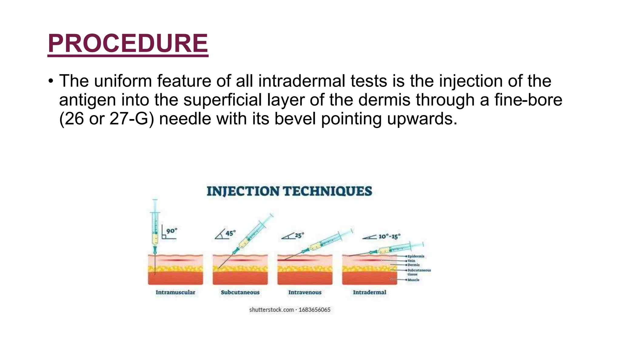
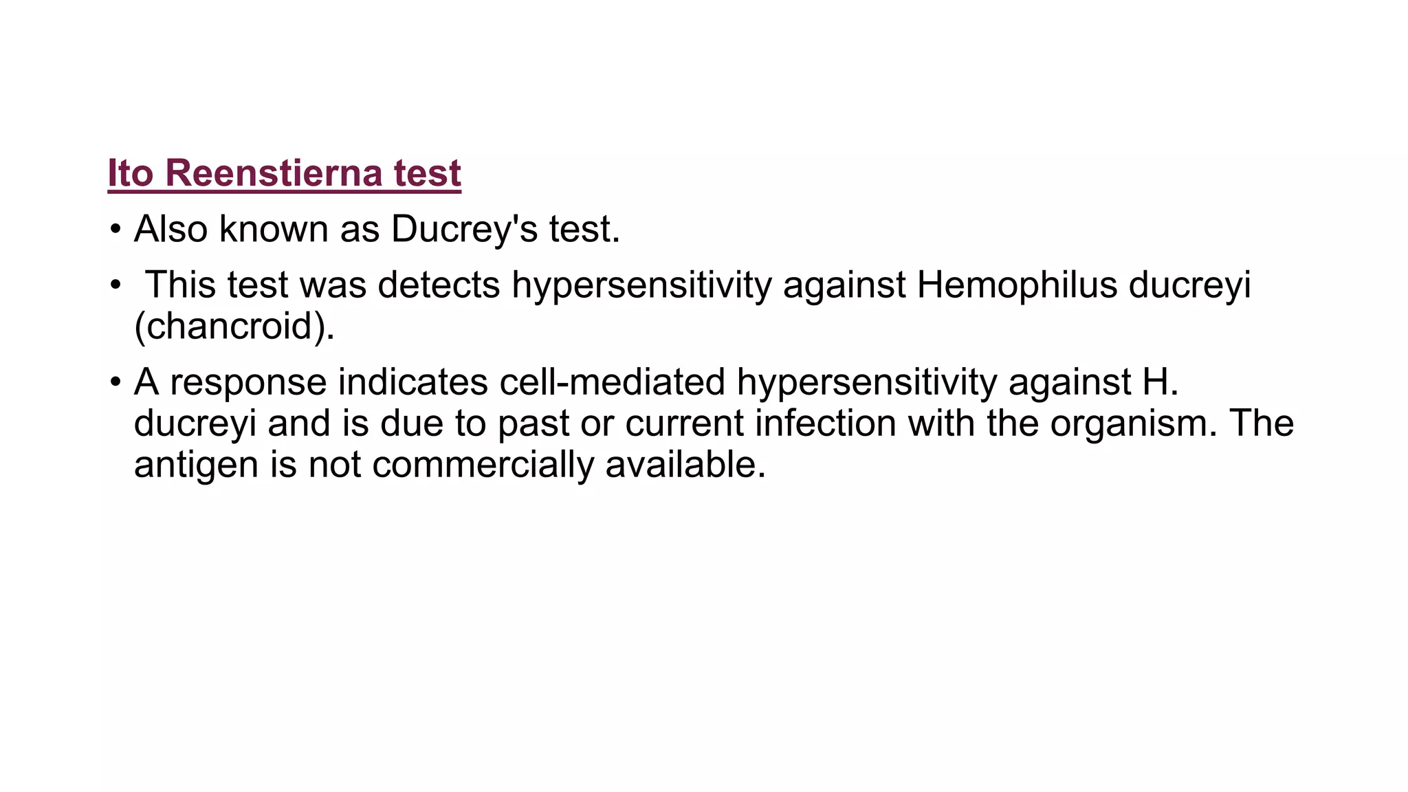
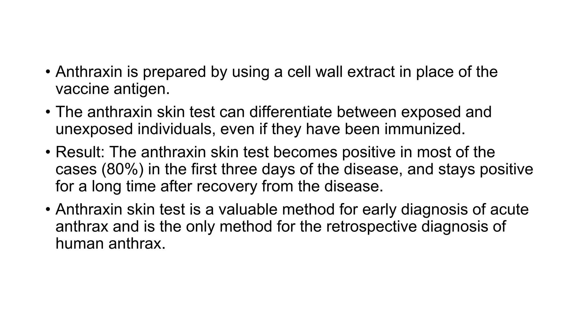
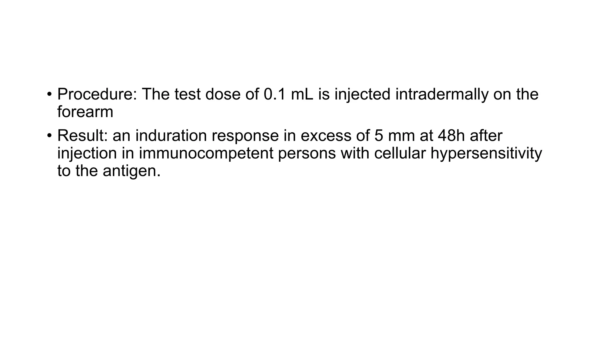
SlideShare a Scribd company logo
INTRADERMAL TESTS
PRESENTER: Dr Vinayak Akki PGY1
MODERATOR: Dr. A. Venkata Krishna Sir (Professor & HOD)
PAPER INCHARGE : Dr. A. Venkata Krishna Sir (Professor & HOD)
GUIDE:Dr. A. Venkata Krishna Sir (Professor & HOD)
OVERVIEW
• Definition
• Procedure
• Interpretation
• IDT for infectious diseases
Bacterial diseases
Fungal diseases
Parasitic diseases
• IDT for non infectious diseases
DEFINITION
• A test for immunity or hypersensitivity made by injecting a
minute amount of diluted antigen into the skin
• Also called as Intracutaneous test
• For the first time used in 1916
• Intradermal tests are widely used to support the diagnosis of
dermatological and nondermatological diseases.
• They are mainly indicated for the detection of immediate (Type I
hypersensitivity) and delayed type hypersensitivity (DTH, Type
IV hypersensitivity) towards exogenous or endogenous
antigens.
Prerequisites
• Before undertaking intradermal test, it is advisable to stop or
avoid systemic steroids or immunosuppressive agents at least
three days before the procedure as moderate to large doses of
corticosteroids may inhibit a DTH reaction.
• Intradermal testing is in general safe with few reactions and
does not appear to result in sensitization, resuscitative
measures should be kept ready as there remains possibility of
anaphylaxis.
• In case of anaphylaxis supplies needed:
• Epinephrine 1:1000
• Systemic corticosteroid
• Systemic antihistamine
• Oxygen
Contraindications
• Severe eczema,dermographism
• Severe asthma.
• Drug intake such as antihistamines, ACE inhibitors,beta
blockers,antidepressants,calcineurin inhibitors as they affect
result interpretation.
• Acute or chronic UV B radiation exposure in the skin test area.
• Pregnancy
Side effects:
• Irritation
• Itching
• Anaphylaxis
• Granuloma formation in TST
• Exacerbation of asthma
PROCEDURE
• The uniform feature of all intradermal tests is the injection of the
antigen into the superficial layer of the dermis through a fine-bore
(26 or 27-G) needle with its bevel pointing upwards.
• The quantity injected may vary from 0.01 to 0.1 ml but conventionally
0.1 ml is universally used.
• Although the tests could be done at any site, the proximal part of the
flexor aspect of the forearm is conventionally used.
• This site can be conveniently exposed and the skin of the proximal
area is more sensitive.
INTERPRETATION OF INTRADERMAL
TESTS
• The optimal time for reading the reaction depends upon the
pharmacological agent used for the test and the type of
immunological reaction to be observed.
• IDT for drug sensitivity are read at 15 minutes.
• IDT for the detection of DTH are read at 48h, although they can be
read as early as 12h and as late as 4 days.
• The size of the induration is more important than erythema while
interpreting Type IV hypersensitivity.
• The measurement of a wheal is made by diameter, more
sophisticated methods like volume measurements and Doppler flow
have been used.
• If the wheal is not circular, an approximation may be made by
averaging maximum/minimum diameters, or more accurately the
area may be calculated by the formula D1*D2*π/4, where D1 and D2
are the maximum and minimum diameters .
• For irregular wheal's, a tracing may be made on squared paper.
Pseudopodia should be noted, but for measurement of diameter they
are ignored.
• Attempts to assess the volume of a wheal are less satisfactory for
routine use
• The size of the wheal is not directly proportional to the dose of the
active agent, but may vary with the total volume of fluid injected.
• For accurate quantitative observations, wheal diameters below 4 mm
or above 15 mm cannot be relied upon.
INTRADERMAL TESTS ARE BROADLY
DIVIDED INTO TWO CATEGORIES
A. INTRADERMAL TESTS USED FOR INFECTIOUS DISEASES.
B. INTRADERMAL TESTS USED FOR NON-INFECTIOUS
DISEASES.
INTRADERMAL TESTS USED FOR INFECTIOUS DISEASES
IDT USED FOR BACTERIAL
INFECTIONS
• Tuberculin test
• Lepromin test
• Frei's test
• Ito Reenstierna test
• Anthraxin test
• Foshay test
• Dick's test
IDT FOR FUNGAL INFECTIONS
• Trichophytin test
• Candidin test
• Coccidioidin test
• Histoplasmin test
IDT FOR PARASITIC INFECTIONS
• Leishmanin test
• Casoni test
• Onchocerca skin test
INTRADERMAL TESTS USED FOR NON INFECTIOUS
DISEASES
• Intradermal sensitivity test for common allergens
• Predictive sensitization tests
• Autologous serum skin testing (ASST)
• Kveim-Siltzbach test
• Pathergy test
• Auto erythrocyte sensitization test
• Histamine test
• Pilocarpine test
INTRADERMAL TESTS USED FOR INFECTIOUS
DISEASES
IDT USED FOR BACTERIAL INFECTIONS
Tuberculin test
• Also known as Pirquet's test/reaction or scarification test.
• Antigen used - Tuberculin, a purified protein extract of bacilli grown on 5%
glycerol broth (ultra filtrate of tubercle bacilli containing over 200 ag shared
with bovine BCG and many NTM)
• Routinely used is PPD equivalent to 1 TU of RT23 strain with tween 80.
• PROCEDURE: 0.1ml is injected intradermally on the flexor aspect of the
forearm and a reading taken after 48h-72h (Mantoux method).
• RESULT: Induration measuring more than 10 mm in diameter is considered
to be a positive response while that measuring less than 5 mm is
considered as negative.
• A positive test indicates past or present infection with M. tuberculosis
(latent or clinical) or vaccination with bacillus Calmette-Guerin (BCG) or
other mycobacteria.
• A tuberculin test represents DTH but this response is not associated
with protective immunity against M.tuberculosis.
• A positive test does not indicate active infection except in children
younger than two years and is directly proportional to the strength of the
reaction.
• An induration of more than 15 mm is usually not due to BCG
vaccination.
• Readings between 6 mm and 9 mm are doubtful and could be because
of an atypical mycobacterial infection.
5 mm considered + for high risk group
which includes – close contacts,
radiographic abnormalities, HIV, on
corticosteroids, on
immunosuppressive drugs.
10 mm considered + for migrants in
endemic areas, health care
workers, homeless, silicosis, DM,
CKD
15 mm considered + for no risk persons
• A negative tuberculin test can also occur due to technical errors.
• A negative tuberculin test indicates non-exposure or decreased or
absent delayed hypersensitivity to M. tuberculosis as in HIV
infection/AIDS, disseminated tuberculosis, primary
immunodeficiency disorders of CMI, lymphoreticular malignancies,
sarcoidosis, etc.
• A repeat test is not advocated before one week as the tuberculin
injected for the first test has a booster effect on the subsequent
dose.
• Small non necrotising response is consistent with protection.
• Strong reaction to tuberculin directly correlates with prevalence of
active Tb.
• Weaker reaction does not correlate with the active Tb and may be
due to other infection or waning of reactivity due to ageing
(reversion).
• TST can act as a triggering factor for granuloma annulare
• TST is recommended in the work up of patients with sarcoidosis
• TST is routinely recommended before starting steroids,
immunosuppressive, immunomodulator drugs and biologicals
• Tuberculin's from atypical mycobacteria or environmental bacteria
have also been prepared.
• They include PPD-B for Battey mycobacteria, PPD-Y for M. kansasii
, SCROFULIN for M scrofulaceum and BURULI for M. ulcerans .
• Lack of specificity is the major drawback in population vaccinated
with BCG
• Now TST is being replaced by IGRA (interferon gamma release
assay) & NAAC ( nucleic acid amplification)
Disease Reactivity
Primary inoculation Tb - At beginning, + later
Scrofuloderma +
Orificial tb Usually +
Ac cutaneous tb -
Metastatic tuberculous abscess Usually + but can be – due to poor GC
Lupus vulgaris Strongly +
Tuberculids Strongly + but bx & culture -
Lichen scrofulosum Strongly + and can even ulcerate
Papulonecrotic tuberculid + and necrotic r/n
Erythema induration of bazin +
Lupus miliaris disseminated faciei +
Tuberculous mastitis +
Lepromin test
• It is a prognostic test which helps in classifying leprosy.
• It cannot be used for diagnostic purpose since it is positive in a
significant number of normal people and is negative towards the
lepromatous pole of the disease.
• Antigens used – 1. Mitsuda lepromin, an autoclaved suspension of
tissue (whole bacilli) obtained from experimentally infected
armadillos (Lepromin A)
2. Dharmendra lepromin, a purified chloroform-ether extracted
suspension of M. leprae (fractionated bacilli with soluble protein
component). More specific.
In India, lepromin antigen is prepared by the Central Leprosy
Teaching and Research Institute, Chengalpattu,Tamil Nadu.
• Result: The response after intradermal injection is biphasic, with an
early Fernandez reaction (48-72 hours – erythema with induration)
and a late Mitsuda reaction( 4 weeks – nodule/ulceration).
• Both responses are manifestations of CMI towards the antigen.
Response Time Reading Inference Remarks
Fernandez
reaction
48-
72h
Erythema or induration <5 mm
Erythema or induration 5-
10mm
Erythema or induration 11-
15mm
Erythema or induration >16mm
Negative
Weakly positive(+)
Moderately
positive(++)
Strongly positive
(+++)
More prominent with
Dharmendra
Lepromin
Measure of existing DTH
Mitsuda
reaction
3-4
weeks
Nothing to see/feel
Papule<3mm
Erythematous papule 4-7 mm
Erythematous papule 7-10 mm
Erythematous papule >10 mm
or
One of any size with ulceration.
Negative
Doubtful
Positive (+)
Positive (++)
Positive (+++)
More prominent with mistuda
Lepromin indicates the ability to
generate a cell mediated
immune response
• In immune competent person early lepromin reaction indicates a
state of sensitisation against M leprae & was regarded as evidence
for prior infection
• Late reaction represents CMI in patients & normal persons
• Incidence of LL is higher in sub clinically infected & Mitsuda negative
persons
• Test is positive in TT & BT with increase in sensitivity in T1R &
negative in other types.
Provides immunological aid in classification
Spectrum TT BT BB BL LL
Reactivity +++ +/++ -/+WEAKLY - VE -VE
• NEGATIVE in histoid leprosy
• Strongly POSITIVE in HIV + leprosy
Limitations :
• Low specificity
• Paucity of human leproma
• Reduced availability of test subjects
MEDINA TEST:
• Similar to lepromin test but antigen is prepared from lesions of lucio
leprosy
• Second generation based tests – soluble antigen lipoarabinomannan
(MLSA-LAM)
• MLCW antigen to test cell proliferation & CMI
Frei's test
• Frei's test was developed in 1925 for lymphogranuloma venereum
(LGV)
• Antigen used - Frei's antigen.
• It indicates delayed hypersensitivity to an intradermal standardized
antigen prepared from chlamydia grown in the yolk sac of a chick
embryo.
• Result: The test is read after 48h and again on the fourth day.
A nodule more than 5mm at fourth day is considered a positive
response.
• A positive test indicates past or present chlamydial infection.
• Frei's test becomes positive two to eight weeks after infection.
• Frei's antigen is common to all chlamydial species and is not
specific to LGV.
• Due to its non specific nature, the test is no longer used.
Ito Reenstierna test
• Also known as Ducrey's test.
• This test was detects hypersensitivity against Hemophilus ducreyi
(chancroid).
• A response indicates cell-mediated hypersensitivity against H.
ducreyi and is due to past or current infection with the organism. The
antigen is not commercially available.
Anthraxin test
• The anthraxin skin test measures DTH to anthrax antigens is used
as a diagnostic tool.
• It has also been recommended by the WHO for evaluation of
immunological memory against anthrax.
• Antigen used - Anthraxin, a cell wall extract from the vegetative, non
capsulated strain of B. anthracis (Sterne strain), consists mainly of a
complex of peptidoglycans and polysaccharides.
• Anthraxin is prepared by using a cell wall extract in place of the
vaccine antigen.
• The anthraxin skin test can differentiate between exposed and
unexposed individuals, even if they have been immunized.
• Result: The anthraxin skin test becomes positive in most of the
cases (80%) in the first three days of the disease, and stays positive
for a long time after recovery from the disease.
• Anthraxin skin test is a valuable method for early diagnosis of acute
anthrax and is the only method for the retrospective diagnosis of
human anthrax.
Foshay test
• The test involves intradermal injection of a suspension of killed
Bartonella henselae , the causative agent of cat-scratch disease.
• Antigen used - The antigen is prepared from the sterile lymph node
material obtained from a patient of cat-scratch disease.
• Result: The appearance of an area of erythema more than 5 mm
diameter after 48h at the injection site is considered a positive
reaction.
• A positive test is a sign of past or present infection.
• The test is of historical importance only, no longer used for diagnosis
because of concerns about the transmission of the organism.
Dick's test
• This test was used to identify children susceptible to scarlet fever,
but is of historical value as scarlet fever is no longer a common or
serious disease.
• The antigen used is pyrogenic exotoxin or Dick's or scarlatinal toxin
of Streptococcus pyogenes . It is also known as erythrogenic toxin
because intradermal injection causes an erythematous reaction in a
susceptible person.
• Procedure: About 0.1 cc of the toxin in the dilution of 1:1000 is
injected intradermally on the forearm.
• Result: the response is read after 24h.
• The reaction is considered positive when there is an erythema more
than 5 mm in diameter and strongly positive if induration also develops.
• A positive reaction develops after 8-12h and reaches its maximum after
24h.
• It is negative in convalescent patients and in insusceptible individuals.
IDT FOR FUNGAL INFECTIONS
Trichophytin test
• This test is performed to detect allergic hypersensitivity towards
dermatophytes.
• Antigen used - Trichophytin antigen is a glycopeptide extracted
(using acetone-ethylene glycol extraction method) from the spores
and mycelia of dermatophytes, mostly Trichophyton
mentagrophytes.
• Procedure: Trichophytin in the concentration of either 10 μg, 1 μg or
0.1 μg in 0.1 ml of normal saline is injected intradermally into the
flexor forearm.
• Result: The test is read after 20 min for an immediate reaction and
after 24 or 72h for a delayed response.
• Erythema, edema or induration may be seen and a papulovesicular
or ulcerative lesion can also occur.
• A wheal larger than 10 mm in diameter at 20 min and induration
more than 5 mm at 72h is considered a positive response.
Candidin test
• Hypersensitivity towards Candida albicans is universal.
• Hence, a test cannot be used to diagnose the infection.
• The candidin test serves as an aid to evaluate the cellular immune response
in patients suspected of having reduced CMI.
• Antigen used - Candidin is used as a recall antigen for detecting DTH by
intradermal testing.
• Candidin is made from the culture filtrate and cells of two strains of Candida
albicans .
• Compared with healthy controls, reactivity to candidin is significantly
reduced in patients with AIDS, Hodgkin's disease and sarcoidosis(tuberculin
sensitivity is more reliable in the latter case).
• Procedure: The test dose of 0.1 mL is injected intradermally on the
forearm
• Result: an induration response in excess of 5 mm at 48h after
injection in immunocompetent persons with cellular hypersensitivity
to the antigen.
Coccidioidin test
• Antigens used - two antigens available: coccidioidin and spherulin.
• Coccidioidin is prepared from autolysates of the mycelial phase (saprophytic
phase) of Coccidioides immitis , and spherulin, a more sensitive reagent,
from the spherule phase (parasitic phase).
• A positive test is helpful in detecting exposure to the fungus Coccidioides
immitis .
• It can help in differentiating the chronic pulmonary disease caused by this
fungus from other chronic cavitary, nodular and fibrotic diseases like
tuberculosis and malignancies.
• The test is negative in patients with disseminated disease or in patients with
thin-walled cavities.
Histoplasmin test
• Histoplasmin skin testing is useful in epidemiological studies, such
as investigations of case cluster or the definition of endemic areas
but is not predictive of histoplasmosis.
• An antigen containing the M and H precipitins of Histoplasma
capsulatum tends to elicit both immediate and delayed
hypersensitivity, and hence more false-positive reactions.
• An antigen that is deficient in the M component can reduce this
problem.
IDT FOR PARASITIC INFECTIONS
Leishmanin test (Montenegro test)
Antigen used - Leishmanin, which contains killed Leishmania.
• A positive reaction consists of a palpable nodule more than 5 mm in
diameter developing in 48 to 72h and indicates DTH, but not
necessarily immunity, to leishmania organisms.
• The test is not species-specific.
• It becomes positive early in the course of cutaneous or
mucocutaneous leishmaniasis (except in diffuse cutaneous
leishmaniasis) and only after recovery from visceral leishmaniasis.
• It is highly sensitive for cutaneous leishmaniasis.
• It is a useful and important tool for epidemiological, immunological,
and diagnostic studies and is an essential component of vaccine
trials.
• However, it cannot distinguish between present and past infection.
Casoni test:
• Done for Hadatid disease or Echinococcosis.
• Intradermal injection of 0.2 mL of fresh sterile hydatid fluid or
hydatid antigen produces in half an hour a 5 cm wheal with
multiple pseudopodia fading in an hour in positive cases.
• Normal saline is used as a control. A rapid slide test has been
proposed for resource poor settings.
Onchocerca skin test
• The intradermal test with products from Onchocerca volvulus is highly
sensitive in the detection of active onchocerciasis.
• However, a few reports indicate decreased CMI to parasite-derived
antigens, delayed skin-test reactivity, and response to unrelated
antigens during Onchocerca volvulus infection.
• Intradermal tests in various skin or systemic infections thus serve an
important role in reaching a diagnosis and predicting the prognosis of
these conditions.
INTRADERMAL TESTS USED FOR NON
INFECTIOUS DISEASES
Intradermal sensitivity test for drug
• Intradermal testing is a rapid, convenient and reproducible method of
detecting drug hypersensitivity (drug-specific IgE antibodies).
• It is commonly performed for penicillin, general and local anaesthetic
agents, tetanus toxoid, iodinated radiocontrast media, insulin,
heterologous sera, collagen chymopapain etc.
• Intradermal testing is in general safe with few reactions and does not
appear to result in sensitization.
• However, resuscitative measures should be kept ready as there
remains possibility of anaphylaxis.
• Penicillin intradermal skin tests should be carried out using major
determinant (benzyl penicilloyl polylysine, PPL)and minor
determinant mixture (benzyl penicillin, benzyl penicilloate and benzyl
penilloate) antigens.
• The test can also be performed using 2-10 units of freshly prepared
penicillin.
• Skin test is read at 20 minutes.
Result:
Negative response no increase in size of original bleb
and no greater reaction than the
control site
Ambiguous response wheal only slightly larger than initial
injection bleb, with or without
accompanying erythematous flare
and slightly larger than the control
site; OR discordance between
duplicates
Positive response itching and significant increase in
size of original blebs to at least 5mm.
Wheal may exceed 20 mm in
diameter and exhibit pseudopods
• Negative intradermal test does not rule out penicillin sensitivity.
• Other agents are tested similarly for detection of immediate and
delayed type of hypersensitivity.
• Intradermal skin tests have no predictive value in non IgE-mediated
reactions such as serum sickness, haemolytic anaemia, drug fever,
interstitial nephritis, contact dermatitis, maculopapular exanthemata or
exfoliative dermatitis.
• Skin testing is contraindicated where there is a history of exfoliative
dermatitis, Stevens-Johnson syndrome or TEN.
Predictive sensitization tests
• Sensitization index is the relative capacity of a given agent to induce
sensitization in a group of humans or animals.
• Predictive sensitization tests are used to compare the sensitizing
properties of new products or chemicals with those of known
substances.
• Both in guinea-pigs and humans, an estimate of the sensitizing
potential can be performed using intradermal route.
• Draize test and Freund's complete adjuvant test are intradermal
methods of testing sensitization potential.
Autologus serum skin testing (ASST)
• Autologous serum skin test is a simple in-vivo clinical test for the detection
of basophil histamine releasing activity and to diagnose chronic autoimmune
urticaria among chronic spontaneous urticaria patients.
• About 25-45% of patients of chronic idiopathic urticaria have autoantibodies
against the high affinity IgE receptor FceRI or IgE that are capable of
histamine release.
• These antibodies, if present in serum, can cause wheal and erythema
following intradermal serum injection. This reaction forms the basis of the
ASST.
• Procedure: The test is performed by injecting 0.05 ml of the patient's own
serum intradermally into the left flexor forearm two inches below the
antecubital crease and a saline control into the right forearm.
• Serum is obtained after withdrawing 5 ml of venous blood and
standing for about 45 min for separation.
• The process can be hastened up by centrifugation of the blood.
• About 5 ml of venous blood was collected in a sterile vacutainer and
allowed to clot at room temperature for 30 min. Serum was
centrifuged at 2000 rpm for 15 min and 0.05 ml of autologous serum
was injected intradermally, in uninvolved skin, using a 1 ml insulin
syringe (30 gauge needle) into the right forearm 2 cm below the
cubital fossa.
• Similarly, 0.05 ml of 0.9% sterile normal saline (negative control) was
injected intradermally proximally into the left forearm, and at a
distance of at least 5 cm, 0.05 ml of histamine (10 μg/ml) was
injected distally into the left forearm as a positive control.
• A serum induced erythematous wheal with a diameter of 1.5 mm
more than the saline induced response within 30 min was taken as
positive
• ASST positivity has been found to correlate well with the severity
and duration of attack of urticaria.
• The cases of idiopathic urticaria which are ASST positive are
designated as autoimmune urticaria.
• Western blot, ELISA, flow cytometry may be useful for screening in
the future
Intradermal allergy testing
• A small amount of diluted allergen is injected into the dermis.
• It is carried out with allergen concentrations 100 to 1000 times less
than that used for skin prick tests. It increases the sensitivity but
decreases the specificity of the test.
• It is most commonly used in testing for drug and venom allergy.
• IDSTs have a very high non-specific reaction rate and are not
recommended for testing with inhalants or foods and food allergens.
• Moreover, intradermal tests carry a higher risk of adverse reactions
than SPTs.
Kveim-Siltzbach test
• It is a valuable intradermal test for the diagnosis of sarcoidosis.
• The test can be utilized to differentiate from other causes of diffuse
pulmonary mottling, uveitis and erythema nodosum.
• The antigen is prepared from the splenic tissue obtained from a
proven case of sarcoidosis either after splenectomy or during
autopsy.
• About 0.1-0.15 ml of this antigen is injected intradermally, a nodule
develops after four to six weeks, which can be biopsied for
histopathological confirmation of the diagnosis.
• False positive reactions were found in an appreciable proportion of
patients with Crohn's disease, ulcerative colitis and tuberculous
lymphadenitis, but only with few batches of commercially available
antigens.
Pathergy test
• Pathergy is the development of a papulopustular lesion around a
puncture site on the skin, 24-48h after the injection of a sterile
substance like normal saline intradermally. This phenomenon forms
the basis of the pathergy test.
• The test is used as a diagnostic criterion for Behcet’s disease.
• Results of the test depend upon the type of needle used; reactivity
varies with the diameter and sharpness of needle.
• The sensitivity and intensity of the reaction is considerably less with
sharp needles and needles with smaller diameter.
• Pathergy is also a reported phenomenon in pyoderma gangrenosum,
in hairy cell leukaemia, Hodgkin's lymphoma and in chronic myeloid
leukaemia treated with interferon alpha.
• Histopathological evaluation of the test is not found to be more
sensitive than the clinical evaluation.
Auto erythrocyte sensitization test
• The intradermal test for the diagnosis of auto erythrocyte
sensitization syndrome is done with washed RBCs of the patient in
the intrascapular region with a saline control on the opposite side.
• Patient develops a painful ecchymotic reaction within two hours at
the site of the injection indicating positive test.
• The control site does not show reaction.
Histamine test
• When histamine is injected intradermally, it causes bright red
histamine flare due to capillary vasodilatation.
• However, this effect is due to axon reflex within dermal nerves.
• The histamine test can be used to test integrity of dermal nerves in
cases of tuberculoid leprosy.
• One drop of histamine acid diphosphate 1 in 1000 (1mg/ml) is
placed on the skin surface, one each on the normal skin and the
other on the suspected patch. Superficial prick is made through the
drop. The histamine solution is wiped off and the area is watched for
5 minutes.
• Response takes little longer on the extremities.
• In the normal skin, a wheal at the site of prick and a flare of 2 cm
diameter surrounding the wheal would be seen (test labeled as
positive).
• The flare disappears in 10 minutes.
• In the patch of leprosy the flare is impaired or absent and only a
wheal is seen (test negative). The flare is feeble and delayed in
indeterminate and borderline leprosy, and absent in a tuberculoid
lesion.
• The response in the skin to histamine depends on the integrity of
nerves of the autonomic nervous system.
• Autonomic nerves are nonmyelinated nerve fibers sheathed only by
Schwann cells and are distributed along with the small dermal blood
vessels.
• In early leprosy, which manifests as localized skin patches, the
Schwann cells are parasitized by M. leprae.
• There is perivascular and perineural inflammation and the functions
of sympathetic nerves of the skin supplying the blood vessels are
impaired, even before the sensory nerves are affected.
• The test is useful only to differentiate hypopigmented macules of
leprosy from those of other skin diseases.
• There will be a normal flare (positive test) in other hypopigmented
macules due to non leprosy conditions like vitiligo, pityriasis alba,
fungal infections, etc.
• The histamine test will also be negative in patients without any skin
changes but only with areas of nerve deficit due to other peripheral
neuropathies.
• In patients with dark skin this test may not be useful because the
flare is not easily visible.
Pilocarpine test
• Pilocarpine intradermal test is used to detect the integrity of dermal
nerves in suspected cases of tuberculoid leprosy.
• About 0.2 ml of 1 in 1000 solution of pilocarpine nitrate is injected
intradermally into the lesion, the injection site is then painted with
tincture of iodine and then dusted with starch powder.
• Sweating, if present, causes blue discoloration of the powder.
• Alternatively, quinizarin powder can be used in place of starch
powder with the advantage that there is no need for painting the test
site with tincture of iodine.
Prausnitz - Kustner (PK) Test
• Used to test people for life threatening allergens such as bee venom
• Test involves transferring serum from the test subject to another
healthy person.
• Serum from allergic patient is injected into healthy person and 24
hrs later suspected antigen is injected intradermally into healthy
person.
• If allergic patient had developed antibodies, this will cause local
reaction in the healthy person when the antibodies mix with
antigen
• A positive PK test appears as wheal and flare demonstrating type 1
hypersensitivity reaction reaction.
• No longer recommended now.
Therapeutic uses of IDT:
Autoinoculation :
• Done in viral warts, by excising ,mincing and implanting
homogeneous tissue in a small dermal pocket in forearm.
• Antibodies thus produced help in clearing the warts
• It’s an effective treatment modality for multiple and recurrent
warts.
Autologous serum therapy:
• Used in Chronic urticaria.
• Repeated injections of autologous whole blood or serum can induce
desensitisation or tolerisation of auto reactive chronic urticaria
patients to pro inflammatory signals expressed in their circulation.
REFERENCES
• Rook’s textbook of dermatology
• IAL textbook of leprosy
• IADVL textbook of dermatology
• IJDVL
• Internet
intradermal tests.pptx

More Related Content

What's hot

Suture care
Suture careSuture care
Suture care
arunmtin
 
suturing and care of drainage
suturing and care of drainagesuturing and care of drainage
suturing and care of drainage
Mahesh Chand
 
Colostomy care
Colostomy careColostomy care
Colostomy care
Sam Asir
 
Suture Removal-Stitch Out Protocol
Suture Removal-Stitch Out ProtocolSuture Removal-Stitch Out Protocol
Suture Removal-Stitch Out Protocol
Salahuddin Chaudhry
 
Topical application
Topical applicationTopical application
Topical application
JayaR62
 
Assisting with Cast application and removal.pptx
Assisting with Cast application and removal.pptxAssisting with Cast application and removal.pptx
Assisting with Cast application and removal.pptx
AbdiWakjira2
 
ABDOMINAL PARACENTESIS
ABDOMINAL PARACENTESISABDOMINAL PARACENTESIS
Dermatitis ppt
Dermatitis pptDermatitis ppt
Dermatitis ppt
ROMAN BAJRANG
 
Drain care
Drain care Drain care
Drain care
wcmc
 
Abdominal paracentesis
Abdominal paracentesis Abdominal paracentesis
Intradermal
IntradermalIntradermal
Bcls
BclsBcls
Surgical wound dressing
Surgical wound dressingSurgical wound dressing
Surgical wound dressing
Slideshare User
 
Insertion of NG tube
Insertion of NG tubeInsertion of NG tube
Insertion of NG tube
KHyati CHaudhari
 
Pre hospital iv maintenance
Pre hospital iv maintenancePre hospital iv maintenance
Pre hospital iv maintenance
VASS Yukon
 
Urinary Catheterization
Urinary CatheterizationUrinary Catheterization
Urinary Catheterization
Ahmad Thanin
 
Wound Dressing
Wound DressingWound Dressing
Wound Dressing
Cikbungazafieya Zawani
 
Wound dressing
Wound dressingWound dressing
Wound dressing
Nelson Munthali
 
Catheterisation
CatheterisationCatheterisation
Catheterisation
Deblina Roy
 
Chest tube drainage
Chest tube drainageChest tube drainage
Chest tube drainage
Mahalakshmi Lakshmanan
 

What's hot (20)

Suture care
Suture careSuture care
Suture care
 
suturing and care of drainage
suturing and care of drainagesuturing and care of drainage
suturing and care of drainage
 
Colostomy care
Colostomy careColostomy care
Colostomy care
 
Suture Removal-Stitch Out Protocol
Suture Removal-Stitch Out ProtocolSuture Removal-Stitch Out Protocol
Suture Removal-Stitch Out Protocol
 
Topical application
Topical applicationTopical application
Topical application
 
Assisting with Cast application and removal.pptx
Assisting with Cast application and removal.pptxAssisting with Cast application and removal.pptx
Assisting with Cast application and removal.pptx
 
ABDOMINAL PARACENTESIS
ABDOMINAL PARACENTESISABDOMINAL PARACENTESIS
ABDOMINAL PARACENTESIS
 
Dermatitis ppt
Dermatitis pptDermatitis ppt
Dermatitis ppt
 
Drain care
Drain care Drain care
Drain care
 
Abdominal paracentesis
Abdominal paracentesis Abdominal paracentesis
Abdominal paracentesis
 
Intradermal
IntradermalIntradermal
Intradermal
 
Bcls
BclsBcls
Bcls
 
Surgical wound dressing
Surgical wound dressingSurgical wound dressing
Surgical wound dressing
 
Insertion of NG tube
Insertion of NG tubeInsertion of NG tube
Insertion of NG tube
 
Pre hospital iv maintenance
Pre hospital iv maintenancePre hospital iv maintenance
Pre hospital iv maintenance
 
Urinary Catheterization
Urinary CatheterizationUrinary Catheterization
Urinary Catheterization
 
Wound Dressing
Wound DressingWound Dressing
Wound Dressing
 
Wound dressing
Wound dressingWound dressing
Wound dressing
 
Catheterisation
CatheterisationCatheterisation
Catheterisation
 
Chest tube drainage
Chest tube drainageChest tube drainage
Chest tube drainage
 

Similar to intradermal tests.pptx

Mendel mantoux-test
Mendel mantoux-testMendel mantoux-test
Mendel mantoux-test
Zaid Azhar
 
Detection of cellular immunity.pptx
Detection of cellular immunity.pptxDetection of cellular immunity.pptx
Detection of cellular immunity.pptx
anitmaryjoselin
 
TREATMENT of tb.pptx
TREATMENT of tb.pptxTREATMENT of tb.pptx
TREATMENT of tb.pptx
SahilVerma19852
 
The mantoux test
The mantoux testThe mantoux test
The mantoux test
Azizah Mh
 
Hand infection - An often ignored problem
Hand infection - An often ignored problemHand infection - An often ignored problem
Hand infection - An often ignored problem
Ahmed Suparno Bahar Moni
 
EPIDEMIOLOGY OF TUBERCULOSIS
EPIDEMIOLOGY OF TUBERCULOSISEPIDEMIOLOGY OF TUBERCULOSIS
EPIDEMIOLOGY OF TUBERCULOSIS
MAHESWARI JAIKUMAR
 
Antibiotics surgery
Antibiotics surgeryAntibiotics surgery
Antibiotics surgery
sauvik2014
 
Antibiotic-Presentation.pptx
Antibiotic-Presentation.pptxAntibiotic-Presentation.pptx
Antibiotic-Presentation.pptx
Reena Kulshrestha
 
Antibiotics treatment & management
Antibiotics treatment & management Antibiotics treatment & management
Antibiotics treatment & management
Baiti Basheer
 
6949_antibioticinsurgery.pptx
6949_antibioticinsurgery.pptx6949_antibioticinsurgery.pptx
6949_antibioticinsurgery.pptx
Gokul Krishnan
 
Antigen & Antibody Interactions
Antigen & Antibody InteractionsAntigen & Antibody Interactions
Antigen & Antibody Interactions
Guddeti Prashanth Kumar
 
Antimicrobial Stewardship 2014
Antimicrobial Stewardship 2014Antimicrobial Stewardship 2014
Antimicrobial Stewardship 2014
BBrauer25
 
Antimicrobial stewardship 2014 (1)
Antimicrobial stewardship 2014 (1)Antimicrobial stewardship 2014 (1)
Antimicrobial stewardship 2014 (1)
BBrauer25
 
Future directions in neonatal sepsis
Future directions in neonatal sepsisFuture directions in neonatal sepsis
Future directions in neonatal sepsis
abdullah alzahrani
 
Pid
PidPid
Pelvic Inflammatory Disease
Pelvic Inflammatory DiseasePelvic Inflammatory Disease
Pelvic Inflammatory Disease
limgengyan
 
Paediatric immunization
Paediatric immunizationPaediatric immunization
Paediatric immunization
Chacha Piru
 
Paediatric immunization
Paediatric immunizationPaediatric immunization
Paediatric immunization
Chacha Piru
 
antitubercular drug susceptibility testing
antitubercular drug susceptibility testingantitubercular drug susceptibility testing
antitubercular drug susceptibility testing
Malathi Murugesan
 
INVESTIGATIONS AND TREATMENT OF TUBERCULOSIS.pptx
INVESTIGATIONS AND TREATMENT OF TUBERCULOSIS.pptxINVESTIGATIONS AND TREATMENT OF TUBERCULOSIS.pptx
INVESTIGATIONS AND TREATMENT OF TUBERCULOSIS.pptx
vanshikauppal1
 

Similar to intradermal tests.pptx (20)

Mendel mantoux-test
Mendel mantoux-testMendel mantoux-test
Mendel mantoux-test
 
Detection of cellular immunity.pptx
Detection of cellular immunity.pptxDetection of cellular immunity.pptx
Detection of cellular immunity.pptx
 
TREATMENT of tb.pptx
TREATMENT of tb.pptxTREATMENT of tb.pptx
TREATMENT of tb.pptx
 
The mantoux test
The mantoux testThe mantoux test
The mantoux test
 
Hand infection - An often ignored problem
Hand infection - An often ignored problemHand infection - An often ignored problem
Hand infection - An often ignored problem
 
EPIDEMIOLOGY OF TUBERCULOSIS
EPIDEMIOLOGY OF TUBERCULOSISEPIDEMIOLOGY OF TUBERCULOSIS
EPIDEMIOLOGY OF TUBERCULOSIS
 
Antibiotics surgery
Antibiotics surgeryAntibiotics surgery
Antibiotics surgery
 
Antibiotic-Presentation.pptx
Antibiotic-Presentation.pptxAntibiotic-Presentation.pptx
Antibiotic-Presentation.pptx
 
Antibiotics treatment & management
Antibiotics treatment & management Antibiotics treatment & management
Antibiotics treatment & management
 
6949_antibioticinsurgery.pptx
6949_antibioticinsurgery.pptx6949_antibioticinsurgery.pptx
6949_antibioticinsurgery.pptx
 
Antigen & Antibody Interactions
Antigen & Antibody InteractionsAntigen & Antibody Interactions
Antigen & Antibody Interactions
 
Antimicrobial Stewardship 2014
Antimicrobial Stewardship 2014Antimicrobial Stewardship 2014
Antimicrobial Stewardship 2014
 
Antimicrobial stewardship 2014 (1)
Antimicrobial stewardship 2014 (1)Antimicrobial stewardship 2014 (1)
Antimicrobial stewardship 2014 (1)
 
Future directions in neonatal sepsis
Future directions in neonatal sepsisFuture directions in neonatal sepsis
Future directions in neonatal sepsis
 
Pid
PidPid
Pid
 
Pelvic Inflammatory Disease
Pelvic Inflammatory DiseasePelvic Inflammatory Disease
Pelvic Inflammatory Disease
 
Paediatric immunization
Paediatric immunizationPaediatric immunization
Paediatric immunization
 
Paediatric immunization
Paediatric immunizationPaediatric immunization
Paediatric immunization
 
antitubercular drug susceptibility testing
antitubercular drug susceptibility testingantitubercular drug susceptibility testing
antitubercular drug susceptibility testing
 
INVESTIGATIONS AND TREATMENT OF TUBERCULOSIS.pptx
INVESTIGATIONS AND TREATMENT OF TUBERCULOSIS.pptxINVESTIGATIONS AND TREATMENT OF TUBERCULOSIS.pptx
INVESTIGATIONS AND TREATMENT OF TUBERCULOSIS.pptx
 

Recently uploaded

Basavarajeeyam - Ayurvedic heritage book of Andhra pradesh
Basavarajeeyam - Ayurvedic heritage book of Andhra pradeshBasavarajeeyam - Ayurvedic heritage book of Andhra pradesh
Basavarajeeyam - Ayurvedic heritage book of Andhra pradesh
Dr. Madduru Muni Haritha
 
#cALL# #gIRLS# In Dehradun ꧁❤8107221448❤꧂#cALL# #gIRLS# Service In Dehradun W...
#cALL# #gIRLS# In Dehradun ꧁❤8107221448❤꧂#cALL# #gIRLS# Service In Dehradun W...#cALL# #gIRLS# In Dehradun ꧁❤8107221448❤꧂#cALL# #gIRLS# Service In Dehradun W...
#cALL# #gIRLS# In Dehradun ꧁❤8107221448❤꧂#cALL# #gIRLS# Service In Dehradun W...
chandankumarsmartiso
 
Ear and its clinical correlations By Dr. Rabia Inam Gandapore.pptx
Ear and its clinical correlations By Dr. Rabia Inam Gandapore.pptxEar and its clinical correlations By Dr. Rabia Inam Gandapore.pptx
Ear and its clinical correlations By Dr. Rabia Inam Gandapore.pptx
Dr. Rabia Inam Gandapore
 
Journal Article Review on Rasamanikya
Journal Article Review on RasamanikyaJournal Article Review on Rasamanikya
Journal Article Review on Rasamanikya
Dr. Jyothirmai Paindla
 
NVBDCP.pptx Nation vector borne disease control program
NVBDCP.pptx Nation vector borne disease control programNVBDCP.pptx Nation vector borne disease control program
NVBDCP.pptx Nation vector borne disease control program
Sapna Thakur
 
Vestibulocochlear Nerve by Dr. Rabia Inam Gandapore.pptx
Vestibulocochlear Nerve by Dr. Rabia Inam Gandapore.pptxVestibulocochlear Nerve by Dr. Rabia Inam Gandapore.pptx
Vestibulocochlear Nerve by Dr. Rabia Inam Gandapore.pptx
Dr. Rabia Inam Gandapore
 
OCT Training Course for clinical practice Part 1
OCT Training Course for clinical practice Part 1OCT Training Course for clinical practice Part 1
OCT Training Course for clinical practice Part 1
KafrELShiekh University
 
Chapter 11 Nutrition and Chronic Diseases.pptx
Chapter 11 Nutrition and Chronic Diseases.pptxChapter 11 Nutrition and Chronic Diseases.pptx
Chapter 11 Nutrition and Chronic Diseases.pptx
Earlene McNair
 
Identification and nursing management of congenital malformations .pptx
Identification and nursing management of congenital malformations .pptxIdentification and nursing management of congenital malformations .pptx
Identification and nursing management of congenital malformations .pptx
MGM SCHOOL/COLLEGE OF NURSING
 
THERAPEUTIC ANTISENSE MOLECULES .pptx
THERAPEUTIC ANTISENSE MOLECULES    .pptxTHERAPEUTIC ANTISENSE MOLECULES    .pptx
THERAPEUTIC ANTISENSE MOLECULES .pptx
70KRISHPATEL
 
Local Advanced Lung Cancer: Artificial Intelligence, Synergetics, Complex Sys...
Local Advanced Lung Cancer: Artificial Intelligence, Synergetics, Complex Sys...Local Advanced Lung Cancer: Artificial Intelligence, Synergetics, Complex Sys...
Local Advanced Lung Cancer: Artificial Intelligence, Synergetics, Complex Sys...
Oleg Kshivets
 
ABDOMINAL TRAUMA in pediatrics part one.
ABDOMINAL TRAUMA in pediatrics part one.ABDOMINAL TRAUMA in pediatrics part one.
ABDOMINAL TRAUMA in pediatrics part one.
drhasanrajab
 
TEST BANK For Community Health Nursing A Canadian Perspective, 5th Edition by...
TEST BANK For Community Health Nursing A Canadian Perspective, 5th Edition by...TEST BANK For Community Health Nursing A Canadian Perspective, 5th Edition by...
TEST BANK For Community Health Nursing A Canadian Perspective, 5th Edition by...
Donc Test
 
Part II - Body Grief: Losing parts of ourselves and our identity before, duri...
Part II - Body Grief: Losing parts of ourselves and our identity before, duri...Part II - Body Grief: Losing parts of ourselves and our identity before, duri...
Part II - Body Grief: Losing parts of ourselves and our identity before, duri...
bkling
 
CHEMOTHERAPY_RDP_CHAPTER 1_ANTI TB DRUGS.pdf
CHEMOTHERAPY_RDP_CHAPTER 1_ANTI TB DRUGS.pdfCHEMOTHERAPY_RDP_CHAPTER 1_ANTI TB DRUGS.pdf
CHEMOTHERAPY_RDP_CHAPTER 1_ANTI TB DRUGS.pdf
rishi2789
 
Osteoporosis - Definition , Evaluation and Management .pdf
Osteoporosis - Definition , Evaluation and Management .pdfOsteoporosis - Definition , Evaluation and Management .pdf
Osteoporosis - Definition , Evaluation and Management .pdf
Jim Jacob Roy
 
Aortic Association CBL Pilot April 19 – 20 Bern
Aortic Association CBL Pilot April 19 – 20 BernAortic Association CBL Pilot April 19 – 20 Bern
Aortic Association CBL Pilot April 19 – 20 Bern
suvadeepdas911
 
Netter's Atlas of Human Anatomy 7.ed.pdf
Netter's Atlas of Human Anatomy 7.ed.pdfNetter's Atlas of Human Anatomy 7.ed.pdf
Netter's Atlas of Human Anatomy 7.ed.pdf
BrissaOrtiz3
 
The Best Ayurvedic Antacid Tablets in India
The Best Ayurvedic Antacid Tablets in IndiaThe Best Ayurvedic Antacid Tablets in India
The Best Ayurvedic Antacid Tablets in India
Swastik Ayurveda
 
Integrating Ayurveda into Parkinson’s Management: A Holistic Approach
Integrating Ayurveda into Parkinson’s Management: A Holistic ApproachIntegrating Ayurveda into Parkinson’s Management: A Holistic Approach
Integrating Ayurveda into Parkinson’s Management: A Holistic Approach
Ayurveda ForAll
 

Recently uploaded (20)

Basavarajeeyam - Ayurvedic heritage book of Andhra pradesh
Basavarajeeyam - Ayurvedic heritage book of Andhra pradeshBasavarajeeyam - Ayurvedic heritage book of Andhra pradesh
Basavarajeeyam - Ayurvedic heritage book of Andhra pradesh
 
#cALL# #gIRLS# In Dehradun ꧁❤8107221448❤꧂#cALL# #gIRLS# Service In Dehradun W...
#cALL# #gIRLS# In Dehradun ꧁❤8107221448❤꧂#cALL# #gIRLS# Service In Dehradun W...#cALL# #gIRLS# In Dehradun ꧁❤8107221448❤꧂#cALL# #gIRLS# Service In Dehradun W...
#cALL# #gIRLS# In Dehradun ꧁❤8107221448❤꧂#cALL# #gIRLS# Service In Dehradun W...
 
Ear and its clinical correlations By Dr. Rabia Inam Gandapore.pptx
Ear and its clinical correlations By Dr. Rabia Inam Gandapore.pptxEar and its clinical correlations By Dr. Rabia Inam Gandapore.pptx
Ear and its clinical correlations By Dr. Rabia Inam Gandapore.pptx
 
Journal Article Review on Rasamanikya
Journal Article Review on RasamanikyaJournal Article Review on Rasamanikya
Journal Article Review on Rasamanikya
 
NVBDCP.pptx Nation vector borne disease control program
NVBDCP.pptx Nation vector borne disease control programNVBDCP.pptx Nation vector borne disease control program
NVBDCP.pptx Nation vector borne disease control program
 
Vestibulocochlear Nerve by Dr. Rabia Inam Gandapore.pptx
Vestibulocochlear Nerve by Dr. Rabia Inam Gandapore.pptxVestibulocochlear Nerve by Dr. Rabia Inam Gandapore.pptx
Vestibulocochlear Nerve by Dr. Rabia Inam Gandapore.pptx
 
OCT Training Course for clinical practice Part 1
OCT Training Course for clinical practice Part 1OCT Training Course for clinical practice Part 1
OCT Training Course for clinical practice Part 1
 
Chapter 11 Nutrition and Chronic Diseases.pptx
Chapter 11 Nutrition and Chronic Diseases.pptxChapter 11 Nutrition and Chronic Diseases.pptx
Chapter 11 Nutrition and Chronic Diseases.pptx
 
Identification and nursing management of congenital malformations .pptx
Identification and nursing management of congenital malformations .pptxIdentification and nursing management of congenital malformations .pptx
Identification and nursing management of congenital malformations .pptx
 
THERAPEUTIC ANTISENSE MOLECULES .pptx
THERAPEUTIC ANTISENSE MOLECULES    .pptxTHERAPEUTIC ANTISENSE MOLECULES    .pptx
THERAPEUTIC ANTISENSE MOLECULES .pptx
 
Local Advanced Lung Cancer: Artificial Intelligence, Synergetics, Complex Sys...
Local Advanced Lung Cancer: Artificial Intelligence, Synergetics, Complex Sys...Local Advanced Lung Cancer: Artificial Intelligence, Synergetics, Complex Sys...
Local Advanced Lung Cancer: Artificial Intelligence, Synergetics, Complex Sys...
 
ABDOMINAL TRAUMA in pediatrics part one.
ABDOMINAL TRAUMA in pediatrics part one.ABDOMINAL TRAUMA in pediatrics part one.
ABDOMINAL TRAUMA in pediatrics part one.
 
TEST BANK For Community Health Nursing A Canadian Perspective, 5th Edition by...
TEST BANK For Community Health Nursing A Canadian Perspective, 5th Edition by...TEST BANK For Community Health Nursing A Canadian Perspective, 5th Edition by...
TEST BANK For Community Health Nursing A Canadian Perspective, 5th Edition by...
 
Part II - Body Grief: Losing parts of ourselves and our identity before, duri...
Part II - Body Grief: Losing parts of ourselves and our identity before, duri...Part II - Body Grief: Losing parts of ourselves and our identity before, duri...
Part II - Body Grief: Losing parts of ourselves and our identity before, duri...
 
CHEMOTHERAPY_RDP_CHAPTER 1_ANTI TB DRUGS.pdf
CHEMOTHERAPY_RDP_CHAPTER 1_ANTI TB DRUGS.pdfCHEMOTHERAPY_RDP_CHAPTER 1_ANTI TB DRUGS.pdf
CHEMOTHERAPY_RDP_CHAPTER 1_ANTI TB DRUGS.pdf
 
Osteoporosis - Definition , Evaluation and Management .pdf
Osteoporosis - Definition , Evaluation and Management .pdfOsteoporosis - Definition , Evaluation and Management .pdf
Osteoporosis - Definition , Evaluation and Management .pdf
 
Aortic Association CBL Pilot April 19 – 20 Bern
Aortic Association CBL Pilot April 19 – 20 BernAortic Association CBL Pilot April 19 – 20 Bern
Aortic Association CBL Pilot April 19 – 20 Bern
 
Netter's Atlas of Human Anatomy 7.ed.pdf
Netter's Atlas of Human Anatomy 7.ed.pdfNetter's Atlas of Human Anatomy 7.ed.pdf
Netter's Atlas of Human Anatomy 7.ed.pdf
 
The Best Ayurvedic Antacid Tablets in India
The Best Ayurvedic Antacid Tablets in IndiaThe Best Ayurvedic Antacid Tablets in India
The Best Ayurvedic Antacid Tablets in India
 
Integrating Ayurveda into Parkinson’s Management: A Holistic Approach
Integrating Ayurveda into Parkinson’s Management: A Holistic ApproachIntegrating Ayurveda into Parkinson’s Management: A Holistic Approach
Integrating Ayurveda into Parkinson’s Management: A Holistic Approach
 

intradermal tests.pptx

  • 1. INTRADERMAL TESTS PRESENTER: Dr Vinayak Akki PGY1 MODERATOR: Dr. A. Venkata Krishna Sir (Professor & HOD) PAPER INCHARGE : Dr. A. Venkata Krishna Sir (Professor & HOD) GUIDE:Dr. A. Venkata Krishna Sir (Professor & HOD)
  • 2. OVERVIEW • Definition • Procedure • Interpretation • IDT for infectious diseases Bacterial diseases Fungal diseases Parasitic diseases • IDT for non infectious diseases
  • 3. DEFINITION • A test for immunity or hypersensitivity made by injecting a minute amount of diluted antigen into the skin • Also called as Intracutaneous test • For the first time used in 1916
  • 4. • Intradermal tests are widely used to support the diagnosis of dermatological and nondermatological diseases. • They are mainly indicated for the detection of immediate (Type I hypersensitivity) and delayed type hypersensitivity (DTH, Type IV hypersensitivity) towards exogenous or endogenous antigens.
  • 5. Prerequisites • Before undertaking intradermal test, it is advisable to stop or avoid systemic steroids or immunosuppressive agents at least three days before the procedure as moderate to large doses of corticosteroids may inhibit a DTH reaction.
  • 6. • Intradermal testing is in general safe with few reactions and does not appear to result in sensitization, resuscitative measures should be kept ready as there remains possibility of anaphylaxis. • In case of anaphylaxis supplies needed: • Epinephrine 1:1000 • Systemic corticosteroid • Systemic antihistamine • Oxygen
  • 7. Contraindications • Severe eczema,dermographism • Severe asthma. • Drug intake such as antihistamines, ACE inhibitors,beta blockers,antidepressants,calcineurin inhibitors as they affect result interpretation. • Acute or chronic UV B radiation exposure in the skin test area. • Pregnancy
  • 8. Side effects: • Irritation • Itching • Anaphylaxis • Granuloma formation in TST • Exacerbation of asthma
  • 9. PROCEDURE • The uniform feature of all intradermal tests is the injection of the antigen into the superficial layer of the dermis through a fine-bore (26 or 27-G) needle with its bevel pointing upwards.
  • 10. • The quantity injected may vary from 0.01 to 0.1 ml but conventionally 0.1 ml is universally used. • Although the tests could be done at any site, the proximal part of the flexor aspect of the forearm is conventionally used. • This site can be conveniently exposed and the skin of the proximal area is more sensitive.
  • 11. INTERPRETATION OF INTRADERMAL TESTS • The optimal time for reading the reaction depends upon the pharmacological agent used for the test and the type of immunological reaction to be observed. • IDT for drug sensitivity are read at 15 minutes. • IDT for the detection of DTH are read at 48h, although they can be read as early as 12h and as late as 4 days. • The size of the induration is more important than erythema while interpreting Type IV hypersensitivity.
  • 12. • The measurement of a wheal is made by diameter, more sophisticated methods like volume measurements and Doppler flow have been used. • If the wheal is not circular, an approximation may be made by averaging maximum/minimum diameters, or more accurately the area may be calculated by the formula D1*D2*π/4, where D1 and D2 are the maximum and minimum diameters . • For irregular wheal's, a tracing may be made on squared paper. Pseudopodia should be noted, but for measurement of diameter they are ignored. • Attempts to assess the volume of a wheal are less satisfactory for routine use
  • 13. • The size of the wheal is not directly proportional to the dose of the active agent, but may vary with the total volume of fluid injected. • For accurate quantitative observations, wheal diameters below 4 mm or above 15 mm cannot be relied upon.
  • 14. INTRADERMAL TESTS ARE BROADLY DIVIDED INTO TWO CATEGORIES A. INTRADERMAL TESTS USED FOR INFECTIOUS DISEASES. B. INTRADERMAL TESTS USED FOR NON-INFECTIOUS DISEASES.
  • 15. INTRADERMAL TESTS USED FOR INFECTIOUS DISEASES IDT USED FOR BACTERIAL INFECTIONS • Tuberculin test • Lepromin test • Frei's test • Ito Reenstierna test • Anthraxin test • Foshay test • Dick's test IDT FOR FUNGAL INFECTIONS • Trichophytin test • Candidin test • Coccidioidin test • Histoplasmin test IDT FOR PARASITIC INFECTIONS • Leishmanin test • Casoni test • Onchocerca skin test
  • 16. INTRADERMAL TESTS USED FOR NON INFECTIOUS DISEASES • Intradermal sensitivity test for common allergens • Predictive sensitization tests • Autologous serum skin testing (ASST) • Kveim-Siltzbach test • Pathergy test • Auto erythrocyte sensitization test • Histamine test • Pilocarpine test
  • 17. INTRADERMAL TESTS USED FOR INFECTIOUS DISEASES
  • 18. IDT USED FOR BACTERIAL INFECTIONS Tuberculin test • Also known as Pirquet's test/reaction or scarification test. • Antigen used - Tuberculin, a purified protein extract of bacilli grown on 5% glycerol broth (ultra filtrate of tubercle bacilli containing over 200 ag shared with bovine BCG and many NTM) • Routinely used is PPD equivalent to 1 TU of RT23 strain with tween 80. • PROCEDURE: 0.1ml is injected intradermally on the flexor aspect of the forearm and a reading taken after 48h-72h (Mantoux method). • RESULT: Induration measuring more than 10 mm in diameter is considered to be a positive response while that measuring less than 5 mm is considered as negative.
  • 19. • A positive test indicates past or present infection with M. tuberculosis (latent or clinical) or vaccination with bacillus Calmette-Guerin (BCG) or other mycobacteria. • A tuberculin test represents DTH but this response is not associated with protective immunity against M.tuberculosis. • A positive test does not indicate active infection except in children younger than two years and is directly proportional to the strength of the reaction. • An induration of more than 15 mm is usually not due to BCG vaccination. • Readings between 6 mm and 9 mm are doubtful and could be because of an atypical mycobacterial infection.
  • 20. 5 mm considered + for high risk group which includes – close contacts, radiographic abnormalities, HIV, on corticosteroids, on immunosuppressive drugs. 10 mm considered + for migrants in endemic areas, health care workers, homeless, silicosis, DM, CKD 15 mm considered + for no risk persons
  • 21. • A negative tuberculin test can also occur due to technical errors. • A negative tuberculin test indicates non-exposure or decreased or absent delayed hypersensitivity to M. tuberculosis as in HIV infection/AIDS, disseminated tuberculosis, primary immunodeficiency disorders of CMI, lymphoreticular malignancies, sarcoidosis, etc. • A repeat test is not advocated before one week as the tuberculin injected for the first test has a booster effect on the subsequent dose.
  • 22. • Small non necrotising response is consistent with protection. • Strong reaction to tuberculin directly correlates with prevalence of active Tb. • Weaker reaction does not correlate with the active Tb and may be due to other infection or waning of reactivity due to ageing (reversion).
  • 23. • TST can act as a triggering factor for granuloma annulare • TST is recommended in the work up of patients with sarcoidosis • TST is routinely recommended before starting steroids, immunosuppressive, immunomodulator drugs and biologicals
  • 24. • Tuberculin's from atypical mycobacteria or environmental bacteria have also been prepared. • They include PPD-B for Battey mycobacteria, PPD-Y for M. kansasii , SCROFULIN for M scrofulaceum and BURULI for M. ulcerans . • Lack of specificity is the major drawback in population vaccinated with BCG • Now TST is being replaced by IGRA (interferon gamma release assay) & NAAC ( nucleic acid amplification)
  • 25. Disease Reactivity Primary inoculation Tb - At beginning, + later Scrofuloderma + Orificial tb Usually + Ac cutaneous tb - Metastatic tuberculous abscess Usually + but can be – due to poor GC Lupus vulgaris Strongly + Tuberculids Strongly + but bx & culture - Lichen scrofulosum Strongly + and can even ulcerate Papulonecrotic tuberculid + and necrotic r/n Erythema induration of bazin + Lupus miliaris disseminated faciei + Tuberculous mastitis +
  • 26.
  • 27. Lepromin test • It is a prognostic test which helps in classifying leprosy. • It cannot be used for diagnostic purpose since it is positive in a significant number of normal people and is negative towards the lepromatous pole of the disease. • Antigens used – 1. Mitsuda lepromin, an autoclaved suspension of tissue (whole bacilli) obtained from experimentally infected armadillos (Lepromin A) 2. Dharmendra lepromin, a purified chloroform-ether extracted suspension of M. leprae (fractionated bacilli with soluble protein component). More specific. In India, lepromin antigen is prepared by the Central Leprosy Teaching and Research Institute, Chengalpattu,Tamil Nadu.
  • 28. • Result: The response after intradermal injection is biphasic, with an early Fernandez reaction (48-72 hours – erythema with induration) and a late Mitsuda reaction( 4 weeks – nodule/ulceration). • Both responses are manifestations of CMI towards the antigen.
  • 29. Response Time Reading Inference Remarks Fernandez reaction 48- 72h Erythema or induration <5 mm Erythema or induration 5- 10mm Erythema or induration 11- 15mm Erythema or induration >16mm Negative Weakly positive(+) Moderately positive(++) Strongly positive (+++) More prominent with Dharmendra Lepromin Measure of existing DTH Mitsuda reaction 3-4 weeks Nothing to see/feel Papule<3mm Erythematous papule 4-7 mm Erythematous papule 7-10 mm Erythematous papule >10 mm or One of any size with ulceration. Negative Doubtful Positive (+) Positive (++) Positive (+++) More prominent with mistuda Lepromin indicates the ability to generate a cell mediated immune response
  • 30. • In immune competent person early lepromin reaction indicates a state of sensitisation against M leprae & was regarded as evidence for prior infection • Late reaction represents CMI in patients & normal persons • Incidence of LL is higher in sub clinically infected & Mitsuda negative persons • Test is positive in TT & BT with increase in sensitivity in T1R & negative in other types.
  • 31. Provides immunological aid in classification Spectrum TT BT BB BL LL Reactivity +++ +/++ -/+WEAKLY - VE -VE • NEGATIVE in histoid leprosy • Strongly POSITIVE in HIV + leprosy
  • 32. Limitations : • Low specificity • Paucity of human leproma • Reduced availability of test subjects
  • 33. MEDINA TEST: • Similar to lepromin test but antigen is prepared from lesions of lucio leprosy • Second generation based tests – soluble antigen lipoarabinomannan (MLSA-LAM) • MLCW antigen to test cell proliferation & CMI
  • 34. Frei's test • Frei's test was developed in 1925 for lymphogranuloma venereum (LGV) • Antigen used - Frei's antigen. • It indicates delayed hypersensitivity to an intradermal standardized antigen prepared from chlamydia grown in the yolk sac of a chick embryo. • Result: The test is read after 48h and again on the fourth day. A nodule more than 5mm at fourth day is considered a positive response.
  • 35. • A positive test indicates past or present chlamydial infection. • Frei's test becomes positive two to eight weeks after infection. • Frei's antigen is common to all chlamydial species and is not specific to LGV. • Due to its non specific nature, the test is no longer used.
  • 36. Ito Reenstierna test • Also known as Ducrey's test. • This test was detects hypersensitivity against Hemophilus ducreyi (chancroid). • A response indicates cell-mediated hypersensitivity against H. ducreyi and is due to past or current infection with the organism. The antigen is not commercially available.
  • 37. Anthraxin test • The anthraxin skin test measures DTH to anthrax antigens is used as a diagnostic tool. • It has also been recommended by the WHO for evaluation of immunological memory against anthrax. • Antigen used - Anthraxin, a cell wall extract from the vegetative, non capsulated strain of B. anthracis (Sterne strain), consists mainly of a complex of peptidoglycans and polysaccharides.
  • 38. • Anthraxin is prepared by using a cell wall extract in place of the vaccine antigen. • The anthraxin skin test can differentiate between exposed and unexposed individuals, even if they have been immunized. • Result: The anthraxin skin test becomes positive in most of the cases (80%) in the first three days of the disease, and stays positive for a long time after recovery from the disease. • Anthraxin skin test is a valuable method for early diagnosis of acute anthrax and is the only method for the retrospective diagnosis of human anthrax.
  • 39. Foshay test • The test involves intradermal injection of a suspension of killed Bartonella henselae , the causative agent of cat-scratch disease. • Antigen used - The antigen is prepared from the sterile lymph node material obtained from a patient of cat-scratch disease. • Result: The appearance of an area of erythema more than 5 mm diameter after 48h at the injection site is considered a positive reaction. • A positive test is a sign of past or present infection. • The test is of historical importance only, no longer used for diagnosis because of concerns about the transmission of the organism.
  • 40. Dick's test • This test was used to identify children susceptible to scarlet fever, but is of historical value as scarlet fever is no longer a common or serious disease. • The antigen used is pyrogenic exotoxin or Dick's or scarlatinal toxin of Streptococcus pyogenes . It is also known as erythrogenic toxin because intradermal injection causes an erythematous reaction in a susceptible person. • Procedure: About 0.1 cc of the toxin in the dilution of 1:1000 is injected intradermally on the forearm.
  • 41. • Result: the response is read after 24h. • The reaction is considered positive when there is an erythema more than 5 mm in diameter and strongly positive if induration also develops. • A positive reaction develops after 8-12h and reaches its maximum after 24h. • It is negative in convalescent patients and in insusceptible individuals.
  • 42. IDT FOR FUNGAL INFECTIONS Trichophytin test • This test is performed to detect allergic hypersensitivity towards dermatophytes. • Antigen used - Trichophytin antigen is a glycopeptide extracted (using acetone-ethylene glycol extraction method) from the spores and mycelia of dermatophytes, mostly Trichophyton mentagrophytes. • Procedure: Trichophytin in the concentration of either 10 μg, 1 μg or 0.1 μg in 0.1 ml of normal saline is injected intradermally into the flexor forearm.
  • 43. • Result: The test is read after 20 min for an immediate reaction and after 24 or 72h for a delayed response. • Erythema, edema or induration may be seen and a papulovesicular or ulcerative lesion can also occur. • A wheal larger than 10 mm in diameter at 20 min and induration more than 5 mm at 72h is considered a positive response.
  • 44. Candidin test • Hypersensitivity towards Candida albicans is universal. • Hence, a test cannot be used to diagnose the infection. • The candidin test serves as an aid to evaluate the cellular immune response in patients suspected of having reduced CMI. • Antigen used - Candidin is used as a recall antigen for detecting DTH by intradermal testing. • Candidin is made from the culture filtrate and cells of two strains of Candida albicans . • Compared with healthy controls, reactivity to candidin is significantly reduced in patients with AIDS, Hodgkin's disease and sarcoidosis(tuberculin sensitivity is more reliable in the latter case).
  • 45. • Procedure: The test dose of 0.1 mL is injected intradermally on the forearm • Result: an induration response in excess of 5 mm at 48h after injection in immunocompetent persons with cellular hypersensitivity to the antigen.
  • 46. Coccidioidin test • Antigens used - two antigens available: coccidioidin and spherulin. • Coccidioidin is prepared from autolysates of the mycelial phase (saprophytic phase) of Coccidioides immitis , and spherulin, a more sensitive reagent, from the spherule phase (parasitic phase). • A positive test is helpful in detecting exposure to the fungus Coccidioides immitis . • It can help in differentiating the chronic pulmonary disease caused by this fungus from other chronic cavitary, nodular and fibrotic diseases like tuberculosis and malignancies. • The test is negative in patients with disseminated disease or in patients with thin-walled cavities.
  • 47. Histoplasmin test • Histoplasmin skin testing is useful in epidemiological studies, such as investigations of case cluster or the definition of endemic areas but is not predictive of histoplasmosis. • An antigen containing the M and H precipitins of Histoplasma capsulatum tends to elicit both immediate and delayed hypersensitivity, and hence more false-positive reactions. • An antigen that is deficient in the M component can reduce this problem.
  • 48. IDT FOR PARASITIC INFECTIONS Leishmanin test (Montenegro test) Antigen used - Leishmanin, which contains killed Leishmania. • A positive reaction consists of a palpable nodule more than 5 mm in diameter developing in 48 to 72h and indicates DTH, but not necessarily immunity, to leishmania organisms. • The test is not species-specific. • It becomes positive early in the course of cutaneous or mucocutaneous leishmaniasis (except in diffuse cutaneous leishmaniasis) and only after recovery from visceral leishmaniasis. • It is highly sensitive for cutaneous leishmaniasis.
  • 49. • It is a useful and important tool for epidemiological, immunological, and diagnostic studies and is an essential component of vaccine trials. • However, it cannot distinguish between present and past infection.
  • 50. Casoni test: • Done for Hadatid disease or Echinococcosis. • Intradermal injection of 0.2 mL of fresh sterile hydatid fluid or hydatid antigen produces in half an hour a 5 cm wheal with multiple pseudopodia fading in an hour in positive cases. • Normal saline is used as a control. A rapid slide test has been proposed for resource poor settings.
  • 51. Onchocerca skin test • The intradermal test with products from Onchocerca volvulus is highly sensitive in the detection of active onchocerciasis. • However, a few reports indicate decreased CMI to parasite-derived antigens, delayed skin-test reactivity, and response to unrelated antigens during Onchocerca volvulus infection. • Intradermal tests in various skin or systemic infections thus serve an important role in reaching a diagnosis and predicting the prognosis of these conditions.
  • 52. INTRADERMAL TESTS USED FOR NON INFECTIOUS DISEASES
  • 53. Intradermal sensitivity test for drug • Intradermal testing is a rapid, convenient and reproducible method of detecting drug hypersensitivity (drug-specific IgE antibodies). • It is commonly performed for penicillin, general and local anaesthetic agents, tetanus toxoid, iodinated radiocontrast media, insulin, heterologous sera, collagen chymopapain etc. • Intradermal testing is in general safe with few reactions and does not appear to result in sensitization. • However, resuscitative measures should be kept ready as there remains possibility of anaphylaxis.
  • 54. • Penicillin intradermal skin tests should be carried out using major determinant (benzyl penicilloyl polylysine, PPL)and minor determinant mixture (benzyl penicillin, benzyl penicilloate and benzyl penilloate) antigens. • The test can also be performed using 2-10 units of freshly prepared penicillin. • Skin test is read at 20 minutes.
  • 55. Result: Negative response no increase in size of original bleb and no greater reaction than the control site Ambiguous response wheal only slightly larger than initial injection bleb, with or without accompanying erythematous flare and slightly larger than the control site; OR discordance between duplicates Positive response itching and significant increase in size of original blebs to at least 5mm. Wheal may exceed 20 mm in diameter and exhibit pseudopods
  • 56. • Negative intradermal test does not rule out penicillin sensitivity. • Other agents are tested similarly for detection of immediate and delayed type of hypersensitivity. • Intradermal skin tests have no predictive value in non IgE-mediated reactions such as serum sickness, haemolytic anaemia, drug fever, interstitial nephritis, contact dermatitis, maculopapular exanthemata or exfoliative dermatitis. • Skin testing is contraindicated where there is a history of exfoliative dermatitis, Stevens-Johnson syndrome or TEN.
  • 57. Predictive sensitization tests • Sensitization index is the relative capacity of a given agent to induce sensitization in a group of humans or animals. • Predictive sensitization tests are used to compare the sensitizing properties of new products or chemicals with those of known substances. • Both in guinea-pigs and humans, an estimate of the sensitizing potential can be performed using intradermal route. • Draize test and Freund's complete adjuvant test are intradermal methods of testing sensitization potential.
  • 58. Autologus serum skin testing (ASST) • Autologous serum skin test is a simple in-vivo clinical test for the detection of basophil histamine releasing activity and to diagnose chronic autoimmune urticaria among chronic spontaneous urticaria patients. • About 25-45% of patients of chronic idiopathic urticaria have autoantibodies against the high affinity IgE receptor FceRI or IgE that are capable of histamine release. • These antibodies, if present in serum, can cause wheal and erythema following intradermal serum injection. This reaction forms the basis of the ASST. • Procedure: The test is performed by injecting 0.05 ml of the patient's own serum intradermally into the left flexor forearm two inches below the antecubital crease and a saline control into the right forearm.
  • 59. • Serum is obtained after withdrawing 5 ml of venous blood and standing for about 45 min for separation. • The process can be hastened up by centrifugation of the blood. • About 5 ml of venous blood was collected in a sterile vacutainer and allowed to clot at room temperature for 30 min. Serum was centrifuged at 2000 rpm for 15 min and 0.05 ml of autologous serum was injected intradermally, in uninvolved skin, using a 1 ml insulin syringe (30 gauge needle) into the right forearm 2 cm below the cubital fossa. • Similarly, 0.05 ml of 0.9% sterile normal saline (negative control) was injected intradermally proximally into the left forearm, and at a distance of at least 5 cm, 0.05 ml of histamine (10 μg/ml) was injected distally into the left forearm as a positive control.
  • 60. • A serum induced erythematous wheal with a diameter of 1.5 mm more than the saline induced response within 30 min was taken as positive
  • 61. • ASST positivity has been found to correlate well with the severity and duration of attack of urticaria. • The cases of idiopathic urticaria which are ASST positive are designated as autoimmune urticaria. • Western blot, ELISA, flow cytometry may be useful for screening in the future
  • 62. Intradermal allergy testing • A small amount of diluted allergen is injected into the dermis. • It is carried out with allergen concentrations 100 to 1000 times less than that used for skin prick tests. It increases the sensitivity but decreases the specificity of the test. • It is most commonly used in testing for drug and venom allergy. • IDSTs have a very high non-specific reaction rate and are not recommended for testing with inhalants or foods and food allergens. • Moreover, intradermal tests carry a higher risk of adverse reactions than SPTs.
  • 63. Kveim-Siltzbach test • It is a valuable intradermal test for the diagnosis of sarcoidosis. • The test can be utilized to differentiate from other causes of diffuse pulmonary mottling, uveitis and erythema nodosum. • The antigen is prepared from the splenic tissue obtained from a proven case of sarcoidosis either after splenectomy or during autopsy. • About 0.1-0.15 ml of this antigen is injected intradermally, a nodule develops after four to six weeks, which can be biopsied for histopathological confirmation of the diagnosis.
  • 64. • False positive reactions were found in an appreciable proportion of patients with Crohn's disease, ulcerative colitis and tuberculous lymphadenitis, but only with few batches of commercially available antigens.
  • 65. Pathergy test • Pathergy is the development of a papulopustular lesion around a puncture site on the skin, 24-48h after the injection of a sterile substance like normal saline intradermally. This phenomenon forms the basis of the pathergy test. • The test is used as a diagnostic criterion for Behcet’s disease. • Results of the test depend upon the type of needle used; reactivity varies with the diameter and sharpness of needle. • The sensitivity and intensity of the reaction is considerably less with sharp needles and needles with smaller diameter.
  • 66. • Pathergy is also a reported phenomenon in pyoderma gangrenosum, in hairy cell leukaemia, Hodgkin's lymphoma and in chronic myeloid leukaemia treated with interferon alpha. • Histopathological evaluation of the test is not found to be more sensitive than the clinical evaluation.
  • 67. Auto erythrocyte sensitization test • The intradermal test for the diagnosis of auto erythrocyte sensitization syndrome is done with washed RBCs of the patient in the intrascapular region with a saline control on the opposite side. • Patient develops a painful ecchymotic reaction within two hours at the site of the injection indicating positive test. • The control site does not show reaction.
  • 68. Histamine test • When histamine is injected intradermally, it causes bright red histamine flare due to capillary vasodilatation. • However, this effect is due to axon reflex within dermal nerves. • The histamine test can be used to test integrity of dermal nerves in cases of tuberculoid leprosy. • One drop of histamine acid diphosphate 1 in 1000 (1mg/ml) is placed on the skin surface, one each on the normal skin and the other on the suspected patch. Superficial prick is made through the drop. The histamine solution is wiped off and the area is watched for 5 minutes. • Response takes little longer on the extremities.
  • 69. • In the normal skin, a wheal at the site of prick and a flare of 2 cm diameter surrounding the wheal would be seen (test labeled as positive). • The flare disappears in 10 minutes. • In the patch of leprosy the flare is impaired or absent and only a wheal is seen (test negative). The flare is feeble and delayed in indeterminate and borderline leprosy, and absent in a tuberculoid lesion.
  • 70. • The response in the skin to histamine depends on the integrity of nerves of the autonomic nervous system. • Autonomic nerves are nonmyelinated nerve fibers sheathed only by Schwann cells and are distributed along with the small dermal blood vessels. • In early leprosy, which manifests as localized skin patches, the Schwann cells are parasitized by M. leprae. • There is perivascular and perineural inflammation and the functions of sympathetic nerves of the skin supplying the blood vessels are impaired, even before the sensory nerves are affected.
  • 71. • The test is useful only to differentiate hypopigmented macules of leprosy from those of other skin diseases. • There will be a normal flare (positive test) in other hypopigmented macules due to non leprosy conditions like vitiligo, pityriasis alba, fungal infections, etc. • The histamine test will also be negative in patients without any skin changes but only with areas of nerve deficit due to other peripheral neuropathies. • In patients with dark skin this test may not be useful because the flare is not easily visible.
  • 72. Pilocarpine test • Pilocarpine intradermal test is used to detect the integrity of dermal nerves in suspected cases of tuberculoid leprosy. • About 0.2 ml of 1 in 1000 solution of pilocarpine nitrate is injected intradermally into the lesion, the injection site is then painted with tincture of iodine and then dusted with starch powder. • Sweating, if present, causes blue discoloration of the powder. • Alternatively, quinizarin powder can be used in place of starch powder with the advantage that there is no need for painting the test site with tincture of iodine.
  • 73. Prausnitz - Kustner (PK) Test • Used to test people for life threatening allergens such as bee venom • Test involves transferring serum from the test subject to another healthy person. • Serum from allergic patient is injected into healthy person and 24 hrs later suspected antigen is injected intradermally into healthy person. • If allergic patient had developed antibodies, this will cause local reaction in the healthy person when the antibodies mix with antigen • A positive PK test appears as wheal and flare demonstrating type 1 hypersensitivity reaction reaction. • No longer recommended now.
  • 74. Therapeutic uses of IDT: Autoinoculation : • Done in viral warts, by excising ,mincing and implanting homogeneous tissue in a small dermal pocket in forearm. • Antibodies thus produced help in clearing the warts • It’s an effective treatment modality for multiple and recurrent warts.
  • 75. Autologous serum therapy: • Used in Chronic urticaria. • Repeated injections of autologous whole blood or serum can induce desensitisation or tolerisation of auto reactive chronic urticaria patients to pro inflammatory signals expressed in their circulation.
  • 76. REFERENCES • Rook’s textbook of dermatology • IAL textbook of leprosy • IADVL textbook of dermatology • IJDVL • Internet