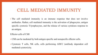
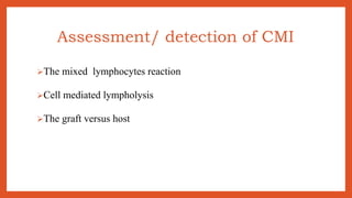
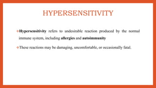
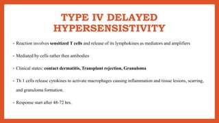
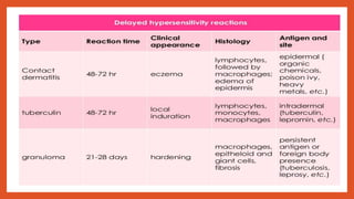
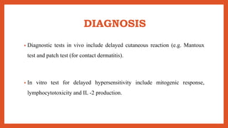
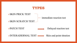
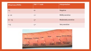
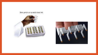
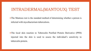
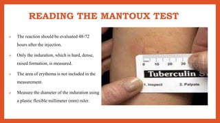
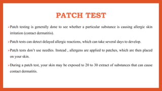
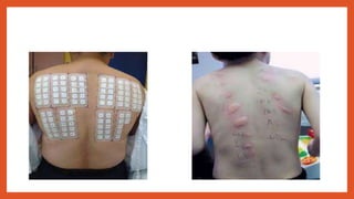
2. CELL MEDIATED IMMUNITY
• The cell mediated immunity is an immune response that does not involve
antibodies. Rather, cell mediated immunity is the activation of phagocytes, antigen
specific cytotoxic T-lymphocytes, and the release of various cytokines response to
an antigen.
Effector cells of CMI
• CMI can be mediated by both antigen specific and nonspecific effector cells.
• Cytotoxic T cells, NK cells, cells performing ADCC (antibody dependent cell
mediated cytotoxicity.
3. Assessment/ detection of CMI
The mixed lymphocytes reaction
Cell mediated lympholysis
The graft versus host
4. HYPERSENSITIVITY
Hypersensitivity refers to undesirable reaction produced by the normal
immune system, including allergies and autoimmunity
These reactions may be damaging, uncomfortable, or occasionally fatal.
5. CLASSIFICATION
• Type I – immediate, atopic, anaphylactic
• Type II – Antibody dependent
• Type III – immune complex
• Type IV – Cell mediated / Delayed type of hypersensitivity
6. TYPE IV DELAYED
HYPERSENSISTIVITY
• Reaction involves sensitized T cells and release of its lymphokines as mediators and amplifiers
• Mediated by cells rather then antibodies
• Clinical states: contact dermatitis, Transplant rejection, Granuloma
• Th 1 cells release cytokines to activate macrophages causing inflammation and tissue lesions, scarring,
and granuloma formation.
• Response start after 48-72 hrs.
7.
8. DIAGNOSIS
• Diagnostic tests in vivo include delayed cutaneous reaction (e.g. Mantoux
test and patch test (for contact dermatitis).
• In vitro test for delayed hypersensitivity include mitogenic response,
lymphocytotoxicity and IL -2 production.
9. SKIN TEST
INTRODUCTION:
Delayed – type hypersensitivity (DTH) skin testing is used clinically for two primary reasons
1. Assess immune competence
2. To determine whether a patient has memory T cells that recognize a particular pathogen.
Testing performed by intradermal (not subcutaneous) injection of sterilely prepared antigens
into the forearm or other easily accessible skin site.
10. • The degree of induration ( swelling due to inflammation) is measured 48 hours
after injection.
• The inflammation result from production of local cytokines and chemokines at
the injection site.
• The DTH response is a true measure of the cellular - dependent arm of the
immune response.
11. TYPES
SKIN PRICK TEST
immediate reaction test
SKIN SCRATCH TEST
PATCH TEST Delayed reaction test
INTERADERMAL TEST Skin end point titration
12. SKIN PRICK / SCRATCH TEST
A skin prick test, also called a puncture or scratch test, checks for immediate allergic reaction to as
many as 40 different at once.
This test is usually done to identify allergies to pollen, mold, pet dander, dust mites and foods.
In adults, the test is usually done on the forearm. Children may be tested on the upper back
Allergy skin tests aren’t painful. This type of testing uses needles (lancets) that barely penetrate the
skin, surface.
You won’t bleed or feel more than mild, momentary discomfort.
13. PROCEDURE
• After cleaning the test site with alcohol,
The nurse draws small marks on your skin and applies a drop of allergen
extract next to each mark.
• He or she then uses a lancet to prick the
extract into the skin’s surface.
• A new lancet is used for each allergen.
14. RESULT
About 15 min after the skin pricks, the nurse observes your skin for signs of allergic
reaction. If you are allergic to one of the substance tested, you’ll develop a raised , red,
itchy bumps (wheal) that may look like a mosquito bite. A nurse will them measure the
bump’s size.
After the nurse record the result, he or she will clean you skin with alcohol to remove
the mark
17. INTRADERMAL(MANTOUX) TEST
The Mantoux test is the standard method of determining whether a person is
infected with mycobacterium tuberculosis.
The local skin reaction to Tuberculin Purified Protein Derivative (PPD)
injected into the skin is used to assess the individual’s sensitivity to
tuberculin protein.
18. MANTOUX TEST
The Mantoux test is given to:
Children aged 3 months to 6 years living at high risk environments.
Infants and children under six years of age with a history of residence or prolonged stay
(more than three months) in a country of high endemic.
There is a history of TB in a household contact in the last five years.
Those who have had close contact with a person with known TB.
19. ADMINISTERING THE MANTOUX
TEST
Tuberculin purified protein derivative (PPD) RT 23 SSI, 2 T.U. (tuberculin unit)
/ 0.1 ml, solution for injection:
• 1 dose of 2 T.U./0.1 ml contains 0.04 micrograms of Tuberculin PPD RT 23.
• 1 dose of 10 T.U./0.1 ml contains 0.2 micrograms of Tuberculin PPD RT 23.
• Store at 2°c - 8°c, protected from light
1 ml graduated syringe fitted with a short bevel 26G (0.45X10mm) needle
20. INJECTION SITE
The test is usually applied on the middle third of
the flexor surface of the forearm, as a reaction may
be weaker near the wrist or the elbow joint.
It is usually applied on the left forearm.
Ensure adequate lighting.
Select an area of healthy skin which is free of
muscle margins, heavy hair, veins, sores, or scars.
Only visibly dirty skin needs to be washed with
soap and water.
21. PROCEDURE
1. Use a 1 ml syringe to aspirate out 0.1 ml of PPD
RT 23.
2. Inject the PPD intradermally on the volar surface
of the forearm.
3. Position the syringe at a 10 ° - 15° to the forearm
and insert just below the epidermis (about 2 mm)
4. Remove the needle quickly, do not massage or
use.
22. • Mark down the site , date and time of injection, on the forearm
after 48 -72 hours, read the test result by
• Marking down the transverse diameter of induration, not
erythema, by Sokal's ballpoint method.
• Measure the largest transverse diameter of induration and note
down in millimeters(mm).
23. READING THE MANTOUX TEST
The reaction should be evaluated 48-72
hours after the injection.
Only the induration, which is hard, dense,
raised formation, is measured.
The area of erythema is not included in the
measurement.
Measure the diameter of the induration using
a plastic flexible millimeter (mm) ruler.
24. PATCH TEST
Patch testing is generally done to see whether a particular substance is causing allergic skin
irritation (contact dermatitis).
Patch tests can detect delayed allergic reactions, which can take several days to develop.
Patch tests don’t use needles. Instead , allergens are applied to patches, which are then placed
on your skin.
During a patch test, your skin may be exposed to 20 to 30 extract of substances that can cause
contact dermatitis.
25.
26. These can include latex, medications, fragrances, preservatives, hair dyes, metals
and resins.
You wear the patches on your arm or back for 48 hours.
During this time, you should avoid bathing and activities that cause heavy
sweating.
The patches are removed when you return to your doctor’s office.
Irritated skin at the patch site may indicate an allergy.
27. T & B CELL ASSAY
Assessment of the cell-mediated immune system is obviously important for the diagnosis and
monitoring of cell-mediated immune deficiencies.
It has application for the diagnosis of infection, monitoring the effects of immunomodulatory therapy,
and assessing the response to vaccination.
List of applications will likely grow as therapies for immune mediated disorders are discovered and
newer technologies come into more common use.
Initial assessment of integrity of cell – mediated immune system is obtained with CBC and differential
count.
From this evaluation, the proportion and absolute number of major cell lineages are determined.
28. T CELL SUBSET ANALYSIS
• Once the percentage of cells is obtained from flow cytometric analysis, the
absolute number of cells in each subset maybe calculated if the total number
of lymphocytes is known.
• Therefore its important that the total and differential white blood count be
accurate.
• Since the reference range of each cell subset has not been established, each
laboratory must establish its own range.
29. • Control populations should include a large sample of healthy men and
women.
• The reference range should be those values that fall within the 5th and 95th
percentile of all values obtained.
• Most frequently measured T-cell subsets in clinical settings are helper T cells
that express CD4 and suppressor T cells that express CD8.
• The ratio of CD4-positive cells and CD8-positive cells can be calculated.
30. • However this ratio may not be accurate since other cells express CD4 or CD8.
• Decrease in CD4 & CD8 positive cell maybe seen in primary and secondary
immunodeficiency.
• In primary immunodeficiency, cellular defect maybe due to conditions itself, where
as in secondary immunodeficiency, an infection causes decrease in some cell types.
31. • T- cell subset quantitation is important in solid organ transplantation to
monitor the effect of immunosuppressive therapy.
• After transplantation, quantitation of T cell subtypes is used to indicate the
success of transplantation.
• Total number of T-cells- CD4 & CD8 positive cell population is important.
32. B-CELL ANALYSIS
• The most useful surface marker on mature B cell is the surface
immunoglobulin(sIg) which is synthesized by B cell and serve as antigen
receptor.
• Flowcytometry can be used to quantitate mature B cell by identifying (sIg), but
cant be used to quantitate precursor B cell as because B cell do not express sIg
on their surface.
33. • sIg is of IgM and IgD classes with varying amount on each cell. Some B cell also
express surface IgG or IgA. Therefore antibody used in flowcytometric analysis should
be a mixture of anti-immunoglobulin heavy chain types to detect all B cells.
• The distribution of fluorescence intensity is the same for kappa & lambda chains.
• Polyspecific fluorescented antisera may also be used to manually quantitate B cells
with fluorescent microscope.
• This manual procedure is not as accurate as those assays done on the flow cytometer.
34. • Normally CD5 is found on T cells. In case of CLL, CD5 is found on surface of neoplastic
B cells.
• CD5 is also present on the surface of B cells following bone marrow transplantation.
• There are several technical problems when using sIg to quantitate B cells, because other
cell types may also have immunoglobulin on their surface.
• Normal B cells, NK cells, activated T cells and phagocytic cells have Fc receptors that can
bind circulating immunoglobulin.
35. • An incubation period of 1 hour at 37 deg. C without serum prior to labelling the cells with
fluoresceinated antiserum will remove immunoglobulin from surface of cells that have not
synthesized the immunoglobulin.
• Its important to enumerate B cells in normal individual and patients with primary
immunodeficiency's, some lymphomas, and neoplasms such as multiple myeloma.
• Some cases such as hypogammaglobinemia, number of B cells is within reference range,
even though serum immunoglobulin concentration is decreased.
36. • Monoclonal antibodies maybe used to detect the presence of clonal proliferation of
B cells.
• Sometime antiserum specific for kappa and lambda light chains is used to detect
clonal proliferation . Clone will express kappa or lambda chains as part of sIg but
not both.
37. Methods to detect t cell and b cell
assay
Flowcytometry
Fluorescent activated cell sorting
mixed lymphocyte culture.