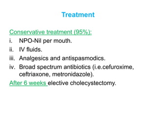
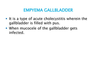
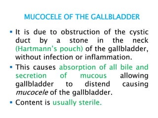
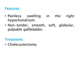
College Call Girls Pune Mira 9907093804 Short 1500 Night 6000 Best call girls...
Acute cholecystitis
1. ACUTE CHOLECYSTITIS
LT COL SM SHAHADAT HOSSAIN
MCPS,FCPS( Surgery),FCPS(Thoracic Surgery)
Adv Trg on Thoracoscopy,CNUH,South Korea
2. ACUTE CHOLECYSTITIS
Commonly, it occurs in a patient with pre-
existing chronic cholecystitis but often also can
occur as a first presentation.
Usual cause is impacted gallstone in the
Hartmann’s pouch, obstructing cystic duct.
3. Causative bacteria
E. coli—most common
Klebsiella, Pseudomonas, Proteus
Strep. faecalis
Salmonella
Clostridium welchii
5. Mode of Infection
Haematogenous through hepatic artery—cystic
artery.
Portal vein.
Through bile.
6. Pathology of Acute Cholecystitis
Gallbladder distended with oedematous friable
wall.
Wall contains dilated vessels.
Areas of necrosis and patchy gangrene may
occur in severe cases.
Mucosa shows ulceration and necrosis.
Lumen contains infected fluid/infected bile or
frank pus.
7. Clinical Features
Sudden onset of pain in the right
hypochondrium, with tenderness, guarding, and
rigidity.
Fever, nausea (25%).
Jaundice.
Tachycardia.
Palpable, tender, smooth, soft gallbladder.
Area of hyperaesthesia between 9th and 11th
ribs right posteriorly (Boas’s sign).
Murphy’s sign: positive.
8. Investigations
Total blood count: neutrophilia.
LFT increased serum bilirubin.
Ultrasound abdomen—reveals presence or
absence of gallstones; and thickening of
gallbladder wall.
Plain X-ray abdomen—10% of gallstones are
radio-opaque.
HIDA radioisotope study; Non-visualisation of
gallbladder.
CT scan to identify perforation, impacted stone,
wall thickness and oedema.
9. Differential diagnosis of radio-opaque
shadow
1. Kidney stone
2. Gallstone
3. Calcified 12th rib tip
4. Phlebolith
5. Pancreatic stone
6. Radio-opaque foreign body
7. Faecolith
8. Calcified lymph node
9. Calcified renal tuberculosis
10. Adrenal tumour—calcification
13. Treatment
Conservative treatment (95%):
i. NPO-Nil per mouth.
ii. IV fluids.
iii. Analgesics and antispasmodics.
iv. Broad spectrum antibiotics (i.e.cefuroxime,
ceftriaxone, metronidazole).
After 6 weeks elective cholecystectomy.
14. EMPYEMA GALLBLADDER
It is a type of acute cholecystitis wherein the
gallbladder is filled with pus.
When mucocele of the gallbladder gets
infected.
15. Treatment
Antibiotics: cefotaxime, quinolones,
ceftriaxone.
Cholecystectomy—an emergency procedure.
Cholecystostomy with foley’s or malecot’s
catheter kept in situ.
After 6 weeks, cholecystectomy is done.
16. MUCOCELE OF THE GALLBLADDER
It is due to obstruction of the cystic
duct by a stone in the neck
(Hartmann’s pouch) of the gallbladder,
without infection or inflammation.
This causes absorption of all bile and
secretion of mucous allowing
gallbladder to distend causing
mucocele of the gallbladder.
Content is usually sterile.
17. Features:
Painless swelling in the right
hypochondrium.
Non-tender, smooth, soft, globular,
palpable gallbladder.
Treatment:
Cholecystectomy