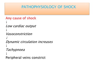
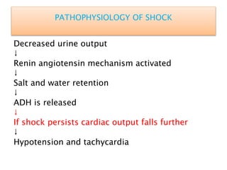
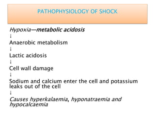
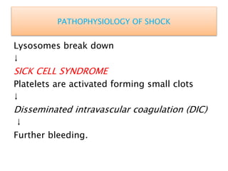
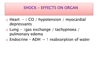
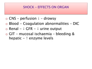
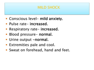
13. PATHOPHYSIOLOGY OF SHOCK
Any cause of shock
↓
Low cardiac output
↓
Vasoconstriction
↓
Dynamic circulation increases
↓
Tachypnoea
↓
Peripheral veins constrict
14. PATHOPHYSIOLOGY OF SHOCK
Decreased urine output
↓
Renin angiotensin mechanism activated
↓
Salt and water retention
↓
ADH is released
↓
If shock persists cardiac output falls further
↓
Hypotension and tachycardia
15. PATHOPHYSIOLOGY OF SHOCK
Hypoxia—metabolic acidosis
↓
Anaerobic metabolism
↓
Lactic acidosis
↓
Cell wall damage
↓
Sodium and calcium enter the cell and potassium
leaks out of the cell
↓
Causes hyperkalaemia, hyponatraemia and
hypocalcaemia
16. PATHOPHYSIOLOGY OF SHOCK
Lysosomes break down
↓
SICK CELL SYNDROME
Platelets are activated forming small clots
↓
Disseminated intravascular coagulation (DIC)
↓
Further bleeding.
17. SHOCK – EFFECTS ON ORGAN
o Heart – ↓ CO / hypotension / myocardial
depressants
o Lung - ↓gas exchange / tachypnoea /
pulmonary edema
o Endocrine – ADH → ↑ reabsorption of water
18. SHOCK – EFFECTS ON ORGAN
o CNS – perfusion ↓ – drowsy
o Blood - Coagulation abnormalities – DIC
o Renal - ↓ GFR - ↓ urine output
o GIT – mucosal ischaemia – bleeding &
hepatic - ↑ enzyme levels
19. STAGES OF SHOCK
Stage1 compensatory shock by
neuroendocrine response to maintain the
perfusion of the vital organs like brain, heart,
kidney, liver.
Stage2 decompensatory shock where there is
persistent shock with severe hypotension,
oliguria, tachycardia.
Stage3 irreversible shock with severe hypoxia
and MODS.
26. MONITORS FOR ORGAN/SYSTEMIC PERFUSION.
Systemic perfusion
I. Base deficit
II. Lactate
III. Mixed venous oxygen saturation
Kidney Urine output
27. MONITORS FOR ORGAN/SYSTEMIC PERFUSION
Muscle
a. Near-infrared spectroscopy
b. Tissue oxygen electrode
Gut
a. Sublingual capnometry
b. Gut mucosal pH
c. Laser Doppler flowmetry
28. MONITORS FOR ORGAN/SYSTEMIC PERFUSION
Brain
a. Conscious level
b. Tissue oxygen electrode
c. Near-infrared spectroscopy
30. PRINCIPLES OF RESUSCITATION
A: Airway
Patent upper airway
B: Breathing
Adequate ventilation and oxygenation
C: Circulation
Placement of adequate IV access
31. FLUID THERAPY IN SHOCK
Crystalloid Solutions
I. Normal saline
II. Ringers Lactate solution
III. Hartmann’s solution
IV. Colloid Solutions
V. Blood transfusion
32. DYNAMIC FLUID RESPONSE
Infusing 250-500ml of Fluid rapidly in 5 - 10
mts.
1. Responders – Improvement
2. Transient responders – revert back
3. Non – responders
33. VASOPRESSORS / INOTROPIC DRUGS
Vasopressors – Phenylephrine / NA
Distributive shock states
Septic shock / Neurogenic
Inotropics - Dobutamine
Cardiogenic shock / Severe septic shock
To increase the cardiac output
34. OTHER TREATMENTS
1. Correction of Acid – base balance
2. Steriods - Hydrocortisone
3. Antibiotics
4. Catheterisation
5. Nasal O2 / Ventilatory support
6. CVP Line
7. Control of Pain
8. ICU – Critical care management
35. SEPTIC SHOCK
• Septic shock may be due to gram-positive
organisms, gram negative organisms, fungi,
viruses or protozoal origin.
• Gram-negative septicaemia/gram-negative
septic shock is called as endotoxic shock.
36. PATHOPHYSIOLOGY OF SEPTIC SHOCK
Toxins/endotoxins from organisms like E. Coli, Klebsiella,
Pseudomonas, and Proteus
↓
Inflammation, cellular activation of macrophages,
neutrophils,
monocytes
↓
Release of cytokines, free radicals
↓
Chemotaxis of cells, endothelial injury, altered
coagulation
cascade—SIRS
↓
37. PATHOPHYSIOLOGY OF SEPTIC SHOCK
Reversible hyperdynamic warm stage of septic
shock with
fever, tachycardia, tachypnoea
↓
Severe circulatory failure with MODS (failure of
lungs, kidneys, liver, heart) with DIC
↓
Hypodynamic, irreversible cold stage of septic
shock.