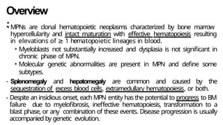
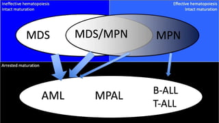
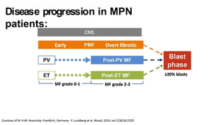
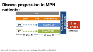
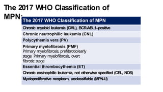
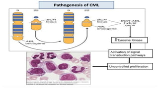
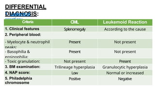
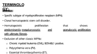
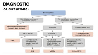
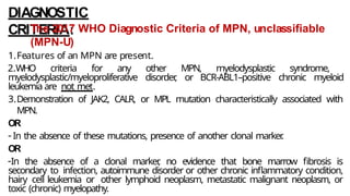
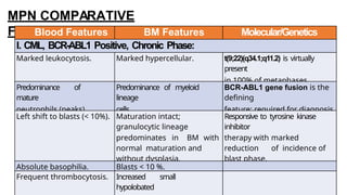
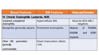
Myeloproliferative Neoplasms
(MPNs)
By:
Ahmed Makboul Ahmed
M.B.B.Ch, M.Sc
Assistant Lecturer
, Clinical Pathology Department, South Egypt Cancer
Institute
Overview
:
- MPNs are clonal hematopoietic neoplasms characterized by bone marrow
hypercellularity and intact maturation with effective hematopoiesis resulting
in elevations of ≥ 1 hematopoietic lineages in blood.
• Myeloblasts not substantially increased and dysplasia is not significant in
chronic phase of MPN.
• Molecular genetic abnormalities are present in MPN and define some
subtypes.
- Splenomegaly and hepatomegaly are common and caused by the
sequestration of excess blood cells, extramedullary hematopoiesis, or both.
- Despite an insidious onset, each MPN entity has the potential to progress to BM
failure due to myelofibrosis, ineffective hematopoiesis, transformation to a
blast phase, or any combination of these events. Disease progression is usually
accompanied by genetic evolution.
Disease progression in MPN
patients:
Disease progression in MPN
patients:
The 2017 WHO Classification of
MPN:The 2017 WHO Classification of MPN
Chronic myeloid leukemia (CML), BCR-ABL1–positive
Chronic neutrophilic leukemia (CNL)
Polycythemia vera (PV)
Primary myelofibrosis (PMF)
Primary myelofibrosis, prefibrotic/early
stage Primary myelofibrosis, overt
fibrotic stage
Essential thrombocythemia (ET)
Chronic eosinophilic leukemia, not otherwise specified (CEL, NOS)
Myeloproliferative neoplasm, unclassifiable (MPN-U)
Chronic Myeloid Leukemia, BCR-ABL1
Positive
TERMINOLO
GY:
Definition:
•Specific subtype of myeloproliferative neoplasm that harbors BCR-ABL1 gene
fusion.
oBCR-ABL1 fusion gene results from reciprocal translocation involving long
arms of chromosomes 9 and 22.
oResults in production of abnormal tyrosine kinase protein.
•3 classically defined phases of disease: Chronic, Accelerated, and Blast:
oChronic: 1 - 9% blasts: indolent.
oAccelerated phase (10-19% blasts as one example): Disease progression.
oBlast phase (≥ 20% blasts): Aggressive disease, often refractory to therapy.
•The natural history of untreated CML is bi- or triphasic: an initial indolent
chronic phase (CP) is followed by an accelerated phase (AP), a blast phase (BP)
ETIOLOGY/
PATHOGENESIS:
t(9;22)(q34;q11.2):
-Reciprocal translocation following
breakpoints at BCR and ABL1 loci.
- Translocation is balanced in majority
of cases.
-t(9;22)(q34;q11.2) fuses 3’
sequences from ABL1 to 5’
sequences from BCR.
ABL1 breakpoint:
• ABL1 gene is located on 9q34.
• ABL1 breakpoint typically
occurs between exon 1 and 2 (a2).
BCR
breakpoints:
At least 3 different breakpoints:
1. Major breakpoint cluster region (M-BCR):
• Encodes a p210 fusion protein.
• M-BCR present in 99% of CML and 40% of adult Ph(+) B-cell ALL cases.
2. Minor breakpoint cluster region (m-BCR):
• m-BCR encode a p190 fusion protein.
• m-BCR present in 90% of pediatric ALL and 60% of adult ALL.
• m-BCR is also present in rare CML cases (1%).
3. Micro breakpoint cluster region (µ-BCR):
• µ-BCR encodes a p230 fusion protein.
• CML cases with this fusion show prominent neutrophils &/or conspicuous
thrombocytosis.
The Philadelphia
Chromosome
The Philadelphia
Chromosome
CLINICAL
FEATURES:
• Age:
oThe medianage at diagnosis is 65 years, but children as young as 3 years
can be affected.
• Gender:
oA slight male predominance is observed.
• Clinical picture:
oPatients are often initially identified due to leukocytosis being found during
routine blood work, because approximately half of the patients at
diagnosis do not have symptoms.
oThe other half complain of fatigue, malaise, weight loss, or night sweats.
• Physical examination:
oSplenomegaly and hepatomegaly are seen in about 50% and 20% of
patients, respectively.
MICROSCOPIC
PATHOLOGY:
CHRONIC PHASE:
Peripheral Blood:
WBCs:
• Granulocytic leukocytosis with left shift to
immaturity.
• Predominance of neutrophils and
myelocytes (2 peaks).
• < 10 % blasts (Usually 1 – 9%).
• Basophilia.
• Often eosinophilia.
• Absolute monocyte count is elevated (> 1 x
109/L); but the percentage is usually < 3%.
• No significant granulocytic
dysplasia or toxic changes.
RBCs:
• No or mild anemia.
• Circulating nucleated red
blood cells.
Platelets:
• Preserved/elevated platelet count. Marked thrombocytosis is
unusual.
• Atypical large plateletsor megakaryocytic
cytoplasmic fragments; megakaryocytic nuclei.
circulatin
g
Bone marrow
aspirate:
Cellularity:
• Hypercellular for age due to marked
granulocytic proliferation.
Erythroid Series:
• Erythroid precursors are reduced
in percentage but show normal
maturation.
Granulocytic Series:
• Usually 1 – 9%.
o 10% or more indicates disease
progression.
• Myeloid:erythroid (M:E) ratio > 10:1.
• Predominanceof neutrophils and
myelocytes (2 peaks).
Megakaryocytic Series:
• Usually increased megakaryocytes.
• Distinctive megakaryocytic morphology; small
and monolobulated (so-called dwarf
megakaryocytes).
Other Findings:
• Sea-blue histiocytes due to increased cell
turnover
. These cells carry BCR-ABL1 because
they are progeny of the affected leukemic
stem cell.
Bone marrow core
biopsy:
Cellularity:
• 95% cellular due to marked granulocytic
predominance.
Erythroid Series:
• Decreased erythroid lineage.
Granulocytic Series:
• Marked granulocytic predominance.
• M:E ratio > 10:1.
• No clusters of blasts.
Megakaryocytic Series:
• Small, monolobulated, (dwarf) megakaryocytes.
Other Findings:
Disease progression:
ACCELERATED PHASE
(AP):
The 2017 WHO criteria for diagnosis of accelerated phase include:
1. Hematologic criteria:
- Persistent or increasing WBC count (>10 x 109/L), unresponsive to therapy.
- Persistent or increasing splenomegaly, unresponsive to therapy.
- Persistent thrombocytosis (>1000 x 109/L), unresponsive to therapy.
- Persistent thrombocytopenia (<100 x 109/L) unrelated to therapy.
- 20% or more basophils in the peripheral blood.
- 10% - 19% blasts in the peripheral blood or bone marrow.
2. Cytogenetic criteria:
- Additional clonal chromosomal abnormalities in Ph+ cells at
diagnosis that include “major
route”abnormalities (second Ph, trisomy 8,
isochromosome 17q, trisomy 19), a complex
karyotype, or abnormalities of 3q26.2.
3. Provisional response to tyrosine kinase inhibitor
criteria:
-Hematologic resistance to the first TKI (or failure to achieve a complete
hematologic response to the first TKI).
or
-Any hematologic, cytogenetic,or molecular indications of resistance to two
sequential TKIs.
or
- Occurrence of two or more mutations in BCR-ABL1 during TKI therapy.
Any one or more of the previous hematologic/cytogenetic criteria or
response to TKI criteria diagnose CML accelerated phase
BLAST PHASE
(BP):
The BP is diagnosed when:
-Blasts equal or are greater than 20% of the peripheral blood WBC or of the nucleated
cells of the BM.
OR
- When there is an extramedullary blast proliferation.
Types of blast phase:
• 70-80% myeloid: (70% granulocytic, 10-20% megakaryocytic, < 10% erythroid,
< 5% monocytic).
• 20-30% lymphoid: (90% B-lymphoblastic leukemia, T-lymphoblastic leukemia
uncommon).
o Presence of lymphoblasts(detection of any lymphoblasts
should raise concern for impending lymphoid blast phase).
• < 5% biphenotypic.
ANCILLARY
TESTS:
1. Immunohistochemistry:
• Role is minimal in chronic-phase CML.
• Blast markers: CD34 &/or CD117, TdT: Assess for increased &/or clustered
blasts.
2. Flow Cytometric Immunophenotyping:
• Plays minimal role in chronic-phase CML.
• Useful in blast lineage determination in accelerated/blast phase.
oAberrant antigen expression on blasts not uncommon.
 Myeloid antigen expression (e.g., CD13, CD33) on lymphoblasts.
 Lymphoid antigen expression on myeloblasts.
• If concern for lymphoblasts, immunophenotyping is warranted.
3. Conventional Cytogenetic Analysis (Karyotyping):
• Should be performed routinely in work-up for
myeloid neoplasm.
• Detection of t(9;22)(q34.1;q11.2):
ot(9;22)(q34.1;q11.2) (or
variant) diagnosis.
&/orBCR-ABL1 gene fusionrequired
for
 t(9;22)(q34.1;q11.2) is most often reciprocal translocation found on
derivative chromosome 22.
of cases (not detected
by
ot(9;22)(q34.1;q11.2) reportedly
cryptic in 5% karyotyping).
 Do FISH or molecular for BCR-ABL1
fusion.
• Clonal evolution in accelerated/blast phase:
oAdditional Philadelphia chromosome, trisomy 8,
isochromosome (17q), and trisomy 19 (major route abnormalities).
The Philadelphia
Chromosome
4. Fluorescence in Situ Hybridization
(FISH):
• Reveals/confirms genetic fusion of
BCR-ABL1.
• Advantage
s:oMore sensitive than conventional
cytogenetics.
oDetects most cryptic rearrangements.
• Disadvantage
s:oRare cryptic rearrangements
may be missed.
oNot sensitive enough for
MRD or early relapse detection.
BC
R
ABL
1
BCR-
ABL
BCR-
ABL
5. Molecular RT-PCR Based
Studies:
a). Detection of BCR-ABL1 transcripts:
• Multiple primer sets to detect different fusion transcripts.
• Different fusion transcripts show variable association with different disease types:
• p210 seen in most cases of CML.
• p190 seen in majority of cases of pediatric B-lymphoblastic leukemia, BCR-ABL1
positive.
• p230 (rare); associated with neutrophilia and may be thrombocytosis.
• Determination of particular fusion transcript (e.g., p210) is useful for MRD
monitoring.
b). ABL1 kinase domain testing:
• Considered in case of TKI resistance to detect mutations in ABL1.
• Prior to mutation testing:
o Confirm TKI treatment compliance.
DIFFERENTIAL
DIAGNOSIS:
1. Leukemoid
reaction:Criteria CML Leukemoid Reaction
1. Clinical features Splenomegaly According to the cause
2. Peripheral blood:
- Myelocyte & neutrophil
peaks:
Present Not present
- Basophilia &
eosinophilia:
Present Not present
- Toxic granulation: Not present Present
3. BM examination: Trilineage hyperplasia Granulocytic hyperplasia
4. NAP score: Low Normal or increased
5. Philadelphia
chromosome
Positive Negative
2. BCR-ABL1(-) Myeloproliferative Neoplasm:
• Key feature distinguishing from CML is lack of BCR-ABL1.
• JAK2 V617F mutation identified in
o> 95% of polycythemia vera.
o50% of essential thrombocythemia and primary myelofibrosis.
oNot seen in classic CML.
• Also seeJAK2 exon 12, CALR, and MPL
mutations in specific subsets ofthese diseases.
• Chronic neutrophilic leukemia: Most cases show CSF3R mutation.
3. Atypical Chronic Myelogenous Leukemia BCR-ABL1
Negative:
• Unfortunate disease name. No relationship to CML.
• BCR-ABL1 Negative
• Dysplasia prominent
• Recurrent SETBP1 mutations
Polycythemia Vera
(PV)
TERMINOLO
GY:
Definition:
• Classic myeloproliferative neoplasm (MPN) characterized by:
o Increased red blood cells (RBCs).
o JAK2 gene gain of function somatic mutation.
Phases of PV:
1.Pre-polycythemic phase with mild erythrocytosis (so-called masked PV): Borderline
to only mild erythrocytosis.
2.Overt polycythemic phase: Associated with significantly increased Hb, Hct, and RBC
mass.
3.Spent phase and post-polycythemic myelofibrosis:
• Decrease in RBC mass; patient often anemic.
• Further enlargement of spleen.
• Marked reticulin and collagen fibrosis of BM.
ETIOLOGY/
PATHOGENESIS:
I. JAK2 V617F mutation:
• Detected in > 95% of PV cases.
• Point mutation in exon 14 of
pseudokinase domain:
o Substitution of a G to T at 1849
position.
o This substitutionleads to
substitutionof valine for
phenylalanine (V617F).
o Mutation involves myeloid lineages
and is absent in lymphocytes.
• Consequences of JAK2 V617F
mutation:
o Gain-of-function somatic
II. JAK2 exon 12 mutations:
• Uncommon, except in JAK2 V617F-negative PV.
• Present in 3% of PV cases.
• JAK2 exon12 mutations appear to result
specifically in an erythrocytosis phenotype.
CLINICAL
FEATURES:
• Age:
oThe average age at diagnosis is 60 years.
• Gender:
oSlight male predominance.
• Clinical picture:
oMany patients are asymptomatic.
oThe diagnosis may be suspected by the findings of plethora and
splenomegaly on examination or abnormalities on a routine blood count
that, in addition to increased HGB and HCT, often include
leukocytosis and/or thrombocytosis.
oThe main causes of morbidity and mortality are due to complications of
blood hyperviscosity, which stems from increases in red cell mass and
MICROSCOPIC
PATHOLOGY:
1. Pre-polycythemic and polycythemic
phases: Peripheral Blood:
WBCs:
• Neutrophilia and, rarely, basophilia may be
present.
• Occasional immature granulocytic cells may be
seen in polycythemic phase.
RBCs:
• Mild to significantly increased
normochromic/normocytic RBCs.
• The RBC indicesare usuallynormal
unless there is concomitant iron
deficiency.
Platelets:
Bone marrow examination:
Cellularity:
• Typically, hypercellular for age.
• Increased subcortical cellularity.
• Panmyelosis: prominent erythroid, granulocytic and megakaryocytic
proliferation.
Erythroid series:
• Erythropoiesis is prominent, often occurs in expanded erythroid islands.
• It demonstrates normoblastic maturation.
Granulocytic series:
• Granulopoiesis may show a shift toward immaturity.
• There is no increase in the percentage of blasts.
• There is no significant dysplasia.
Megakaryocytic series:
• Increased number of megakaryocytes with variably hyperlobated forms.
2. Spent phase and post-polycythemic
myelofibrosis: Peripheral Blood:
• Peripheral blood with anemia &/or
leukoerythroblastic picture.
• Prominent anisopoikilocytosis, including teardrop
forms and nucleated RBCs.
• Immature granulocytic cells but no significant
dysplastic cells.
Bone marrow examination:
• Cellularity: Variable
cellularity hypocellular for
age.
but
often
• Erythroid & Granulocytic
Series:
Decreased erythropoiesis and
granulopoiesis.
• Megakaryocytic Series: Megakaryocytes
generally similar to those seen in
antecedent PV.
• Other Findings:
oProminent reticulin and collagen fibrosis.
oOsteosclerosis may be seen in
late-stage disease.
ANCILLARY
TESTS:
1. Flow Cytometric Immunophenotyping:
• No consistent
immnuophenotypic
abnormality transformation.
described in absence of
leukemic
2. Conventional Cytogenetic Analysis (Karyotyping):
• Common cytogenetic abnormalities:
o Trisomy 8, trisomy 9, del(20q), del(13q), and del(9p).
o Complex cytogenetic abnormalities in post-polycythemic myelofibrosis.
• Cytogenetic abnormalities in PV patients with transformation to myelodysplastic
syndrome (MDS) or blast phase:
o Detected in virtually all patients with transformation to MDS/blast phase.
o Include those commonly seen in therapy-related MDS/acute myeloid leukemia.
3. Molecular Genetic Testing:
JAK2 V617F mutation:
• Gain-of-function mutation.
• Mutation occurs in all cells of myeloid lineages.
• Present in 95% of patients with PV.
• JAK2 V617F mutation is homozygous in most PV cases.
JAK2 exon 12 mutations:
• Relatively specific for JAK2 V617F-negative PV.
• 4% frequency among all patients with PV.
• JAK2 exon 12 mutations are often heterozygous.
• Associated with predominantly erythroid myelopoiesis and younger
age at diagnosis.
4. Other Laboratory tests:
Serum erythropoietin (EPO):
• Serum EPO levels are typically decreased in PV, in contrast to elevated
levels usually found in secondary polycythemia.
• Measurement of EPO levels is an important study that should be
performed early in the workup of polycythemia.
• A normal EPO level does not necessarily exclude PV or secondary
erythrocytosis.
DIAGNOSTIC
CRITERIA:
The 2017 WHO Diagnostic Criteria for Polycythemia
Vera (PV) Major Criteria:
1.Hemoglobin > 16.5 g/dL in men, >
16.0 g/dL in women; or hematocrit > 49% in
men, > 48% in women;
or increased red cell mass (More than
25% above mean predicted value).
2. BM biopsy showing hypercellularity for age
with trilineage growth (panmyelosis) including
prominent erythroid, granulocytic, and megakaryocytic proliferation with
pleomorphic, mature megakaryocytes (differences in size).
3.Presence of JAK2 V617F or JAK2 exon 12 mutation.
Minor Criterion:
- Subnormal serum erythropoietin level.
The 2017 WHO Diagnostic Criteria for Post-polycythemia vera Myelofibrosis:
Required Criteria:
1.Documentation of a previous diagnosis of WHO-defined polycythemia vera.
2.Bone marrow fibrosis grade 2-3 (on 0-3 scale).
Additional Criteria (two are required):
1. Anemia or sustained loss of either phlebotomy (in the absence of cytoreductive therapy) or cytoreductive
treatment
requirement for erythrocytosis.
2.Leukoerythroblastic picture in PB smear.
3.Increasing splenomegaly:
 defined as either an increase in palpable splenomegaly of > 5 cm from baseline (distance from the
left costal margin) or the appearance of newly palpable splenomegaly.
4.Development of > 1 of 3 constitutional symptoms:
 > 10% weight loss in 6 months.

DIAGNOSTIC
ALGORITHM:
Essential Thrombocythemia
(ET)
TERMINOLO
GY:
Definitions:
• Specific subtype of myeloproliferative neoplasm (MPN).
• Hematopoietic proliferation essentially restricted to megakaryocytic
lineage.
• Exclusion of other classic MPNs:
oChronic myeloid leukemia, BCR-ABL1 positive.
oPolycythemia vera.
oPrimary myelofibrosis, early phase.
ETIOLOGY/
PATHOGENESIS:
Molecular Mutations:
3 driver mutations are identified:
I. JAK2 V617F mutation:
• 1st driver mutation to be identified.
II. MPL W515L mutation:
• 2nd driver mutation to be identified.
• 3-5% of ET cases harbor this mutation.
• Substitution of a G to T at nucleotide 1544.
• Results in substitution of amino acid tryptophan to leucine (W515L).
• General outcome of mutation is promotionof constitutive,
cytokine-independent activation of JAK/ STAT signaling pathway.
III. CALR mutation:
• 3rd driver mutation to be identified.
• 20-25% of ET cases harbor this mutation (CALR + ET).
• 2 most common types of mutations:
oType 1 mutation: A 52-bp deletion (Most common type).
oType 2 mutation: A 5-bp insertion.
• Abnormal function of CALR-mutated protein:
oSuggestion that mutant calreticulin activates JAK-STAT pathway.
oResults in excessive platelet production.
• JAK2, CALR, and MPL mutations are most often mutually exclusive.
oPresence of 1 of these mutations distinguishes reactive disorder from
neoplasm.
oThese mutations do not distinguish PMF from PV or ET.
CLINICAL
FEATURES:
• Age:
oThe median age at diagnosis is 60 years.
• Gender:
oSlight female predominance.
• Clinical picture:
oPatients are usually asymptomatic.
oPatients may exhibit symptoms such as headaches, blurred vision, dizziness, and
erythromelalgia (redness, burning, pain of distal ends of toes and fingers) due to
thrombotic occlusion of the microvasculature.
oCatastrophic large vessel thrombosis includes stroke, myocardial infarction, deep
venous thrombosis (including splanchnic vein thrombosis), and peripheral arterial
thrombosis.
oBleeding is less common than thrombosis.
oSplenomegaly is not as prominent as that of the other MPNs, and it occurs in 15–
MICROSCOPIC
PATHOLOGY:
Peripheral Blood:
WBCs:
• The WBCcount and
leukocyte differential are usually
normal.
• Mild leukocytosis is sometimes
seen.
• White blood cell morphology
unremarkable.
• No leucoerythroblastic reaction.
• Neutrophilia uncommon.
• No significant left shift
in granulocytic series.
RBCs:
•Red blood cell morphology
unremarkable.
•Significant anisopoikilocytosis is
uncommon and teardrop-shaped RBCs
are not observed.
Platelets:
•Variable thrombocytosis but ≥ 450 x
10⁹/L.
•Striking platelet anisocytosis:
oSmall and large platelets.
oHypogranular forms may
be seen. oCirculating
Bone marrow:
Cellularity:
• Normal cellularity to mildly hypercellular.
Erythroid and Granulocytic Series:
• Generally, no significant granulocytic or erythroid
proliferation.
• Occasionally may encounter mild granulocytic
hyperplasia.
oConsider early phase of primary myelofibrosis.
• Blasts < 5%.
Megakaryocytic Series:
• Striking megakaryocytic proliferation:
oIncreased numbers.
oLoosely clustered.
oMarkedly enlarged, hyperlobated.
oET megakaryocytes are largest of all BM
disorders.
Other Findings:
• Absent/minimal reticulin fibrosis.
BM aspirate shows the phenomenon of “pseudoparticle”
formation that may occur as a result of marked
thrombocytosis and extensive platelet clumping in vitro. These
particles mimic BMparticles but have less cellular element.
In ET, BM aspirate typically reveals intact granulopoiesis and
erythropoiesis with unremarkable morphology. The very
large hyperlobated megakaryocytes are characteristic.
BM core biopsy shows normal bone and mild hypercellularity.
Increased megakaryocytes form loose clusters and demonstrate
hyperlobulation.
BM core biopsy features of ET. Increased megakaryocytes form
loose clusters and demonstrate hyperlobulation.
ANCILLARY
TESTS:
1. Histochemistry:
• Reticulin:
oAbsent to minimal fibrosis.
oProgressive fibrosis in rare cases.
2. Flow Cytometric Immunophenotyping:
• Blasts < 5% in typical ET.
• Rare cases of leukemic transformation of ET.
oBlasts > 20%; usually myeloid phenotype.
3. Conventional Cytogenetic Analysis (Karyotyping):
• Usually normal karyotype (in 90% of cases).
• Cytogenetic abnormalities are detected in less than 10% of ET cases at
diagnosis.
4. Molecular Genetic Testing:
• JAK2 V617F mutations present in 60%
of cases.
• JAK2 exon 12 mutations are absent.
• CALR mutations in 20-25% of cases.
• MPL mutations present in 3-5% of
cases.
DIFFERENTIAL
DIAGNOSIS:
1. Reactive/Secondary
thrombocytosis:
2. Pre-fibrotic
PMF:
Megakaryocytes in
ET
Megakaryocytes in
prePMF
DIAGNOSTIC
CRITERIA:
The 2017 WHO Diagnostic Criteria of Essential Thrombocythemia
(ET) Major Criteria:
1.Platelet count > 450 x 109/L.
2.BM biopsy showing:
• Proliferation mainly of the megakaryocyte lineage with increased numbers of enlarged,
mature megakaryocytes with hyperlobulated nuclei.
• No significant increase or shift toward immaturity in neutrophil granulopoiesis or
erythropoiesis and very rarely minor (grade 1) increase in reticulin fibers.
3.Not meeting the WHO criteria for BCR-ABL1+ CML, PV, PMF, MDS, or other myeloid
neoplasms.
4.Presence of JAK2, CALR, or MPL mutation.
Minor Criterion:
- Presence of a clonal marker or absence of evidence for reactive thrombocytosis.
Diagnosis of ET requires meeting all four major criteria or the first three major
criteria and the minor criterion.
DIAGNOSTIC
ALGORITHM:
Pathologic Interpretation Pearls:
 Sustained thrombocytosis: Platelet count ≥ 450 x 10⁹/L.
 Reactive thrombocytosis excluded.
 Red blood cell and white blood cell cytology and counts
unremarkable.
 Enlarged megakaryocytes with hyperlobulation.
 Absent/minimal fibrosis.
 Clonal:
o JAK2 V617F positivity in 60%.
o CALR mutations in 20-25%.
SUMMAR
Y:
Primary Myelofibrosis
(PMF)
TERMINOLO
GY:
Definition:
• Specific subtype of myeloproliferative neoplasm (MPN).
• Clonal hematopoietic stem cell disorder.
• Hematopoietic proliferation that shows
predominantly megakaryocytic and granulocytic proliferation
with ultimate fibrosis.
• Exclusion of other classic MPNs:
o Chronic myeloid leukemia (CML), BCR-ABL1 positive.
o Polycythemia vera (PV).
o Essential thrombocythemia (ET).
ETIOLOGY/
PATHOGENESIS:
Molecular Mutations:
3 driver mutations are identified:
I. JAK2 V617F mutation:
• 1st driver mutation to be identified.
II. MPL W515L mutation:
• 2nd driver mutation to be identified.
• 6-7% of PMF cases harbor this mutation.
• Substitution of a G to T at nucleotide 1544.
• Results in substitution of amino acid tryptophan to leucine (W515L).
• General outcome of mutation is promotionof constitutive,
cytokine-independent activation of JAK/ STAT signaling pathway.
III. CALR mutation:
• 3rd driver mutation to be identified.
• 20-25% of PMF cases harbor this mutation.
• 2 most common types of mutations:
oType 1 mutation: A 52-bp deletion (Most common type).
oType 2 mutation: A 5-bp insertion.
• Abnormal function of CALR-mutated protein:
oSuggestion that mutant calreticulin activates JAK-STAT pathway.
oResults in excessive platelet production.
• JAK2, CALR, and MPL mutations are most often mutually exclusive.
oPresence of 1 of these mutations distinguishes reactive disorder from
neoplasm.
oThese mutations do not distinguish PMF from PV or ET.
Triple negative PMF:
•10-15% of PMF casesarenegative for all 3
mutations: JAK2, CALR, and MPL.
•Associated with poor prognosis.
Phases of primary myelofibrosis
(PMF):
There are 2 phases of PMF:
I. Prefibrotic phase (prePMF)
It is characterized by:
- Marked
thrombocytosis in the
peripheral blood.
- Hypercellular BM with
granulocytic and
atypical megakaryocytic
proliferation.
-Absent or only slight reticulin
fibrosis.
- Minimal if any EMH.
II. Fibrotic phase (Overt
PMF)
It is characterized by:
- Variable BM cellularity.
-Reticulin or collagen
fibrosis, osteosclerosis.
ofte
n
-Prominent hepatosplenomegaly
due to EMH.
-Leucoerythroblastic reaction in
the peripheral blood.
CLINICAL
FEATURES:
• Age:
oThe median age at diagnosis is 67 years. About 5% of cases present before 40 years.
• Gender:
oNo significant prediliction.
• Clinical picture:
oPatients are usually asymptomatic.
oPatients may exhibit symptoms such as headaches, blurred vision, dizziness, and
erythromelalgia (redness, burning, pain of distal ends of toes and fingers) due to
thrombotic occlusion of the microvasculature.
oCatastrophic large vessel thrombosis includes stroke, myocardial infarction, deep
venous thrombosis (including splanchnic vein thrombosis), and peripheral arterial
thrombosis.
oBleeding is less common than thrombosis.
oSplenomegaly is not as prominent as that of the other MPNs, and it occurs in 15–
CLINICAL FEATURES:
MICROSCOPIC
PATHOLOGY:
Peripheral blood:
I. Prefibrotic phase (prePMF)
RBCs: Modest anemia.
WBCs: Mild leukocytosis.
Platelets: Moderate to marked
thrombocytosis. Blood film
morphology:
-The most striking finding is often the
marked increase in platelets.
- Mild neutrophilia with a left shift may be
seen.
- No significant dysplasia.
- Variable, often subtle, basophilia.
II. Fibrotic phase (Overt PMF)
There is gradual worsening of hematologic
parameters as the disease progresses.
RBCs: More severe anemia than PrePMF.
WBCs:
- Mild leukocytosis.
-Severe leucopenia may occur as BM failure
becomes more prominent as a result of
increasing fibrosis.
Platelets: Lower platelet count than
PrePMF. Blood film morphology:
The classic findings of patients with
overt PMF are:
-Leucoerythroblastic reaction with numerous
teardrop- shaped RBCs.
- Bizarre, abnormal platelets.
- Blasts occasionally account for
5% or more. Blast
percentages of 10% to 19% in PB indicate
BM
Examination:
I. Pre-fibrotic phase (PrePMF):
Cellularity:
- In prePMF, BM is hypercellular.
Erythroid series:
- Erythropoiesis is reduced in most cases.
Granulocytic series:
- There is an increased number of neutrophils.
-Although there may be a left shift in granulopoiesis, neutrophils at the metamyelocyte through segmented stages usually
predominate.
- The percentage of myeloblasts is not increased.
Megakaryocytic series:
- There is an increased number of atypical megakaryocytes.
- Megakaryocytes in PMF are morphologically more atypical than in any other MPN:
• They vary from small to large. Some have an abnormal nuclear-cytoplasmic ratio and disorganized, plump, cloudlike, or
balloonlike nuclear lobation.
Reticulin stain:
- Reticulin fibers vary in quantity and thickness but are often not increased in prePMF, except focally around blood vessels.
Pre-
fibrotic
II. Fibrotic phase (Overt PMF):
Cellularity:
- As prePMF progresses to the fibrotic stage, the marrow cellularity decreases.
Erythroid and Granulocytic series: depressed.
Megakaryocytic series:
• Megakaryocytic atypia:
• Dense clusters.
• Hyperchromatic and bizarre nuclei.
• Cloud-like nuclei.
Reticulin fibrosis: Reticulin or even overt collagen fibrosis of the marrow becomes
more obvious.
Other findings:
• Osteosclerosis.
• Moderate to marked reticulin/collagen fibrosis.
• Dilated sinuses.
Fibrot
ic
MF-0: Scattered linear reticulin
with no intersections (cross-
overs) corresponding to
normal BM
MF-1: Loose network of
reticulin with many
intersections, especially in
perivascular areas
ANCILLARY
TESTS:
1. Histochemistry (Reticulin
stain):
Semiquantitative Grading of MF
MF-2: Diffuse and dense
↑ in
reticulin with extensive
intersections, occasionally with
focal bundles of collagen
and/or focal
osteosclerosis.
MF-3: Diffuse and dense ↑ in
reticulin with extensive
intersections and coarse
bundles of collagen, often with
osteosclerosis
2. Molecular Genetic Testing:
• JAK2 V617F mutations present in 60% of cases.
• JAK2 exon 12 mutations are absent.
• CALR mutations in 25% of cases.
• MPL mutations present in 6-7% of cases.
• 10 – 15% of cases are negative for JAK2 V617F, CALR and MPL (triple
negative PMF).
Molecular Approach in BCR-ABL
negative MPN:
oThese include: PV, ET and PMF.
1.BCR-ABL1 fusion must be ruled out to exclude chronic myelogenous
leukemia.
2.Cases of suspected PV would be considered for JAK2 V617F and JAK2 exon 12
mutations.
3.If a diagnosis of ET or PMF is favored, mutations may be sought in MPL,
JAK2 (V617F), or CALR.
 Because thesemutations are considered
mutually exclusive, a stepwise reflex algorithm may
be used:
 Based on frequency, best order is JAK2 V617F > CALR > MPL.
4.Next generation sequencing assessment of allof
DIAGNOSTIC
CRITERIA:
The 2017 WHO Diagnostic Criteria of Pre-fibrotic/Early PMF (PrePMF):
Major criteria:
1.Megakaryocytic proliferation and atypia, without reticulin fibrosis > grade 1, accompanied by
increased age- adjusted BM cellularity, granulocytic proliferation and often decreased erythropoiesis
2.Not meeting WHO criteria of BCR/ABL+ CML, PV, ET, MDS or other myeloid neoplasms.
3.Presence of JAK2, MPL or CALR mutation or in the absence of these mutations, presence of another
clonal marker or absence of minor reactive BM reticulin fibrosis.
Minor criteria:
Presence of at least one of the following, confirmed in two consecutive determinations:
a.Anemia not attributed to a comorbid condition.
b.Leukocytosis ≥ 11 x 109/L.
c.Palpable splenomegaly.
d.LDH increased to above upper normal limit of institutional reference range.
Diagnosis of prePMF requires meeting all three major criteria and at least one minor criterion.
The 2017 WHO Diagnostic Criteria of Overt Primary
Myelofibrosis
Major criteria:
1.Megakaryocytic proliferation and atypia, accompanied by either reticulin and/or collagen fibrosis
grades 2 or 3.
2.Not meeting WHO criteria of BCR/ABL+ CML, PV, ET, MDS or other myeloid neoplasms.
3.Presence of JAK2, MPL or CALR mutation or in the absence of these mutations, presence of
another clonal marker or absence of minor reactive BM reticulin fibrosis.
Minor criteria:
Presence of at least one of the following, confirmed in two consecutive determinations:
a.Anemia not attributed to a comorbid condition.
b.Leukocytosis ≥11 x 109/L.
c.Palpable splenomegaly.
d.LDH increased to above upper normal limit of institutional reference range.
e.Leucoerythroblastosis.
Diagnosis of Overt PMF requires meeting all three major criteria and at least one minor criterion.
Chronic Neutrophilic Leukemia
(CNL)
TERMINOLO
GY:
Definition:
• Specific subtype of myeloproliferative neoplasm (MPN).
• Clonal hematopoietic stem cell disorder manifesting predominantly in granulocytic
lineage.
• Characterized by:
o Persistent peripheral blood neutrophilia.
o Persistent BM granulocytic proliferation.
o There is no BCR-ABL1 fusion gene.
o Hepatosplenomegaly common.
o Activating mutation of CSF3R gene. Mutation present in most cases.
o Reactive conditions resulting in neutrophilia must be
excluded in cases lacking clonal genetic marker
.
o Other myeloid
neoplasms
must be systematicallyexcluded in
cases
lacking CSF3R
mutation.
CLINICAL
FEATURES:
• Age:
oThe average age at diagnosis is 60 years.
• Gender:
oSlight male predominance.
• Clinical picture:
oMajority of patients asymptomatic.
oIncidental detection of leukocytosis
on CBC. oHepatomegaly.
oSplenomegaly: the most consistent
clinical finding.
oWeight loss.
oEasy bruising.
MICROSCOPIC
PATHOLOGY:
Peripheral Blood:
•WBC ≥ 25.0 x 10⁹/L.
•Neutrophilia and increased
band forms (≥ 80% of
WBC).
•Toxic changes with prominent granules
may be seen. Must exclude reactive
condition.
•Absence of dysplasia.
•No significant basophilia.
•Immature granulocytes constitute <
10% of WBC.
•Blasts rarely noted.
Bone marrow:
• Cellularity: Hypercellular; > 90%; due to granulocytic predominance.
• Erythroid series: Reduced in percentage but show normoblastic
maturation.
• Granulocytic Series:
oThe percentage of blasts and promyelocytes in BM is not increased at
diagnosis (Blasts < 5%).
oThere is an increase in the percentage of myelocytes,
metamyelocytes, bands, and segmented neutrophils.
oThere is no significant dysplasia.
oBasophilia and eosinophilia are generally not observed.
• Megakaryocytic Series: Normal.
DIAGNOSTIC
CRITERIA:
The 2017 WHO Diagnostic Criteria of Chronic Neutrophilic Leukemia (CNL)
1. Peripheral blood white blood cell count ≥ 25 x 109/L
- Segmented neutrophils plus band forms are > 80% of white blood cells.
- Neutrophil precursors (promyelocytes, myelocytes, metamyelocytes) are <10% of white blood
cells.
- Myeloblasts rarely observed.
- Monocyte count <1 x 10⁹/L.
- No dysgranulopoiesis.
2. Presence of CSF3R T6181 or other activating CSF3R mutation.
OR
- In the absence of a CSFR3R mutation, persistent neutrophilia (at least 3 months) and no
identifiable cause of physiological or reactive neutrophilia, including absence of a plasma cell
neoplasm or
, if present, demonstration of clonality of myeloid cells by cytogenetic or
3. Hypercellular bone marrow:
- Neutrophilic granulocytes increased in percentage and number
.
- Neutrophil maturation appears normal.
- Myeloblasts < 5% of nucleated bone marrow cells.
4. Not meeting the WHO criteria for any other myeloid neoplasm:
- Specifically, no BCR-ABL1; no rearrangement of PDGFRA, PDGFRB, or FGFR1;
no PCM1-JAK2, ETV6-JAK2, or BCR-JAK2.
All criteria must be met for the diagnosis of chronic neutrophilic
leukemia to be made.
DIAGNOSTIC
ALGORITHM:
Chronic Eosinophilic Leukemia
(CEL, NOS)
TERMINOLOGY
:
Definition:
• Clonal hematopoietic myeloproliferative neoplasm.
oManifests as sustained proliferation of eosinophils and precursors.
CLINICAL ISSUES:
Incidence:
• Rare.
• Difficult to determine accurately.
• Significant degree of diagnostic overlap with idiopathic hypereosinophilic
syndrome.
Clinical Presentation:
• Asymptomatic:
oIncidental identification of eosinophilia on complete blood
cell count.
• Symptomatic:
oFever
.
oFatigue
.
oPrurit
us.
oGastr
ointesti
nal
MICROSCOPIC
PATHOLOGY:
Peripheral Blood:
•Eosinophilia:
oMostly mature eosinophils.
oOccasional eosinophil
precursors.
oEosinophil count ≥ 1.5 x 10⁹/L.
•Other features:
• Neutrophilia common.
• Blasts < 20% (usually > 2%).
• No significant population of mast
cells.
• No circulating lymphoma cells.
Bone Marrow:
• Cellularity:
oUsually hypercellular due to
eosinophilia.
• Erythroid and Megakaryocytic Series:
oIntact erythropoiesis and
megakaryopoiesis.
• Granulocytic Series:
oEosinophilia.
oBlasts < 20% (usually >
5%).
oDysplasia is uncommon.
oExclude
myelodysplasia with
associate
d
oCharcot-Leyden crystals may be
ANCILLARY
TESTS:
1. Immunohistochemistry:
• Assess for mast cell disease:
oTryptase and CD25.
oMast cells may be spindled and individually distributed; not in aggregates.
• Assess for Hodgkin/non-Hodgkin lymphoma:
oIf morphology in BM negative:
 No further testing except for consideration of CD30 for occult T-cell
lymphoma.
oIf morphology in BM positive:
 No further testing if morphologic features
similarto lymphoma diagnosed in extramedullary site.
2. Conventional Cytogenetics Analysis (Karyotyping):
•Document clonality if present.
•Exclude recurring genetic abnormality diagnostic of different neoplasm e.g.
ot(9;22)(q34.1;q11.2); BCR-ABL1 fusion in chronic myeloid leukemia.
oRearrangement of PDGFRB (5q31~33),FGFR1 (8p11.23-p11.22), and
PCM1-
JAK2;t(8;9)(p22;q24.1).
oCBFB-MYH11 in AML with inv(16).
3. Fluorescent in Situ Hybridization (FISH):
•Perform testing for FIP1L1-PDGFRA fusion since cytogenetically cryptic.
•Investigate possible PDGFRB, FGFR1, and
PCM1-JAK2 abnormalities by chromosomal analysis.
DIAGNOSTIC
CRITERIA:
The 2017 WHO Diagnostic Criteria of CEL
1.Eosinophilia (eosinophil count 1.5
≥ x 109/L).
2.WHO criteria for BCR-ABL1-positive CML, polycythemia vera, essential
thrombocythemia, PMF, CNL, chronic myelomonocytic leukemia, and BCR-ABL1-
negative atypical CML are not met.
3.No rearrangement of PDGFRA, PDGFRB, or FGFR1, and no PCM1-JAK2, ETV6-JAK2, or
BCR-JAK2 fusion.
4.Blast cells constitute <20% of the cells in the peripheral blood and bone marrow, and
inv(16)(p13.1q22), t(16;16) (p13.1;q22), t(8;21)(q22;q22.1), and otherdiagnostic features of
AML are absent.
5.There is a clonal cytogenetic or molecular genetic abnormality or blast cells account
for 2%
≥ of cells in the peripheral blood or ≥5% in the bone marrow.
All criteria must be met for diagnosis of CEL.
MPN, Unclassifiable
(MPN-U)
TERMINOLO
GY:
Definition:
• The designation MPN, unclassifiable, should be applied only to:
oCases that have definite clinical, laboratory, and morphologic features of
an MPN but fail to meet the criteria for any of the specific MPN entities.
OR
oCases present with features that overlap two or more of the MPN
categories.
• The designation MPN, unclassifiable, should not be used if:
oLaboratory data necessary for classification are incomplete or were never
obtained.
oThe size or quality of BM specimen is inadequate for complete evaluation.
oThe patient has received prior growth factor or cytotoxic therapy.
DIAGNOSTIC
CRITERIA:
The 2017 WHO Diagnostic Criteria of MPN, unclassifiable
(MPN-U)
1.Features of an MPN are present.
2.WHO criteria for any other MPN, myelodysplastic syndrome,
myelodysplastic/myeloproliferative disorder
, or BCR-ABL1–positive chronic myeloid
leukemia are not met.
3.Demonstration of JAK2, CALR, or MPL mutation characteristically associated with
MPN.
OR
- In the absence of these mutations, presence of another clonal marker
.
OR
-In the absence of a clonal marker
, no evidence that bone marrow fibrosis is
secondary to infection, autoimmune disorder or other chronic inflammatory condition,
hairy cell leukemia or other lymphoid neoplasm, metastatic malignant neoplasm, or
toxic (chronic) myelopathy.
SUMMAR
Y
MPN COMPARATIVE
FEATURES:
Blood Features BM Features Molecular/Genetics
I. CML, BCR-ABL1 Positive, Chronic Phase:
Marked leukocytosis. Marked hypercellular. t(9;22)(q34.1;q11.2) is virtually
present
in 100% of metaphases.
Predominance of
mature
neutrophils (peaks).
Predominance of myeloid
lineage
cells.
BCR-ABL1 gene fusion is the
defining
feature; required for diagnosis.
Left shift to blasts (< 10%). Maturation intact;
granulocytic lineage
predominates in BM with
normal maturation and
without dysplasia.
Responsive to tyrosine kinase
inhibitor
therapy with marked
reduction of incidence of
blast phase.
Absolute basophilia. Blasts < 10 %.
Frequent thrombocytosis. Increased small
hypolobated
Blood Features BM Features Molecular/Genetics
II. Polycythemia Vera, Chronic Phase:
Erythrocytosis. Hypercellular
. Must be BCR-ABL1
negative.
Variable leukocytosis
and
nRBCs.
Maturation intact;
panmyelosis.
o JAK2 V617F in >95% of
cases.
o JAK2 exon 12 in the
remainder.
Variable
basophilia;
thrombocytosis.
Blasts <2%. JAK2 inhibitor therapy is
potential
treatment option.
Dysplasia absent. Increased, usually
Blood Features BM Features Molecular/Genetics
III. Essential Thrombocythemia:
Thrombocytosis:
sustained.
Variable cellularity (may be
normal).
Must be BCR-ABL1
negative.
Variable WBC and
basophil
count.
Increased, markedly
hyperlobated
megakaryocytes.
o JAK2 V617F in 50% of
cases.
o CALR mutation in 40%.
o MPL W151L/K
mutation in
small subset.
RBCs parameters usually
normal.
Other lineages
unremarkable.
Some cases are triple
negative.
Blasts <2%. Mutation status linked to
Blood Features BM Features Molecular/Genetics
IV. Primary Myelofibrosis, Early to Overt Fibrotic Phases:
Leukocytosis and
thrombocytosis
common in early disease.
Marked hypercellularity;
granulocytic
lineage predominance;
cellularity decreases as
fibrosis progresses.
Must be BCR-ABL1 negative.
Leucoerythroblastic blood
picture
common in fibrotic phase.
Dilated sinuses with
intrasinusoidal
megakaryocytes.
o JAK2 V617F in 60% of
cases.
o CALR mutation in 40%.
o MPL W151L/K mutation
in small subset.
Teardrop-shaped RBCs in
fibrotic
phase.
Myeloid predominance with
intact
maturation
Some cases are triple
negative.
Variable basophilia. Blasts < 10 %. Mutationstatus linked to
Blood Features BM Features Molecular/Genetics
V. Chronic Neutrophilic Leukemia
Sustained unexplained
neutrophilia.
Hypercellular BM. Must be BCR-ABL1
negative.
May see toxic changes. Myeloid lineage
predominates.
CSF3R mutation in
most
cases; may be
defining event.
Absent basophilia. Intact maturation.
Other CBC parameters
generally
Blasts <2%.
Blood Features BM Features Molecular/Genetics
VI. Chronic Eosinophilic Leukemia, NOS
Sustained unexplained
eosinophilia.
Hypercellular BM. Must be BCR-ABL1
negative.
Basophilia generally absent. Prominent eosinophils. Absence of PDGFRA,
PDGFRB and FGFR1
required.
Other CBC parameters
generally
unremarkable.
Intact maturation, blasts
<2%.

myeloproliferative neoplasmsss kdnfkdnfkdnf

  • 1.
    Myeloproliferative Neoplasms (MPNs) By: Ahmed MakboulAhmed M.B.B.Ch, M.Sc Assistant Lecturer , Clinical Pathology Department, South Egypt Cancer Institute
  • 2.
    Overview : - MPNs areclonal hematopoietic neoplasms characterized by bone marrow hypercellularity and intact maturation with effective hematopoiesis resulting in elevations of ≥ 1 hematopoietic lineages in blood. • Myeloblasts not substantially increased and dysplasia is not significant in chronic phase of MPN. • Molecular genetic abnormalities are present in MPN and define some subtypes. - Splenomegaly and hepatomegaly are common and caused by the sequestration of excess blood cells, extramedullary hematopoiesis, or both. - Despite an insidious onset, each MPN entity has the potential to progress to BM failure due to myelofibrosis, ineffective hematopoiesis, transformation to a blast phase, or any combination of these events. Disease progression is usually accompanied by genetic evolution.
  • 7.
  • 8.
  • 9.
    The 2017 WHOClassification of MPN:The 2017 WHO Classification of MPN Chronic myeloid leukemia (CML), BCR-ABL1–positive Chronic neutrophilic leukemia (CNL) Polycythemia vera (PV) Primary myelofibrosis (PMF) Primary myelofibrosis, prefibrotic/early stage Primary myelofibrosis, overt fibrotic stage Essential thrombocythemia (ET) Chronic eosinophilic leukemia, not otherwise specified (CEL, NOS) Myeloproliferative neoplasm, unclassifiable (MPN-U)
  • 10.
    Chronic Myeloid Leukemia,BCR-ABL1 Positive
  • 11.
    TERMINOLO GY: Definition: •Specific subtype ofmyeloproliferative neoplasm that harbors BCR-ABL1 gene fusion. oBCR-ABL1 fusion gene results from reciprocal translocation involving long arms of chromosomes 9 and 22. oResults in production of abnormal tyrosine kinase protein. •3 classically defined phases of disease: Chronic, Accelerated, and Blast: oChronic: 1 - 9% blasts: indolent. oAccelerated phase (10-19% blasts as one example): Disease progression. oBlast phase (≥ 20% blasts): Aggressive disease, often refractory to therapy. •The natural history of untreated CML is bi- or triphasic: an initial indolent chronic phase (CP) is followed by an accelerated phase (AP), a blast phase (BP)
  • 12.
    ETIOLOGY/ PATHOGENESIS: t(9;22)(q34;q11.2): -Reciprocal translocation following breakpointsat BCR and ABL1 loci. - Translocation is balanced in majority of cases. -t(9;22)(q34;q11.2) fuses 3’ sequences from ABL1 to 5’ sequences from BCR. ABL1 breakpoint: • ABL1 gene is located on 9q34. • ABL1 breakpoint typically occurs between exon 1 and 2 (a2).
  • 13.
    BCR breakpoints: At least 3different breakpoints: 1. Major breakpoint cluster region (M-BCR): • Encodes a p210 fusion protein. • M-BCR present in 99% of CML and 40% of adult Ph(+) B-cell ALL cases. 2. Minor breakpoint cluster region (m-BCR): • m-BCR encode a p190 fusion protein. • m-BCR present in 90% of pediatric ALL and 60% of adult ALL. • m-BCR is also present in rare CML cases (1%). 3. Micro breakpoint cluster region (µ-BCR): • µ-BCR encodes a p230 fusion protein. • CML cases with this fusion show prominent neutrophils &/or conspicuous thrombocytosis.
  • 14.
  • 15.
  • 17.
    CLINICAL FEATURES: • Age: oThe medianageat diagnosis is 65 years, but children as young as 3 years can be affected. • Gender: oA slight male predominance is observed. • Clinical picture: oPatients are often initially identified due to leukocytosis being found during routine blood work, because approximately half of the patients at diagnosis do not have symptoms. oThe other half complain of fatigue, malaise, weight loss, or night sweats. • Physical examination: oSplenomegaly and hepatomegaly are seen in about 50% and 20% of patients, respectively.
  • 18.
    MICROSCOPIC PATHOLOGY: CHRONIC PHASE: Peripheral Blood: WBCs: •Granulocytic leukocytosis with left shift to immaturity. • Predominance of neutrophils and myelocytes (2 peaks). • < 10 % blasts (Usually 1 – 9%). • Basophilia. • Often eosinophilia. • Absolute monocyte count is elevated (> 1 x 10⁹/L); but the percentage is usually < 3%. • No significant granulocytic dysplasia or toxic changes.
  • 19.
    RBCs: • No ormild anemia. • Circulating nucleated red blood cells. Platelets: • Preserved/elevated platelet count. Marked thrombocytosis is unusual. • Atypical large plateletsor megakaryocytic cytoplasmic fragments; megakaryocytic nuclei. circulatin g
  • 20.
    Bone marrow aspirate: Cellularity: • Hypercellularfor age due to marked granulocytic proliferation. Erythroid Series: • Erythroid precursors are reduced in percentage but show normal maturation. Granulocytic Series: • Usually 1 – 9%. o 10% or more indicates disease progression. • Myeloid:erythroid (M:E) ratio > 10:1. • Predominanceof neutrophils and myelocytes (2 peaks).
  • 21.
    Megakaryocytic Series: • Usuallyincreased megakaryocytes. • Distinctive megakaryocytic morphology; small and monolobulated (so-called dwarf megakaryocytes). Other Findings: • Sea-blue histiocytes due to increased cell turnover . These cells carry BCR-ABL1 because they are progeny of the affected leukemic stem cell.
  • 22.
    Bone marrow core biopsy: Cellularity: •95% cellular due to marked granulocytic predominance. Erythroid Series: • Decreased erythroid lineage. Granulocytic Series: • Marked granulocytic predominance. • M:E ratio > 10:1. • No clusters of blasts. Megakaryocytic Series: • Small, monolobulated, (dwarf) megakaryocytes. Other Findings:
  • 23.
    Disease progression: ACCELERATED PHASE (AP): The2017 WHO criteria for diagnosis of accelerated phase include: 1. Hematologic criteria: - Persistent or increasing WBC count (>10 x 109/L), unresponsive to therapy. - Persistent or increasing splenomegaly, unresponsive to therapy. - Persistent thrombocytosis (>1000 x 109/L), unresponsive to therapy. - Persistent thrombocytopenia (<100 x 109/L) unrelated to therapy. - 20% or more basophils in the peripheral blood. - 10% - 19% blasts in the peripheral blood or bone marrow. 2. Cytogenetic criteria: - Additional clonal chromosomal abnormalities in Ph+ cells at diagnosis that include “major route”abnormalities (second Ph, trisomy 8, isochromosome 17q, trisomy 19), a complex karyotype, or abnormalities of 3q26.2.
  • 24.
    3. Provisional responseto tyrosine kinase inhibitor criteria: -Hematologic resistance to the first TKI (or failure to achieve a complete hematologic response to the first TKI). or -Any hematologic, cytogenetic,or molecular indications of resistance to two sequential TKIs. or - Occurrence of two or more mutations in BCR-ABL1 during TKI therapy. Any one or more of the previous hematologic/cytogenetic criteria or response to TKI criteria diagnose CML accelerated phase
  • 25.
    BLAST PHASE (BP): The BPis diagnosed when: -Blasts equal or are greater than 20% of the peripheral blood WBC or of the nucleated cells of the BM. OR - When there is an extramedullary blast proliferation. Types of blast phase: • 70-80% myeloid: (70% granulocytic, 10-20% megakaryocytic, < 10% erythroid, < 5% monocytic). • 20-30% lymphoid: (90% B-lymphoblastic leukemia, T-lymphoblastic leukemia uncommon). o Presence of lymphoblasts(detection of any lymphoblasts should raise concern for impending lymphoid blast phase). • < 5% biphenotypic.
  • 26.
    ANCILLARY TESTS: 1. Immunohistochemistry: • Roleis minimal in chronic-phase CML. • Blast markers: CD34 &/or CD117, TdT: Assess for increased &/or clustered blasts. 2. Flow Cytometric Immunophenotyping: • Plays minimal role in chronic-phase CML. • Useful in blast lineage determination in accelerated/blast phase. oAberrant antigen expression on blasts not uncommon.  Myeloid antigen expression (e.g., CD13, CD33) on lymphoblasts.  Lymphoid antigen expression on myeloblasts. • If concern for lymphoblasts, immunophenotyping is warranted.
  • 27.
    3. Conventional CytogeneticAnalysis (Karyotyping): • Should be performed routinely in work-up for myeloid neoplasm. • Detection of t(9;22)(q34.1;q11.2): ot(9;22)(q34.1;q11.2) (or variant) diagnosis. &/orBCR-ABL1 gene fusionrequired for  t(9;22)(q34.1;q11.2) is most often reciprocal translocation found on derivative chromosome 22. of cases (not detected by ot(9;22)(q34.1;q11.2) reportedly cryptic in 5% karyotyping).  Do FISH or molecular for BCR-ABL1 fusion. • Clonal evolution in accelerated/blast phase: oAdditional Philadelphia chromosome, trisomy 8, isochromosome (17q), and trisomy 19 (major route abnormalities).
  • 28.
  • 29.
    4. Fluorescence inSitu Hybridization (FISH): • Reveals/confirms genetic fusion of BCR-ABL1. • Advantage s:oMore sensitive than conventional cytogenetics. oDetects most cryptic rearrangements. • Disadvantage s:oRare cryptic rearrangements may be missed. oNot sensitive enough for MRD or early relapse detection. BC R ABL 1 BCR- ABL BCR- ABL
  • 30.
    5. Molecular RT-PCRBased Studies: a). Detection of BCR-ABL1 transcripts: • Multiple primer sets to detect different fusion transcripts. • Different fusion transcripts show variable association with different disease types: • p210 seen in most cases of CML. • p190 seen in majority of cases of pediatric B-lymphoblastic leukemia, BCR-ABL1 positive. • p230 (rare); associated with neutrophilia and may be thrombocytosis. • Determination of particular fusion transcript (e.g., p210) is useful for MRD monitoring. b). ABL1 kinase domain testing: • Considered in case of TKI resistance to detect mutations in ABL1. • Prior to mutation testing: o Confirm TKI treatment compliance.
  • 31.
    DIFFERENTIAL DIAGNOSIS: 1. Leukemoid reaction:Criteria CMLLeukemoid Reaction 1. Clinical features Splenomegaly According to the cause 2. Peripheral blood: - Myelocyte & neutrophil peaks: Present Not present - Basophilia & eosinophilia: Present Not present - Toxic granulation: Not present Present 3. BM examination: Trilineage hyperplasia Granulocytic hyperplasia 4. NAP score: Low Normal or increased 5. Philadelphia chromosome Positive Negative
  • 33.
    2. BCR-ABL1(-) MyeloproliferativeNeoplasm: • Key feature distinguishing from CML is lack of BCR-ABL1. • JAK2 V617F mutation identified in o> 95% of polycythemia vera. o50% of essential thrombocythemia and primary myelofibrosis. oNot seen in classic CML. • Also seeJAK2 exon 12, CALR, and MPL mutations in specific subsets ofthese diseases. • Chronic neutrophilic leukemia: Most cases show CSF3R mutation.
  • 34.
    3. Atypical ChronicMyelogenous Leukemia BCR-ABL1 Negative: • Unfortunate disease name. No relationship to CML. • BCR-ABL1 Negative • Dysplasia prominent • Recurrent SETBP1 mutations
  • 35.
  • 36.
    TERMINOLO GY: Definition: • Classic myeloproliferativeneoplasm (MPN) characterized by: o Increased red blood cells (RBCs). o JAK2 gene gain of function somatic mutation. Phases of PV: 1.Pre-polycythemic phase with mild erythrocytosis (so-called masked PV): Borderline to only mild erythrocytosis. 2.Overt polycythemic phase: Associated with significantly increased Hb, Hct, and RBC mass. 3.Spent phase and post-polycythemic myelofibrosis: • Decrease in RBC mass; patient often anemic. • Further enlargement of spleen. • Marked reticulin and collagen fibrosis of BM.
  • 37.
    ETIOLOGY/ PATHOGENESIS: I. JAK2 V617Fmutation: • Detected in > 95% of PV cases. • Point mutation in exon 14 of pseudokinase domain: o Substitution of a G to T at 1849 position. o This substitutionleads to substitutionof valine for phenylalanine (V617F). o Mutation involves myeloid lineages and is absent in lymphocytes. • Consequences of JAK2 V617F mutation: o Gain-of-function somatic
  • 38.
    II. JAK2 exon12 mutations: • Uncommon, except in JAK2 V617F-negative PV. • Present in 3% of PV cases. • JAK2 exon12 mutations appear to result specifically in an erythrocytosis phenotype.
  • 39.
    CLINICAL FEATURES: • Age: oThe averageage at diagnosis is 60 years. • Gender: oSlight male predominance. • Clinical picture: oMany patients are asymptomatic. oThe diagnosis may be suspected by the findings of plethora and splenomegaly on examination or abnormalities on a routine blood count that, in addition to increased HGB and HCT, often include leukocytosis and/or thrombocytosis. oThe main causes of morbidity and mortality are due to complications of blood hyperviscosity, which stems from increases in red cell mass and
  • 40.
    MICROSCOPIC PATHOLOGY: 1. Pre-polycythemic andpolycythemic phases: Peripheral Blood: WBCs: • Neutrophilia and, rarely, basophilia may be present. • Occasional immature granulocytic cells may be seen in polycythemic phase. RBCs: • Mild to significantly increased normochromic/normocytic RBCs. • The RBC indicesare usuallynormal unless there is concomitant iron deficiency. Platelets:
  • 41.
    Bone marrow examination: Cellularity: •Typically, hypercellular for age. • Increased subcortical cellularity. • Panmyelosis: prominent erythroid, granulocytic and megakaryocytic proliferation. Erythroid series: • Erythropoiesis is prominent, often occurs in expanded erythroid islands. • It demonstrates normoblastic maturation. Granulocytic series: • Granulopoiesis may show a shift toward immaturity. • There is no increase in the percentage of blasts. • There is no significant dysplasia. Megakaryocytic series: • Increased number of megakaryocytes with variably hyperlobated forms.
  • 43.
    2. Spent phaseand post-polycythemic myelofibrosis: Peripheral Blood: • Peripheral blood with anemia &/or leukoerythroblastic picture. • Prominent anisopoikilocytosis, including teardrop forms and nucleated RBCs. • Immature granulocytic cells but no significant dysplastic cells.
  • 44.
    Bone marrow examination: •Cellularity: Variable cellularity hypocellular for age. but often • Erythroid & Granulocytic Series: Decreased erythropoiesis and granulopoiesis. • Megakaryocytic Series: Megakaryocytes generally similar to those seen in antecedent PV. • Other Findings: oProminent reticulin and collagen fibrosis. oOsteosclerosis may be seen in late-stage disease.
  • 45.
    ANCILLARY TESTS: 1. Flow CytometricImmunophenotyping: • No consistent immnuophenotypic abnormality transformation. described in absence of leukemic 2. Conventional Cytogenetic Analysis (Karyotyping): • Common cytogenetic abnormalities: o Trisomy 8, trisomy 9, del(20q), del(13q), and del(9p). o Complex cytogenetic abnormalities in post-polycythemic myelofibrosis. • Cytogenetic abnormalities in PV patients with transformation to myelodysplastic syndrome (MDS) or blast phase: o Detected in virtually all patients with transformation to MDS/blast phase. o Include those commonly seen in therapy-related MDS/acute myeloid leukemia.
  • 46.
    3. Molecular GeneticTesting: JAK2 V617F mutation: • Gain-of-function mutation. • Mutation occurs in all cells of myeloid lineages. • Present in 95% of patients with PV. • JAK2 V617F mutation is homozygous in most PV cases. JAK2 exon 12 mutations: • Relatively specific for JAK2 V617F-negative PV. • 4% frequency among all patients with PV. • JAK2 exon 12 mutations are often heterozygous. • Associated with predominantly erythroid myelopoiesis and younger age at diagnosis.
  • 47.
    4. Other Laboratorytests: Serum erythropoietin (EPO): • Serum EPO levels are typically decreased in PV, in contrast to elevated levels usually found in secondary polycythemia. • Measurement of EPO levels is an important study that should be performed early in the workup of polycythemia. • A normal EPO level does not necessarily exclude PV or secondary erythrocytosis.
  • 48.
    DIAGNOSTIC CRITERIA: The 2017 WHODiagnostic Criteria for Polycythemia Vera (PV) Major Criteria: 1.Hemoglobin > 16.5 g/dL in men, > 16.0 g/dL in women; or hematocrit > 49% in men, > 48% in women; or increased red cell mass (More than 25% above mean predicted value). 2. BM biopsy showing hypercellularity for age with trilineage growth (panmyelosis) including prominent erythroid, granulocytic, and megakaryocytic proliferation with pleomorphic, mature megakaryocytes (differences in size). 3.Presence of JAK2 V617F or JAK2 exon 12 mutation. Minor Criterion: - Subnormal serum erythropoietin level.
  • 49.
    The 2017 WHODiagnostic Criteria for Post-polycythemia vera Myelofibrosis: Required Criteria: 1.Documentation of a previous diagnosis of WHO-defined polycythemia vera. 2.Bone marrow fibrosis grade 2-3 (on 0-3 scale). Additional Criteria (two are required): 1. Anemia or sustained loss of either phlebotomy (in the absence of cytoreductive therapy) or cytoreductive treatment requirement for erythrocytosis. 2.Leukoerythroblastic picture in PB smear. 3.Increasing splenomegaly:  defined as either an increase in palpable splenomegaly of > 5 cm from baseline (distance from the left costal margin) or the appearance of newly palpable splenomegaly. 4.Development of > 1 of 3 constitutional symptoms:  > 10% weight loss in 6 months. 
  • 50.
  • 51.
  • 52.
    TERMINOLO GY: Definitions: • Specific subtypeof myeloproliferative neoplasm (MPN). • Hematopoietic proliferation essentially restricted to megakaryocytic lineage. • Exclusion of other classic MPNs: oChronic myeloid leukemia, BCR-ABL1 positive. oPolycythemia vera. oPrimary myelofibrosis, early phase.
  • 53.
    ETIOLOGY/ PATHOGENESIS: Molecular Mutations: 3 drivermutations are identified: I. JAK2 V617F mutation: • 1st driver mutation to be identified. II. MPL W515L mutation: • 2nd driver mutation to be identified. • 3-5% of ET cases harbor this mutation. • Substitution of a G to T at nucleotide 1544. • Results in substitution of amino acid tryptophan to leucine (W515L). • General outcome of mutation is promotionof constitutive, cytokine-independent activation of JAK/ STAT signaling pathway.
  • 54.
    III. CALR mutation: •3rd driver mutation to be identified. • 20-25% of ET cases harbor this mutation (CALR + ET). • 2 most common types of mutations: oType 1 mutation: A 52-bp deletion (Most common type). oType 2 mutation: A 5-bp insertion. • Abnormal function of CALR-mutated protein: oSuggestion that mutant calreticulin activates JAK-STAT pathway. oResults in excessive platelet production. • JAK2, CALR, and MPL mutations are most often mutually exclusive. oPresence of 1 of these mutations distinguishes reactive disorder from neoplasm. oThese mutations do not distinguish PMF from PV or ET.
  • 55.
    CLINICAL FEATURES: • Age: oThe medianage at diagnosis is 60 years. • Gender: oSlight female predominance. • Clinical picture: oPatients are usually asymptomatic. oPatients may exhibit symptoms such as headaches, blurred vision, dizziness, and erythromelalgia (redness, burning, pain of distal ends of toes and fingers) due to thrombotic occlusion of the microvasculature. oCatastrophic large vessel thrombosis includes stroke, myocardial infarction, deep venous thrombosis (including splanchnic vein thrombosis), and peripheral arterial thrombosis. oBleeding is less common than thrombosis. oSplenomegaly is not as prominent as that of the other MPNs, and it occurs in 15–
  • 56.
    MICROSCOPIC PATHOLOGY: Peripheral Blood: WBCs: • TheWBCcount and leukocyte differential are usually normal. • Mild leukocytosis is sometimes seen. • White blood cell morphology unremarkable. • No leucoerythroblastic reaction. • Neutrophilia uncommon. • No significant left shift in granulocytic series.
  • 57.
    RBCs: •Red blood cellmorphology unremarkable. •Significant anisopoikilocytosis is uncommon and teardrop-shaped RBCs are not observed. Platelets: •Variable thrombocytosis but ≥ 450 x 10⁹/L. •Striking platelet anisocytosis: oSmall and large platelets. oHypogranular forms may be seen. oCirculating
  • 58.
    Bone marrow: Cellularity: • Normalcellularity to mildly hypercellular. Erythroid and Granulocytic Series: • Generally, no significant granulocytic or erythroid proliferation. • Occasionally may encounter mild granulocytic hyperplasia. oConsider early phase of primary myelofibrosis. • Blasts < 5%.
  • 59.
    Megakaryocytic Series: • Strikingmegakaryocytic proliferation: oIncreased numbers. oLoosely clustered. oMarkedly enlarged, hyperlobated. oET megakaryocytes are largest of all BM disorders. Other Findings: • Absent/minimal reticulin fibrosis.
  • 60.
    BM aspirate showsthe phenomenon of “pseudoparticle” formation that may occur as a result of marked thrombocytosis and extensive platelet clumping in vitro. These particles mimic BMparticles but have less cellular element. In ET, BM aspirate typically reveals intact granulopoiesis and erythropoiesis with unremarkable morphology. The very large hyperlobated megakaryocytes are characteristic.
  • 61.
    BM core biopsyshows normal bone and mild hypercellularity. Increased megakaryocytes form loose clusters and demonstrate hyperlobulation. BM core biopsy features of ET. Increased megakaryocytes form loose clusters and demonstrate hyperlobulation.
  • 62.
    ANCILLARY TESTS: 1. Histochemistry: • Reticulin: oAbsentto minimal fibrosis. oProgressive fibrosis in rare cases. 2. Flow Cytometric Immunophenotyping: • Blasts < 5% in typical ET. • Rare cases of leukemic transformation of ET. oBlasts > 20%; usually myeloid phenotype. 3. Conventional Cytogenetic Analysis (Karyotyping): • Usually normal karyotype (in 90% of cases). • Cytogenetic abnormalities are detected in less than 10% of ET cases at diagnosis.
  • 63.
    4. Molecular GeneticTesting: • JAK2 V617F mutations present in 60% of cases. • JAK2 exon 12 mutations are absent. • CALR mutations in 20-25% of cases. • MPL mutations present in 3-5% of cases.
  • 64.
  • 65.
  • 66.
  • 67.
    DIAGNOSTIC CRITERIA: The 2017 WHODiagnostic Criteria of Essential Thrombocythemia (ET) Major Criteria: 1.Platelet count > 450 x 109/L. 2.BM biopsy showing: • Proliferation mainly of the megakaryocyte lineage with increased numbers of enlarged, mature megakaryocytes with hyperlobulated nuclei. • No significant increase or shift toward immaturity in neutrophil granulopoiesis or erythropoiesis and very rarely minor (grade 1) increase in reticulin fibers. 3.Not meeting the WHO criteria for BCR-ABL1+ CML, PV, PMF, MDS, or other myeloid neoplasms. 4.Presence of JAK2, CALR, or MPL mutation. Minor Criterion: - Presence of a clonal marker or absence of evidence for reactive thrombocytosis. Diagnosis of ET requires meeting all four major criteria or the first three major criteria and the minor criterion.
  • 68.
  • 69.
    Pathologic Interpretation Pearls: Sustained thrombocytosis: Platelet count ≥ 450 x 10⁹/L.  Reactive thrombocytosis excluded.  Red blood cell and white blood cell cytology and counts unremarkable.  Enlarged megakaryocytes with hyperlobulation.  Absent/minimal fibrosis.  Clonal: o JAK2 V617F positivity in 60%. o CALR mutations in 20-25%. SUMMAR Y:
  • 70.
  • 71.
    TERMINOLO GY: Definition: • Specific subtypeof myeloproliferative neoplasm (MPN). • Clonal hematopoietic stem cell disorder. • Hematopoietic proliferation that shows predominantly megakaryocytic and granulocytic proliferation with ultimate fibrosis. • Exclusion of other classic MPNs: o Chronic myeloid leukemia (CML), BCR-ABL1 positive. o Polycythemia vera (PV). o Essential thrombocythemia (ET).
  • 72.
    ETIOLOGY/ PATHOGENESIS: Molecular Mutations: 3 drivermutations are identified: I. JAK2 V617F mutation: • 1st driver mutation to be identified. II. MPL W515L mutation: • 2nd driver mutation to be identified. • 6-7% of PMF cases harbor this mutation. • Substitution of a G to T at nucleotide 1544. • Results in substitution of amino acid tryptophan to leucine (W515L). • General outcome of mutation is promotionof constitutive, cytokine-independent activation of JAK/ STAT signaling pathway.
  • 73.
    III. CALR mutation: •3rd driver mutation to be identified. • 20-25% of PMF cases harbor this mutation. • 2 most common types of mutations: oType 1 mutation: A 52-bp deletion (Most common type). oType 2 mutation: A 5-bp insertion. • Abnormal function of CALR-mutated protein: oSuggestion that mutant calreticulin activates JAK-STAT pathway. oResults in excessive platelet production. • JAK2, CALR, and MPL mutations are most often mutually exclusive. oPresence of 1 of these mutations distinguishes reactive disorder from neoplasm. oThese mutations do not distinguish PMF from PV or ET.
  • 74.
    Triple negative PMF: •10-15%of PMF casesarenegative for all 3 mutations: JAK2, CALR, and MPL. •Associated with poor prognosis.
  • 75.
    Phases of primarymyelofibrosis (PMF): There are 2 phases of PMF: I. Prefibrotic phase (prePMF) It is characterized by: - Marked thrombocytosis in the peripheral blood. - Hypercellular BM with granulocytic and atypical megakaryocytic proliferation. -Absent or only slight reticulin fibrosis. - Minimal if any EMH. II. Fibrotic phase (Overt PMF) It is characterized by: - Variable BM cellularity. -Reticulin or collagen fibrosis, osteosclerosis. ofte n -Prominent hepatosplenomegaly due to EMH. -Leucoerythroblastic reaction in the peripheral blood.
  • 76.
    CLINICAL FEATURES: • Age: oThe medianage at diagnosis is 67 years. About 5% of cases present before 40 years. • Gender: oNo significant prediliction. • Clinical picture: oPatients are usually asymptomatic. oPatients may exhibit symptoms such as headaches, blurred vision, dizziness, and erythromelalgia (redness, burning, pain of distal ends of toes and fingers) due to thrombotic occlusion of the microvasculature. oCatastrophic large vessel thrombosis includes stroke, myocardial infarction, deep venous thrombosis (including splanchnic vein thrombosis), and peripheral arterial thrombosis. oBleeding is less common than thrombosis. oSplenomegaly is not as prominent as that of the other MPNs, and it occurs in 15–
  • 77.
  • 78.
    MICROSCOPIC PATHOLOGY: Peripheral blood: I. Prefibroticphase (prePMF) RBCs: Modest anemia. WBCs: Mild leukocytosis. Platelets: Moderate to marked thrombocytosis. Blood film morphology: -The most striking finding is often the marked increase in platelets. - Mild neutrophilia with a left shift may be seen. - No significant dysplasia. - Variable, often subtle, basophilia.
  • 79.
    II. Fibrotic phase(Overt PMF) There is gradual worsening of hematologic parameters as the disease progresses. RBCs: More severe anemia than PrePMF. WBCs: - Mild leukocytosis. -Severe leucopenia may occur as BM failure becomes more prominent as a result of increasing fibrosis. Platelets: Lower platelet count than PrePMF. Blood film morphology: The classic findings of patients with overt PMF are: -Leucoerythroblastic reaction with numerous teardrop- shaped RBCs. - Bizarre, abnormal platelets. - Blasts occasionally account for 5% or more. Blast percentages of 10% to 19% in PB indicate
  • 80.
    BM Examination: I. Pre-fibrotic phase(PrePMF): Cellularity: - In prePMF, BM is hypercellular. Erythroid series: - Erythropoiesis is reduced in most cases. Granulocytic series: - There is an increased number of neutrophils. -Although there may be a left shift in granulopoiesis, neutrophils at the metamyelocyte through segmented stages usually predominate. - The percentage of myeloblasts is not increased. Megakaryocytic series: - There is an increased number of atypical megakaryocytes. - Megakaryocytes in PMF are morphologically more atypical than in any other MPN: • They vary from small to large. Some have an abnormal nuclear-cytoplasmic ratio and disorganized, plump, cloudlike, or balloonlike nuclear lobation. Reticulin stain: - Reticulin fibers vary in quantity and thickness but are often not increased in prePMF, except focally around blood vessels.
  • 81.
  • 82.
    II. Fibrotic phase(Overt PMF): Cellularity: - As prePMF progresses to the fibrotic stage, the marrow cellularity decreases. Erythroid and Granulocytic series: depressed. Megakaryocytic series: • Megakaryocytic atypia: • Dense clusters. • Hyperchromatic and bizarre nuclei. • Cloud-like nuclei. Reticulin fibrosis: Reticulin or even overt collagen fibrosis of the marrow becomes more obvious. Other findings: • Osteosclerosis. • Moderate to marked reticulin/collagen fibrosis. • Dilated sinuses.
  • 83.
  • 84.
    MF-0: Scattered linearreticulin with no intersections (cross- overs) corresponding to normal BM MF-1: Loose network of reticulin with many intersections, especially in perivascular areas ANCILLARY TESTS: 1. Histochemistry (Reticulin stain): Semiquantitative Grading of MF
  • 85.
    MF-2: Diffuse anddense ↑ in reticulin with extensive intersections, occasionally with focal bundles of collagen and/or focal osteosclerosis. MF-3: Diffuse and dense ↑ in reticulin with extensive intersections and coarse bundles of collagen, often with osteosclerosis
  • 86.
    2. Molecular GeneticTesting: • JAK2 V617F mutations present in 60% of cases. • JAK2 exon 12 mutations are absent. • CALR mutations in 25% of cases. • MPL mutations present in 6-7% of cases. • 10 – 15% of cases are negative for JAK2 V617F, CALR and MPL (triple negative PMF).
  • 87.
    Molecular Approach inBCR-ABL negative MPN: oThese include: PV, ET and PMF. 1.BCR-ABL1 fusion must be ruled out to exclude chronic myelogenous leukemia. 2.Cases of suspected PV would be considered for JAK2 V617F and JAK2 exon 12 mutations. 3.If a diagnosis of ET or PMF is favored, mutations may be sought in MPL, JAK2 (V617F), or CALR.  Because thesemutations are considered mutually exclusive, a stepwise reflex algorithm may be used:  Based on frequency, best order is JAK2 V617F > CALR > MPL. 4.Next generation sequencing assessment of allof
  • 88.
    DIAGNOSTIC CRITERIA: The 2017 WHODiagnostic Criteria of Pre-fibrotic/Early PMF (PrePMF): Major criteria: 1.Megakaryocytic proliferation and atypia, without reticulin fibrosis > grade 1, accompanied by increased age- adjusted BM cellularity, granulocytic proliferation and often decreased erythropoiesis 2.Not meeting WHO criteria of BCR/ABL+ CML, PV, ET, MDS or other myeloid neoplasms. 3.Presence of JAK2, MPL or CALR mutation or in the absence of these mutations, presence of another clonal marker or absence of minor reactive BM reticulin fibrosis. Minor criteria: Presence of at least one of the following, confirmed in two consecutive determinations: a.Anemia not attributed to a comorbid condition. b.Leukocytosis ≥ 11 x 109/L. c.Palpable splenomegaly. d.LDH increased to above upper normal limit of institutional reference range. Diagnosis of prePMF requires meeting all three major criteria and at least one minor criterion.
  • 89.
    The 2017 WHODiagnostic Criteria of Overt Primary Myelofibrosis Major criteria: 1.Megakaryocytic proliferation and atypia, accompanied by either reticulin and/or collagen fibrosis grades 2 or 3. 2.Not meeting WHO criteria of BCR/ABL+ CML, PV, ET, MDS or other myeloid neoplasms. 3.Presence of JAK2, MPL or CALR mutation or in the absence of these mutations, presence of another clonal marker or absence of minor reactive BM reticulin fibrosis. Minor criteria: Presence of at least one of the following, confirmed in two consecutive determinations: a.Anemia not attributed to a comorbid condition. b.Leukocytosis ≥11 x 109/L. c.Palpable splenomegaly. d.LDH increased to above upper normal limit of institutional reference range. e.Leucoerythroblastosis. Diagnosis of Overt PMF requires meeting all three major criteria and at least one minor criterion.
  • 90.
  • 91.
    TERMINOLO GY: Definition: • Specific subtypeof myeloproliferative neoplasm (MPN). • Clonal hematopoietic stem cell disorder manifesting predominantly in granulocytic lineage. • Characterized by: o Persistent peripheral blood neutrophilia. o Persistent BM granulocytic proliferation. o There is no BCR-ABL1 fusion gene. o Hepatosplenomegaly common. o Activating mutation of CSF3R gene. Mutation present in most cases. o Reactive conditions resulting in neutrophilia must be excluded in cases lacking clonal genetic marker . o Other myeloid neoplasms must be systematicallyexcluded in cases lacking CSF3R mutation.
  • 92.
    CLINICAL FEATURES: • Age: oThe averageage at diagnosis is 60 years. • Gender: oSlight male predominance. • Clinical picture: oMajority of patients asymptomatic. oIncidental detection of leukocytosis on CBC. oHepatomegaly. oSplenomegaly: the most consistent clinical finding. oWeight loss. oEasy bruising.
  • 93.
    MICROSCOPIC PATHOLOGY: Peripheral Blood: •WBC ≥25.0 x 10⁹/L. •Neutrophilia and increased band forms (≥ 80% of WBC). •Toxic changes with prominent granules may be seen. Must exclude reactive condition. •Absence of dysplasia. •No significant basophilia. •Immature granulocytes constitute < 10% of WBC. •Blasts rarely noted.
  • 94.
    Bone marrow: • Cellularity:Hypercellular; > 90%; due to granulocytic predominance. • Erythroid series: Reduced in percentage but show normoblastic maturation. • Granulocytic Series: oThe percentage of blasts and promyelocytes in BM is not increased at diagnosis (Blasts < 5%). oThere is an increase in the percentage of myelocytes, metamyelocytes, bands, and segmented neutrophils. oThere is no significant dysplasia. oBasophilia and eosinophilia are generally not observed. • Megakaryocytic Series: Normal.
  • 96.
    DIAGNOSTIC CRITERIA: The 2017 WHODiagnostic Criteria of Chronic Neutrophilic Leukemia (CNL) 1. Peripheral blood white blood cell count ≥ 25 x 109/L - Segmented neutrophils plus band forms are > 80% of white blood cells. - Neutrophil precursors (promyelocytes, myelocytes, metamyelocytes) are <10% of white blood cells. - Myeloblasts rarely observed. - Monocyte count <1 x 10⁹/L. - No dysgranulopoiesis. 2. Presence of CSF3R T6181 or other activating CSF3R mutation. OR - In the absence of a CSFR3R mutation, persistent neutrophilia (at least 3 months) and no identifiable cause of physiological or reactive neutrophilia, including absence of a plasma cell neoplasm or , if present, demonstration of clonality of myeloid cells by cytogenetic or
  • 97.
    3. Hypercellular bonemarrow: - Neutrophilic granulocytes increased in percentage and number . - Neutrophil maturation appears normal. - Myeloblasts < 5% of nucleated bone marrow cells. 4. Not meeting the WHO criteria for any other myeloid neoplasm: - Specifically, no BCR-ABL1; no rearrangement of PDGFRA, PDGFRB, or FGFR1; no PCM1-JAK2, ETV6-JAK2, or BCR-JAK2. All criteria must be met for the diagnosis of chronic neutrophilic leukemia to be made.
  • 98.
  • 99.
  • 100.
    TERMINOLOGY : Definition: • Clonal hematopoieticmyeloproliferative neoplasm. oManifests as sustained proliferation of eosinophils and precursors. CLINICAL ISSUES: Incidence: • Rare. • Difficult to determine accurately. • Significant degree of diagnostic overlap with idiopathic hypereosinophilic syndrome.
  • 101.
    Clinical Presentation: • Asymptomatic: oIncidentalidentification of eosinophilia on complete blood cell count. • Symptomatic: oFever . oFatigue . oPrurit us. oGastr ointesti nal
  • 102.
    MICROSCOPIC PATHOLOGY: Peripheral Blood: •Eosinophilia: oMostly matureeosinophils. oOccasional eosinophil precursors. oEosinophil count ≥ 1.5 x 10⁹/L. •Other features: • Neutrophilia common. • Blasts < 20% (usually > 2%). • No significant population of mast cells. • No circulating lymphoma cells.
  • 103.
    Bone Marrow: • Cellularity: oUsuallyhypercellular due to eosinophilia. • Erythroid and Megakaryocytic Series: oIntact erythropoiesis and megakaryopoiesis. • Granulocytic Series: oEosinophilia. oBlasts < 20% (usually > 5%). oDysplasia is uncommon. oExclude myelodysplasia with associate d oCharcot-Leyden crystals may be
  • 104.
    ANCILLARY TESTS: 1. Immunohistochemistry: • Assessfor mast cell disease: oTryptase and CD25. oMast cells may be spindled and individually distributed; not in aggregates. • Assess for Hodgkin/non-Hodgkin lymphoma: oIf morphology in BM negative:  No further testing except for consideration of CD30 for occult T-cell lymphoma. oIf morphology in BM positive:  No further testing if morphologic features similarto lymphoma diagnosed in extramedullary site.
  • 105.
    2. Conventional CytogeneticsAnalysis (Karyotyping): •Document clonality if present. •Exclude recurring genetic abnormality diagnostic of different neoplasm e.g. ot(9;22)(q34.1;q11.2); BCR-ABL1 fusion in chronic myeloid leukemia. oRearrangement of PDGFRB (5q31~33),FGFR1 (8p11.23-p11.22), and PCM1- JAK2;t(8;9)(p22;q24.1). oCBFB-MYH11 in AML with inv(16). 3. Fluorescent in Situ Hybridization (FISH): •Perform testing for FIP1L1-PDGFRA fusion since cytogenetically cryptic. •Investigate possible PDGFRB, FGFR1, and PCM1-JAK2 abnormalities by chromosomal analysis.
  • 106.
    DIAGNOSTIC CRITERIA: The 2017 WHODiagnostic Criteria of CEL 1.Eosinophilia (eosinophil count 1.5 ≥ x 109/L). 2.WHO criteria for BCR-ABL1-positive CML, polycythemia vera, essential thrombocythemia, PMF, CNL, chronic myelomonocytic leukemia, and BCR-ABL1- negative atypical CML are not met. 3.No rearrangement of PDGFRA, PDGFRB, or FGFR1, and no PCM1-JAK2, ETV6-JAK2, or BCR-JAK2 fusion. 4.Blast cells constitute <20% of the cells in the peripheral blood and bone marrow, and inv(16)(p13.1q22), t(16;16) (p13.1;q22), t(8;21)(q22;q22.1), and otherdiagnostic features of AML are absent. 5.There is a clonal cytogenetic or molecular genetic abnormality or blast cells account for 2% ≥ of cells in the peripheral blood or ≥5% in the bone marrow. All criteria must be met for diagnosis of CEL.
  • 107.
  • 108.
    TERMINOLO GY: Definition: • The designationMPN, unclassifiable, should be applied only to: oCases that have definite clinical, laboratory, and morphologic features of an MPN but fail to meet the criteria for any of the specific MPN entities. OR oCases present with features that overlap two or more of the MPN categories. • The designation MPN, unclassifiable, should not be used if: oLaboratory data necessary for classification are incomplete or were never obtained. oThe size or quality of BM specimen is inadequate for complete evaluation. oThe patient has received prior growth factor or cytotoxic therapy.
  • 109.
    DIAGNOSTIC CRITERIA: The 2017 WHODiagnostic Criteria of MPN, unclassifiable (MPN-U) 1.Features of an MPN are present. 2.WHO criteria for any other MPN, myelodysplastic syndrome, myelodysplastic/myeloproliferative disorder , or BCR-ABL1–positive chronic myeloid leukemia are not met. 3.Demonstration of JAK2, CALR, or MPL mutation characteristically associated with MPN. OR - In the absence of these mutations, presence of another clonal marker . OR -In the absence of a clonal marker , no evidence that bone marrow fibrosis is secondary to infection, autoimmune disorder or other chronic inflammatory condition, hairy cell leukemia or other lymphoid neoplasm, metastatic malignant neoplasm, or toxic (chronic) myelopathy.
  • 110.
  • 111.
    MPN COMPARATIVE FEATURES: Blood FeaturesBM Features Molecular/Genetics I. CML, BCR-ABL1 Positive, Chronic Phase: Marked leukocytosis. Marked hypercellular. t(9;22)(q34.1;q11.2) is virtually present in 100% of metaphases. Predominance of mature neutrophils (peaks). Predominance of myeloid lineage cells. BCR-ABL1 gene fusion is the defining feature; required for diagnosis. Left shift to blasts (< 10%). Maturation intact; granulocytic lineage predominates in BM with normal maturation and without dysplasia. Responsive to tyrosine kinase inhibitor therapy with marked reduction of incidence of blast phase. Absolute basophilia. Blasts < 10 %. Frequent thrombocytosis. Increased small hypolobated
  • 112.
    Blood Features BMFeatures Molecular/Genetics II. Polycythemia Vera, Chronic Phase: Erythrocytosis. Hypercellular . Must be BCR-ABL1 negative. Variable leukocytosis and nRBCs. Maturation intact; panmyelosis. o JAK2 V617F in >95% of cases. o JAK2 exon 12 in the remainder. Variable basophilia; thrombocytosis. Blasts <2%. JAK2 inhibitor therapy is potential treatment option. Dysplasia absent. Increased, usually
  • 113.
    Blood Features BMFeatures Molecular/Genetics III. Essential Thrombocythemia: Thrombocytosis: sustained. Variable cellularity (may be normal). Must be BCR-ABL1 negative. Variable WBC and basophil count. Increased, markedly hyperlobated megakaryocytes. o JAK2 V617F in 50% of cases. o CALR mutation in 40%. o MPL W151L/K mutation in small subset. RBCs parameters usually normal. Other lineages unremarkable. Some cases are triple negative. Blasts <2%. Mutation status linked to
  • 114.
    Blood Features BMFeatures Molecular/Genetics IV. Primary Myelofibrosis, Early to Overt Fibrotic Phases: Leukocytosis and thrombocytosis common in early disease. Marked hypercellularity; granulocytic lineage predominance; cellularity decreases as fibrosis progresses. Must be BCR-ABL1 negative. Leucoerythroblastic blood picture common in fibrotic phase. Dilated sinuses with intrasinusoidal megakaryocytes. o JAK2 V617F in 60% of cases. o CALR mutation in 40%. o MPL W151L/K mutation in small subset. Teardrop-shaped RBCs in fibrotic phase. Myeloid predominance with intact maturation Some cases are triple negative. Variable basophilia. Blasts < 10 %. Mutationstatus linked to
  • 115.
    Blood Features BMFeatures Molecular/Genetics V. Chronic Neutrophilic Leukemia Sustained unexplained neutrophilia. Hypercellular BM. Must be BCR-ABL1 negative. May see toxic changes. Myeloid lineage predominates. CSF3R mutation in most cases; may be defining event. Absent basophilia. Intact maturation. Other CBC parameters generally Blasts <2%.
  • 116.
    Blood Features BMFeatures Molecular/Genetics VI. Chronic Eosinophilic Leukemia, NOS Sustained unexplained eosinophilia. Hypercellular BM. Must be BCR-ABL1 negative. Basophilia generally absent. Prominent eosinophils. Absence of PDGFRA, PDGFRB and FGFR1 required. Other CBC parameters generally unremarkable. Intact maturation, blasts <2%.