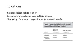
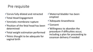
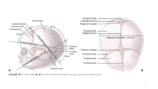
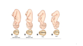
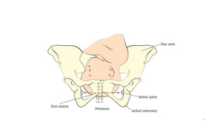
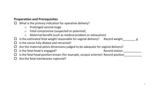
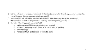
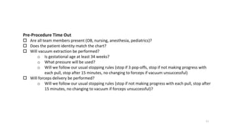
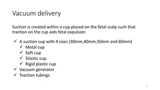
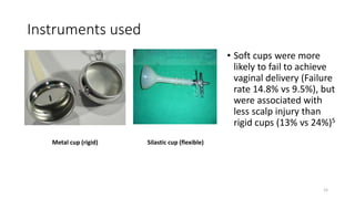
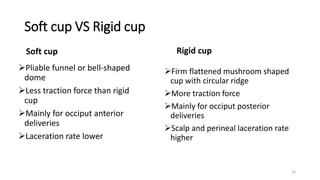
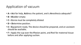
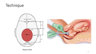
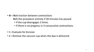
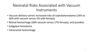
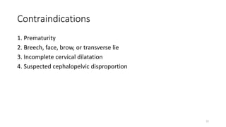
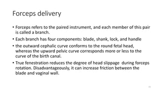
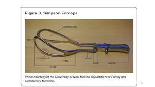
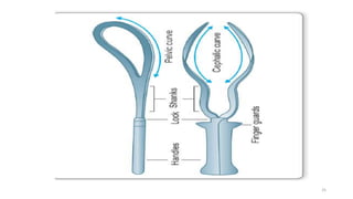
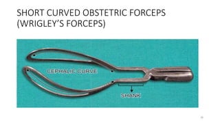
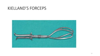
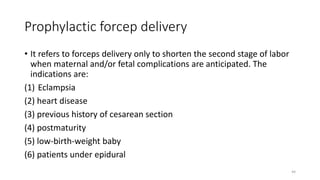
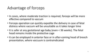
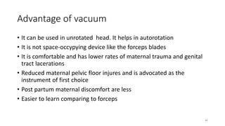
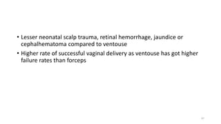
2. Operative vaginal delivery
• Operative vaginal delivery (OVD) is birth accomplished with assistance
from forceps or a vacuum-cup device.
• Once these are applied to the fetal head, outward traction generates
forces that augment maternal pushing to deliver the fetus
1
3. Indications
• Prolonged second stage of labor
• Suspicion of immediate or potential fetal distress
• Shortening of the second stage of labor for maternal benefit
2
4. Pre requisite
Cervix fully dilated and retracted
Fetal Head Engagement
Amniotic membrane rupture
Position of the fetal head has been
determined
Fetal weight estimation performed
Pelvis thought to be adequate for
vaginal birth
Maternal bladder has been
emptied
Adequate Anaesthesia
Consent
Willingness to abandon the
procedure if difficulties occur,
including a plan for proceeding to
cesarean delivery if needed
3
8. Contraindications
• Extreme fetal prematurity
• Fetal demineralizing disease (eg, osteogenesis imperfecta). Fetal
bleeding diathesis
• Unengaged head
• Unknown fetal position
• Brow or face presentation
• Suspected fetal-pelvic disproportion
7
9. Preparation and performance checklist for
operative vaginal delivery
• Using the ACOG bulletin 219 (pre-requisite and indications of
operative vaginal delivery) 2020, SMFM formulated checklist for
operative vaginal delivery under 2 heading
• Preparation and Performance
• Documentation sample
8
14. Vacuum delivery
Suction is created within a cup placed on the fetal scalp such that
traction on the cup aids fetal expulsion
13
A suction cup with 4 sizes (30mm,40mm,50mm and 60mm)
Metal cup
Soft cup
Silastic cup
Rigid plastic cup
Vacuum generator
Traction tubings
16. Instruments used
Metal cup (rigid) Silastic cup (flexible)
• Soft cups were more
likely to fail to achieve
vaginal delivery (Failure
rate 14.8% vs 9.5%), but
were associated with
less scalp injury than
rigid cups (13% vs 24%)5
15
17. Soft cup VS Rigid cup
Soft cup
Pliable funnel or bell-shaped
dome
Less traction force than rigid
cup
Mainly for occiput anterior
deliveries
Laceration rate lower
Rigid cup
Firm flattened mushroom shaped
cup with circular ridge
More traction force
Mainly for occiput posterior
deliveries
Scalp and perineal laceration rate
higher
16
18. Application of vacuum
• A = Ask for help, Address the patient, and is Anesthesia adequate?
• B = Bladder empty
• C = Cervix must be completely dilated
• D = Determine position.
• E = Equipment ready. The device should be prepared, and an assistant
should be available.
• F = Apply the cup over the Flexion point, and Feel for maternal tissue
before and after applying suction.
17
20. • G = Gentle traction
• Traction should be applied steadily and at right angles to the plane of
the cup.
• As the fetal head flexes, passes under the symphysis, and begins to
extend, the vacuum handle will rise from an approximately horizontal
position to an almost vertical position.
• In most cases, traction should be applied only during contractions and
combined with maternal pushing efforts.
19
21. • H = Halt traction between contractions
Halt the procedure entirely if 20 minutes has passed
• if the cup disengages 3 times
• if there is no progress in 3 consecutive contractions.
20
• I = Evaluate for Incision
• J = Remove the vacuum cup when the Jaw is delivered
22. Neonatal Risks Associated with Vacuum
Instruments
• Vacuum delivery carries increased risks of cephalohematoma (14% to
16% with vacuum versus 2% with forceps)
• Retinal hemorrhage (38% vacuum versus 17% forceps), and jaundice.
• Subgaleal hematoma
• Intracranial hemorrhage
21
24. Forceps delivery
• Forceps refers to the paired instrument, and each member of this pair
is called a branch.
• Each branch has four components: blade, shank, lock, and handle
• the outward cephalic curve conforms to the round fetal head,
whereas the upward pelvic curve corresponds more or less to the
curve of the birth canal.
• True fenestration reduces the degree of head slippage during forceps
rotation. Disadvantageously, it can increase friction between the
blade and vaginal wall.
23
30. HOW TO IDENTIFY THE BLADES?
• When articulated: Place the instrument in front of the pelvis with the
tip of the blades pointing upwards and the concave side of the pelvic
curve forward. The blade which corresponds to the left of the
maternal pelvis is the left blade and that to the right side is the right
blade.
• When isolated
(1) The tip should point upwards
(2) the cephalic curve is to be directed inwards and the pelvic curve
forwards.
29
31. Technique
A = Ask for help, Address the patient, and is Anesthesia adequate? An
epidural block or pudendal block are most effective; local anesthesia can be
considered.
B = Bladder empty? Ensure the bladder is not full, as this may lead to
shoulder dystocia and also may lead to bladder injury with an instrument
delivery. If needed, the clinician should perform a straight catheterization.
C = Cervix must be completely dilated.
D = Determine position of the fetal head. Think about shoulder Dystocia.
E = Equipment ready
30
34. 33
• The blades are constructed so that their cephalic curve is closely adapted
to the sides of the fetal head .
• The fetal head is perfectly grasped only when the long axis of the blades
corresponds to the occipitomental diameter
35. 1. The posterior fontanel should be midway between the shanks and 1
cm above the plane of the shanks.
• This ensures the proper flexion of the head to present the narrowest
diameter to the pelvis.
• If the posterior fontanel is higher than 1 cm above the plane of the
shanks, then traction will cause extension of the head, present
greater fetal diameters to the pelvis, and make the delivery more
difficult.
2. The fenestrations should be barely palpable and admit no more than a
fingertip.
• If more than a fingertip is felt, then the blades are not insertedfar
enough to be below the fetal malar eminence and will dig into the
fetal cheeks, potentially causing injury.
34
36. 3. The lambdoidal sutures should be above and equidistant from the
upper or superior surface of each blade.
• This ensures the sagittal suture is in the midline in between the
blades, where it should be to ensure proper forceps application
• When correctly applied (biparietal placement), the blades should be
articulated with ease
• Minor difficulty in locking can be corrected by depressing the handles
on the perineum
• In case of major difficulty, the blades are to be removed, the causes are
to be sought for and the blades are to be reinserted
• The handles should never be forced to lock them
35
37. Difficulty during
• During application of the blades:
(1) Incompletely dilated cervix
(2) Unrotated or nonengaged head
36
(1) Application in unrotated head
(2) Improper insertion of the blade
(not far enough in)
(3) failure to depress the handle
against the perineum
(4) entanglement of the cord or fetal
parts inside the blades.
• Locking:
38. G- Gentle traction and H- Handle elevation
• The birth canal curves through the pelvis from the inlet through the
outlet, and the curve is often described as an arc or a J-shape
• The direction of traction on forceps blades should always be in the
same axis as the pelvic curve for any given station of the head
37
39. • When the fetus is at a +3 (of 5) station, this downward and outward force
will be in the axis of traction and will bring the head down under the
symphysis.
• After the head has come down under the symphysis, the axis of traction
begins to stem upward as the head begins to extend under the symphysis.
Difficulty in traction
(1) Undiagnosed occipitoposterior position
(2) faulty cephalic application
(3) wrong direction of traction
38
40. • The Pajot maneuver consists of having one of the clinician’s hands pulling
on the forceps handles in the same direction that the handles extend
outward and away from the patient
• The other hand should be placed on the shanks, from above or below,
and a downward pull exerted
• Thus, there are two vectors of force: one roughly horizontal outward and
one roughly vertical downward. These vectors summate to a direction of
force that is outward and downward
39
41. I = Evaluate for Incision for episiotomy when the perineum distends.
• Episiotomy may be needed if there is not adequate room for the
physician to safely guide the forceps into the vagina.
• However, episiotomy is usually not indicated and it increases the risk
of anal sphincter lacerations.
J = Remove forceps when the Jaw is reachable.
• The forceps are removed in the reverse order of their application.
• The right blade is removed first by following the curve of the blade up
and over the head anteriorly.
• Then the left blade is removed in a similar fashion.
40
42. Delivery of an OP Position Fetus with
Forceps
• Forceps are applied in usual manner.
• The mechanisms of labor are different with this position extension will
not occur, and further flexion of the head is limited by the symphysis
pubis.
• Horizontal traction is applied to the forceps until the top of the nose
appears beneath the symphysis
• Slow upward motion then exposes the occiput, followed by downward
pressure to deliver the face.
41
43. Maternal complication
• Injury: Vaginal laceration or sulcus tear, cervical tear, extension of
episiotomy to involve the vaginal vault, complete perineal tear
• Nerve injury: Femoral (L2, 3, 4), lumbosacral trunk (L4, 5) with midforceps
delivery
• Postpartum hemorrhage may be—(i) traumatic or (ii) atonic, requiring
blood transfusion or (iii) both, may cause shock
• Infections
• Pelvic floor disorders : Painful perineal scars, dyspareunia, low backache,
genital prolapse, urinary incontinence and anal sphincter dysfunction
42
45. Prophylactic forcep delivery
• It refers to forceps delivery only to shorten the second stage of labor
when maternal and/or fetal complications are anticipated. The
indications are:
(1) Eclampsia
(2) heart disease
(3) previous history of cesarean section
(4) postmaturity
(5) low-birth-weight baby
(6) patients under epidural
44
46. Advantage of forceps
• In cases, where moderate traction is required, forceps will be more
effective compared to vaccum
• Forceps operation can quickly expedite the delivery in case of fetal
distress where vaccum will be unsuitable as it takes longer time
• It is safer at any gestational age baby (even < 36 weeks). The fetal
head remains inside the protective cage
• It can be employed in anterior face or in after-coming head of breech
presentation, where vaccuum is contraindicated
45
47. Advantage of vacuum
• It can be used in unrotated head. It helps in autorotation
• It is not space-occypying device like the forceps blades
• It is comfortable and has lower rates of maternal trauma and genital
tract lacerations
• Reduced maternal pelvic floor injures and is advocated as the
instrument of first choice
• Post partum maternal discomfort are less
• Easier to learn comparing to forceps
46
48. • Lesser neonatal scalp trauma, retinal hemorrhage, jaundice or
cephalhematoma compared to ventouse
• Higher rate of successful vaginal delivery as ventouse has got higher
failure rates than forceps
47
49. References
• Williams obstetrics 26e
• Arias Practice of High Risk Pregnancy and Delivery 4e
• ACOG Practice Bulletin 219 : Operative vaginal delivery
• SMFM Special Statement: Operative vaginal delivery: checklists for
performance and documentation
• Uptodate
48
Non re asurring NST, Thick MSL, Abruptio
Severe or acute pulmonary compromise, decompensation
from intrapartum infection, neurological disease, and serious cardiac
disorders
Regional anaesthesia is preferred for mid cavity IVD, that is when the head is engaged but the station is above 1 2 cm but below the ischial spines.10 For low cavity IVD when the vertex is beyond 1 2cm but not reached the pelvic floor, regional or pudendal block anaesthesia with local infiltration of the perineum is acceptable
(The head is engaged when the widest diameter [the biparietal diameter] has reached or passed through the pelvic inlet. This typically occurs when the leading bony part has reached or passed through the ischial spines)
The safety of forceps or vacuum birth has not been established in disorders that result in demineralization of the skull. There is a theoretic risk for intracranial bleeding, extracranial bleeding, and other brain injuries due to cranial deformation or fracture from these instruments.
In the United States, vacuum extractor is the preferred term, whereas in Europe it is commonly called a ventouse
A Cochrane analysis of data from nine trials including 1375 women concluded that……..
Rigid cups tended to be more suitable for occiput posterior, occiput transverse, and difficult occiput anterior position deliveries given their ability to stay attached despite strong traction, while soft cups appeared to be more appropriate for uncomplicated occiput anterior extractions where less traction is needed and thus the excess risk of scalp injury could be avoided. Most failures were due to cup detachment.
that the rate of cephalohematomas and subgaleal hemorrhage was similar between soft and rigid cups
Rate of cephalohematomas and subgaleal hemorrhage similar with both , soft cup------------reusable,
Success rate higher with metal cup but greater rates of scalp injuries including cephalohematomas
flexion point: when apply pressure to it head flexes if you apply about anterior fontanelle head may get extenddd and cause trauma
0.2 kg/cm2 every 2 minutes until a total negative pressure of 0.8 kg/cm2 is reached
Lower the pressure if contraction in present (prevent fetal scalp bleeding)
Third- and fourth-degree perineal lacerations are grouped as obstetrical anal sphincter injuries (OASIS).